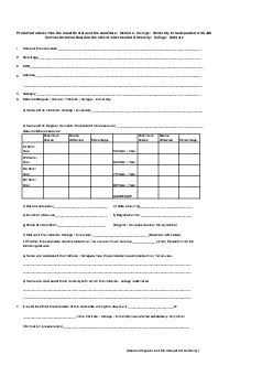
headache dizziness nausea vomiting extreme tiredness and fever The patient had been taking prednisone for a relapse of chronic ulcerative colitis On examination the physician noted that the patient had ID: 742596
Download Presentation The PPT/PDF document "Naegleria Fowleri A 45-year-old man ..." is the property of its rightful owner. Permission is granted to download and print the materials on this web site for personal, non-commercial use only, and to display it on your personal computer provided you do not modify the materials and that you retain all copyright notices contained in the materials. By downloading content from our website, you accept the terms of this agreement.
Slide1
Naegleria
Fowleri
Slide2
A 45-year-old man who had been feeling unwell for several months visited his internist complaining of
headache, dizziness, nausea, vomiting, extreme tiredness, and fever
. The patient had been taking prednisone for a relapse of chronic ulcerative colitis. Slide3
On examination, the physician noted that the patient had
nuchal rigidity and appeared confused
. He performed a lumbar puncture. CSF was sent to the laboratory for bacterial and viral cultures. The Gram stain showed
many neutrophils but no bacteria
. Slide4
Nuchal rigidity – inability to flex forward the neck musclesSlide5
To rule out amebic encephalitis, the physician asked that a wet mount be prepared from the patient
’
s CSF. Microscopic examination of this preparation revealed
motile amebic trophozoites
. Cultures were negative for bacteria and viruses. Slide6
A biopsy specimen containing the parasite causing this patient
’
s infection is shown
inSlide7Slide8
QUESTIONS
1. Which ameba would you expect to be causing this patient
’
s infection? What is the name of this infection? Slide9
Answer
Acanthamoeba spp.
granulomatous amebic encephalitis (GAE)
Once infected, a person may suffer with headaches, stiff neck, nausea and vomiting, tiredness, confusion, lack of attention to people and surroundings, loss of balance and bodily control, seizures, and hallucinations. Signs and symptoms progresses over
several weeks
and death usually occurs.Slide10
QUESTIONS
2. Which ameba may cause a more serious and acute CNS infection and may be confused with this parasite? Slide11
Answer
Naegleria fowleri
Infection with
Naegleria
causes the disease
primary amebic meningoencephalitis (PAM)
, a brain inflammation, which leads to the destruction of brain tissue.
Initial signs and symptoms of PAM include headache, fever, nausea, vomiting, and stiff neck. As the ameba causes more extensive destruction of brain tissue this leads to confusion, lack of attention to people and surroundings, loss of balance and bodily control, seizures, hallucinations. The disease progresses rapidly and infection usually results in death within
3 to 7 days
. Slide12
QUESTIONS
3. How can you distinguish between these amebae? Slide13Slide14Slide15
QUESTIONS
4. How do the infections caused by these two parasites differ? Slide16Slide17
Answer
PAM occurs in persons who are generally healthy prior to infection. Central nervous system involvement arises from organisms that penetrate the nasal passages and enter the brain through the cribriform plate. The organisms can multiply in the tissues of the central nervous system and may be isolated from spinal fluid. In untreated cases death occurs within 1 week of the onset of symptoms. Slide18Slide19
Answer
GAE occurs in persons who are immunodeficient in some way; the organisms cause a granulomatous encephalitis that leads to death in several weeks to a year after the appearance of symptoms. The primary infection site is thought to be the lungs, and the organisms in the brain are generally associated with blood vessels, suggesting vascular dissemination. Prior to 1985 amoebae had been reported isolated from diseased eyes only rarely; cases were associated with trauma to the eye. In 1985-1986, 24 eye cases were reported to CDC and most of these occurred in wearers of contact lenses. It has been demonstrated that many of these infections resulted from the use of home-made saline solutions with the contact lenses.Slide20
QUESTIONS
5.
How is the laboratory diagnosis of this infection made? Slide21
Answer
In Naegleria infections, the diagnosis can be made by microscopic examination of
cerebrospinal fluid (CSF)
. A wet mount may detect
motile trophozoites
, and a Giemsa-stained smear will show trophozoites with typical morphology.Slide22
Answer
In Acanthamoeba infections, the diagnosis can be made from microscopic examination of stained smears of
biopsy specimens (brain tissue, skin, cornea) or of corneal scrapings
, which may detect
trophozoites and cysts
. Cultivation of the causal organism, and its identification by direct immunofluorescent antibody, may also prove useful.Slide23
QUESTIONS
6. Does the ameba causing CNS infection in this patient cause other types of infections? Slide24
Answer
Acanthamoeba
can enter the
skin
through a
cut, wound, or through the nostrils
. Once inside the body, amebas can travel to the
lungs
and through the bloodstream to other parts of the body, especially the
central nervous system (brain and spinal cord)
.Slide25
Answer
Through improper storage, handling, and disinfection of contact lenses,
Acanthamoeba
can enter the
eye
and cause keratitis resulting
in superficial
corneal abrasions.Slide26
KeratitisSlide27
QUESTIONS
7. Which other free-living ameba, recently placed in the same genus as this parasite, causes a CNS infection in humans? Slide28
Answer
Acanthamoeba culbertsoni
Acanthamoeba polyphaga
eyes infection
Acanthamoeba castellanii
Acanthamoeba palestinensis
CNS infection
Acanthamoeba astronyxis
CNS infection
Acanthamoeba hatchetti
eyes infection
Acanthamoeba rhysodesSlide29
QUESTIONS
8. Why is there no satisfactory treatment available to treat this infection? Slide30
Answer
Treatment with
sulfamethazine
may be effective in controling
Acanthamoeba spp.
The following agents have been used to successfully eliminate the amoebic infection in the eye:
ketoconazole, microconazole, and propamidine isothionate
; however,
penetrating keratoplasty
has been necessary to restore useful vision.Slide31
Keratoplasty
– surgery to the cornea
Penetrating
keratoplasty
– corneal transplant Slide32
Naegleria fowleri
Morphology , Biology and Life cycle:
flagellate form
*
Life cycle stage consist
: -motile
trophozoite
:
-
nonmotile
cysts
ameboid
form
*
Reproduction
: simple binary fission
*
Ameboid
form
:
found in tissue , forms a single pseudopod,
dimensions 7 by 20
μ
m,
With a nucleus contain a large central
karyosome
*
Flagellate form
:
with two flagella, pear-shaped, do not
divide
*
Cyst form
:
uninucleate
, circular 7-10
μ
m
in diameter, nucleus is similar to
troph
.
Slide33
Naegleria formsSlide34
Naegleria cyst & trophozoiteSlide35
Life cycle Slide36
Symptoms and pathogeesis
Primary Amebic Meningoencephalitis(P.A.M
.) :
Symptoms
;
headache, fever, nausea
and vomiting accompanied by signs
of meningitis with involvement of the
olfactory, frontal, temporal, and
cerebral areas
Death
: occurs early; the entire
clinical course seldom extends
beyond 3 to 6 days.Slide37
Acanthamoeba
( Hartmanella
) spp.
Morhology, Biology and Life cycle:
These amebae are similar in appearance to the ameboid stage of
Naegleria but have no flagellate stage.
Cyst & Trophozoite may be found in tissue, but cysts are never seen in
Naegleria
infections.
Pseudopods are acanth formsSlide38
Acanthamoeba trophozoiteSlide39
Thanks for your attention