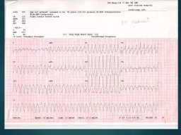
/
 Ventricular Tachycardia in Structural Heart Disease  Ventricular Tachycardia in Structural Heart Disease

Ventricular Tachycardia in Structural Heart Disease - PowerPoint Presentation

tatiana-dople
tatiana-dople . @tatiana-dople
Follow
348 views
Uploaded On 2020-04-06

Ventricular Tachycardia in Structural Heart Disease - PPT Presentation

Dr Sanmath Shetty K DM Cardiology Resident Calicut Medical College Overview Premature Ventricular Complexes PVCs VT in coronary artery disease VT in Dilated Cardiomyopathy Bundle Branch Reentrant BBR ID: 776093

patients ventricular qrs scd patients ventricular qrs scd disease conduction heart mortality cardiac icd therapy due class risk arrhythmias

Share:

Link:

Embed:

Download Presentation from below link

Download Presentation The PPT/PDF document " Ventricular Tachycardia in Structural H..." is the property of its rightful owner. Permission is granted to download and print the materials on this web site for personal, non-commercial use only, and to display it on your personal computer provided you do not modify the materials and that you retain all copyright notices contained in the materials. By downloading content from our website, you accept the terms of this agreement.


Presentation Transcript

Slide1

Ventricular Tachycardia in Structural Heart Disease

Dr

Sanmath

Shetty K

DM Cardiology Resident

Calicut Medical College

Slide2

Overview

Premature Ventricular Complexes (PVCs

)

VT in coronary artery

disease

VT in Dilated

Cardiomyopathy

Bundle Branch Reentrant (BBR)

VT

Arrhythmogenic

right ventricular dysplasia (ARVD)

VT in Hypertrophic Cardiomyopathy

VT long after repair of congenital heart

disease

VT in patients with LV assist devices

Slide3

Premature Ventricular Complexes (PVCs)

Premature impulses (complexes) that originate in the ventricles.Usually benign. Mechanisms:Extrasystoles: More frequent; induced by a mechanism related to the preceeding QRS complex. Most commonly due to reentry; less often induced by post potentials (triggered activity).Fixed or nearly fixed coupling interval. Parasystoles:Less frequent; independent of baseline rhythm.Due to presence of unidirectional entrance block in the parasystolic focus.Varying coupling intervals, interectopic intervals are multiples of each other and presence of fusion complexes.

Slide4

Extrasystole

-

Trigeminy

Parasystole

Slide5

Lown Classification 1971According to prognostic significance (Holter ECG)

Grade 0: No PVC

Grade 1: < 30/

hr

Grade 2: > 30/

hr

Grade 3: Polymorphic PVC

Grade 4a: In pairs

Grade 4b: Runs of monomorphic VT

Grade 5: R on T phenomenon

Slide6

Electrocardiographic forms of presentation

Usually shows a compensatory pause(BC=2xAB)PVC usually fails to discharge SA node.PVC discharges SA node, non complete compensatory pause(BC<2xAB).At slow sinus rates, it enters AV junction leaving it in refractory period but does not prevent the next sinus impulse from being conducted towards the ventricles ( with a longer PR interval)--- interpolated PVC.

Slide7

Morphologies of PVCs

In individuals with no evidence of heart disease: High voltage, unnotched QRS complexes.ST segment depression when QRS positive and vice versa.T wave has asymmetrical branches.In individuals with heart disease:QRS complexes present notches and slurrings and are of low voltage.Symmetrical T waves.

Slide8

Site of origin

RV

ectopics

: LBBB pattern

LV

ectopics

: RBBB pattern

Superior axis: location in or near posterior division of left BB.

Rightward axis: location in or near anterior division of left BB.

Ectopics

from base: positive QRS complexes in precordial leads.

Ectopics

from apex: negative QRS complexes in precordial leads.

QRS duration depends on:

Site of origin

Characteristics of tissues activated by the premature impulse

Coupling time (QRS wider with short coupling interval)

Slide9

VT in structural heart disease

History (

eg

: history of CAD, heart failure, cardiac surgery).

Physical examination

ECG:

Baseline: abnormal Q waves, fragmented QRS complexes, IVCD, poor R wave progression.

During VT: slurring of initial forces, lower amplitude and notching of QRS complexes.

Slide10

VT in structural heart diseaseMechanisms

VT arises distal to the bifurcation of the His bundle in the specialized conduction system, ventricular muscle, or combinations of

both.

Disorders

of impulse formation

Enhanced

automaticity

Triggered activity

Disorders

of impulse conduction

Re-entry

(circus movements)

Slide11

VT in coronary artery disease

Incidence of VT varies according to the type of ACS.

GUSTO- 1 trial: 41,000 patients with STEMI treated with thrombolysis.

VT

3.5

%.

Pooled analysis of 4 major trials in patients with UA/NSTEMI:

VT- 0.8

%.

Al-

Khatib

SM, Granger CB, Huang Y, et al: Sustained ventricular arrhythmias among patients with acute coronary syndromes with no ST-segment elevation: Incidence, predictors, and outcomes. Circulation 2002;106:309.

Clinical presentation – tolerated sustained VT to SCD.

Slide12

SMVT within first 2 days of MI – 3% of cases

Associated with increased in hospital mortality as against those without arrhythmias.

Mortality not increased at 1 year in

30

day survivors.

D

uring

subacute

/ healing phase of MI ( > 2 days)

Associated with reduced LVEF and is a predictor of worse prognosis.

SMVT within 3 months following MI – 40-50% mortality at 2 years.

Predictors of increased mortality-

Anterior wall MI

Frequent episodes of sustained and/or

nonsustained

VT

Heart failure

Multivessel

coronary disease, particularly in individuals with residual ischemia.

D

uring

chronic phase:

Median time: 3 years; can first occur

upto

10-15 years after MI.

Annual mortality : 5 – 15% .

Slide13

Mechanisms of VT in CAD

All arrhythmia mechanisms can converge in VT associated with CAD.

Reentry: VT associated with MI scar.

Automaticity: VT arising from ischemic border during acute ischemia.

Trigerred

activity: VT arising during ischemia due to delayed or early after depolarization.

Slide14

During acute ischemia

Acute ischemia activates ATP sensitive K channels causing increase in extracellular K along with acidosis and hypoxia in cardiac muscle.

Increased extracellular K

greater resting depolarization

decreased conduction velocity

shortening of action potential duration

prolongation of effective refractory period (

postrepolarization

refractoriness)

Increase

in extracellular K depolarizes the RMP causing increase in tissue excitability

.

Injury current flows between ischemic and non ischemic cells at border zone promoting focal activity in normal tissue

.

Polymorphic VT due to

microentry

.

Single reentrant

wavefront

splits into multiple wavelets when it enters surrounding

nonischemic

tissue (shorter effective refractory period).

Slide15

Healing phases of MI

95% of these VTs due to reentry.Two conditions essential for reentry:Unidirectional block of conduction.Circuit cycle longer than any of the refractory periods throughout the cycle.Unidirectional block:Anatomical : discontinuities in ventricular muscle, branching strands of slow conduction or tissue discontinuation due to gap junction abnormalities present in the areas of MI scar.Functional : due to dispersion of refractoriness.

Slide16

The substrate for VT develops gradually over 2 weeks following a MI.

r

emains

indefinitely once formed.

Triggers:

Surges in autonomic tone

Electrolyte imbalance

Acute ischemia

Acute heart failure decompensation

Slide17

MI Scar-Related Sustained Monomorphic VT Circuit

Slide18

Double loop “figure of 8” model

Isthmus: region of slow conduction within the scar.Target site for ablation.Proximal and distal isthmus sites are the entrance and exit respectively.The exit site is the point where the activation wavefront leaves the circuit to depolarize the ventricles.Determines VT morphology.Outer loop connects entry and exit point by a lateral pathway around the border of the scar. Inner loop connects by a protected pathway within the scar.Bystander sites are passively activated ; not integral to the circuit.

Slide19

Clinical presentation

Mild symptoms (palpitations).

Symptoms of

hypoperfusion

(light headedness, altered sensorium,

presyncope

, syncope).

Exacerbation of angina and heart failure.

Sudden collapse.

Hemodynamic consequences depend on:

Ventricular rate

Duration of VT

Presence and extent of LV dysfunction

Loss of

atrioventricular

synchrony

Slide20

ECG features suggesting VT related to old MI

Presence of Q waves (

qR

, QR or

Qr

) in related leads.

Notched or wide QRS complexes.

Low QRS voltage.

Multiple ventricular tachycardia morphologies

.

Slide21

Identifying the site of origin of VT in CAD

ECG tends to locate reentry circuit exit rather than site of VT origin.

Helpful tool to guide mapping and ablation during EP.

Localisation

must be done in 3 axis:

Septal vs lateral walls

Superior vs inferior walls

Apical vs basal regions

Slide22

Identifying the site of origin of VT in CADLateral vs Septal

Lateral wall VTs:RBBB patternWider QRSSequential activation of 2 ventricles.Septal VTs:LBBB patternNarrower QRSParallel activation of both ventricles.Early engagement of HPS.

Slide23

Identifying the site of origin of VT in CADSuperior vs Inferior walls

QRS axis in inferior leads.Superior axis: QRS negative in inferior leads.Inferior axis: QRS positive in inferior leads.IWMI: 80% have superior axis.AWMI: 55% have superior axis. 45% have inferior axis.

Slide24

Identifying the site of origin of VT in CADBasal vs Apical regions

QRS polarity in precordial leads.VTs from base: positive concordant pattern.VTs from apex: negative concordant pattern.

Slide25

Epicardial origin

Rare in post MI VT, less than 2% of all cases.More common in DCM (one third) and Chagas disease (70%).Epicardial origin of ventricular activation widens the initial part of QRS complex – pseudo delta wave.ECG intervals of ventricular activation that suggest an epicardial origin of the VT:pseudo–delta wave (measured from the earliest ventricular activation to the earliest fast deflection in any precordial lead) of 34 milliseconds or more [sensitivity- 83% and specificity- 95%]intrinsicoid deflection time in V2 (measured from the earliest ventricular activation to the peak of the R wave in V2) of more than 85 milliseconds [sensitivity- 87% and specificity- 90%]shortest RS complex duration (measured from the earliest ventricular activation to the nadir of the first S wave in any precordial lead) of 121 milliseconds or more [sensitivity- 76% and specificity- 85%]QRS duration is more than 200 milliseconds.

Slide26

Principles of Management

Acute Management:

VTs with hemodynamic compromise: DC version

.

Medical management:

Amiodarone

drug of choice.

Procainamide and

sotalol

are alternatives.

Lidocaine less effective in the absence of ischemia.

Beta blockers offer additional benefit

.

Treatment of underlying conditions

(

eg

: acute ischemia, decompensated heart failure, electrolyte abnormalities)

Slide27

Long term management

Slide28

Long term management

Prevention of

SCD- ICD implantation.

Adjunctive antiarrhythmic therapy

Reduce the frequency of ventricular arrhythmia in patients with unacceptably frequent ICD therapy

Reduce the rate of VT so that it

is better

tolerated hemodynamically and more amenable to pace termination or low-energy cardioversion

Suppress other arrhythmias (e.g., sinus tachycardia, AF,

nonsustained

VT) that cause symptoms or interfere with ICD

function resulting

in inappropriate

discharges.

Catheter ablation of post-MI VT: 2 indications

Recurrent VT causing frequent ICD shocks and refractory to antiarrhythmic medications

VT storm or incessant VT refractory to antiarrhythmic medications.

Slide29

Slide30

Long term managementSecondary Prevention of SCD

Slide31

Long term managementPrimary Prevention of SCD in Ventricular Arrhythmias

TRIAL

CONTROL

NO OF PTS

POPULATION

MEAN

FOLLOW UP(

mths

)

MORTALITY%

CONTROL

MORTALITY%

ICD

P VALUE

MADIT

Anti arrhythmic therapy

196

Prior

MI; LVEF < 35%, asymptomatic NSVT

27

39

16

0.02

CABG-PATCH

Anti arrhythmic therapy

900

For CABG: LVEF < 35%. Positive

SAECG

32

21

22

0.64

MUSTT

Conventional therapy

704

Prior MI; LVEF < 40%; NSVT, inducible VT on EP study

39

48

24

0.001

MADIT II

Conventional therapy

1232

Prior MI; LVEF < 30%

20

20

14

0.007

DINAMIT

Conventional therapy

674

Recent

MI (within 6-40 d), LVEF < 35%; impaired heart rate variability

39

18

17

0.66

Slide32

Guidelines for ICD in CAD Secondary prevention

ICD therapy is indicated in patients who are survivors

of cardiac

arrest due to VF or hemodynamically unstable

sustained VT

after evaluation to define the cause of the event

and to

exclude any completely reversible causes.

(Class I; LOE A)

Patients experiencing cardiac arrest due to VF 48

hrs

after MI must be optimally evaluated and treated for ischemia.

Evidence of ischemia – complete coronary revascularization.

ICD if revascularization is not possible and there is significant LV dysfunction.

Slide33

Guidelines for ICD in CAD Primary prevention

Class I

:

P

atients

with LVEF less than

or equal

to 35% due to prior MI who are at least 40 days

post-MI and

are in NYHA functional Class II or III. (

LOE: A).

P

atients

with LV dysfunction due

to prior

MI who are at least 40 days post-MI, have an LVEF less than or equal to 30%, and are in NYHA functional Class I. (

LOE:

A

).

P

atients

with

nonsustained

VT

due to

prior MI, LVEF less than or equal to 40%, and inducible VF

or sustained

VT at electrophysiological study. (

LOE: B).

Slide34

VT in Dilated Cardiomyopathy

Multiform VPCs, ventricular pairs, NSVT- 80%-95% DCM patients.

Ventricular arrhythmias more frequent and complex as LV function deteriorates.

NSVT 15%-20% in NYHA I/II to 50%-70% in NYHA IV.

VT may arise in the myocardium or may be through

macroentrant

circuit ( BBR- VT).

BBR-VT ---

Responsible for VT in up to 41% of

DCM.

Slide35

Slide36

Predictors of arrhythmia and mortality

Clinical predictorsSeverity of LV dysfunctionAs CHF symptoms worsen,Risk of total mortality, sudden death and CHF death increases.Ratio of sudden death to CHF death decreases.Once pt develop class IV symptoms, EF less valuable in predicting mortality.Syncope 1 yr SCD rates increases from 12% to 45% when syncope is present.

Slide37

Laboratory values:

Low serum sodium

High plasma norepinephrine, renin and ANP,BNP levels.

ECG predictors:

LBBB

First and second degree AV block

Predictive testing with EP in DCM patients not associated with CAD is controversial.

Presence of polymorphic VT on EPS does not predict risk for SCD.

Induction of sustained monomorphic VT identifies high risk population.

Lack of inducible VT does not predict freedom from sudden death.

Slide38

Effect of HF therapy on ventricular arrhythmias

Beta blockers: substantial part of the survival benefit seen is due to a significant reduction in SCD.

Slide39

Effect of HF therapy on ventricular arrhythmias

ACEI and ARBs: improved survival; conflicting data with reduction in SCD.

CONSENSUS, SOLVD, SAVE – little or no reduction in SCD.

V-

HeFT

, TRACE, AIRE – significant reduction in SCD.

Aldosterone antagonists: Reduce overall mortality and SCD in advanced HF.

Reduction in aldosterone effect on the heart

Maintenance of higher potassium levels

Digoxin and other inotropes:

Proarrhythmic

effect

DIG trial: no net mortality benefit, apparent increase in mortality from arrhythmias ( not statistically significant).

Slide40

Antiarrhythmics:

Amiodarone

:

Initial trials GESICA: mortality benefit.

SCD-

HeFT

: mortality not reduced compared to placebo.

Recommended only for reducing the frequency of shocks in patients with recurrent ventricular arrhythmias (Class

IIa

).

Slide41

Primary prevention with ICD

Slide42

ICD

Primary prevention of SCD:

Patients

with

nonischemic

DCM who

have an LVEF less than or equal to 35% and who are

in NYHA

functional Class II or

III. ( Class I; LOE B).

Patients

with

unexplained syncope

, significant LV dysfunction, and

nonischemic

DCM

. ( Class

IIa

; LOE C).

Patients

with

nonischemic

DCM

who have an LVEF of less than or equal to

35% and

who are in NYHA functional Class I

. (Class

IIb

; LOE C).

Secondary prevention:

ICD is the preferred treatment in DCM patients with resuscitated cardiac arrest from VT/VF.

Slide43

Bundle Branch Reentrant (BBR) VT

Only reentrant VT with a well-defined reentry circuit.The right bundle branch (RB) and left bundle branch (LB) obligatory limbs of the circuit.Connected proximally by the His bundle (HB) and distally by the ventricular septal myocardium.Cannot be induced in patients with normal His Purkinje system (HPS)Electrophysiological properties of normal HPS- very fast conduction velocity and a relatively long refractory period precludes formation of a stable circuit.

Slide44

Epidemiology

6

% of induced sustained monomorphic VT.

Additional myocardial VTs in 25% patients.

Commonly seen in patients with DCM.

DCM anatomic substrate in 45% of BBR-VT ; 41% of all VTs in

DCM patients

is BBR-VT.

Also seen in

Ischemic cardiomyopathy (incidence 4.5 - 6%).

Valvular

heart disease

Aortic or mitral valve surgery can facilitate BBR-VT- close proximity of HPS to

valvular

annuli.

Ebstein’s

anomaly.

Hypertrophic cardiomyopathy.

Myotonic

dystrophy.

Conduction anomalies associated with sodium blockade with

flecainide

.

Slide45

C

hanges from normal physiology for BBR to be sustained:

Anatomically longer reentrant pathway (dilated heart)

Slow conduction in HPS (HPS disease)

Sufficient prolongation of conduction time to allow expiration of refractory period of HPS.

Slide46

Types of BBR-VT

LBBB morphology is commoner.Type A and C are classical BBR-VTs. Type B is most commonly seen in CAD especially those with AWMI with LAF or LPF block.

Slide47

Clinical presentation

Typically unstable.

Very rapid ventricular rates (200-300/min) and poor underlying ventricular function.

75% present with syncope or cardiac arrest.

Baseline ECG

:

NSR or Atrial fibrillation.

Nonspecific IVCD and PR prolongation – most common ECG abnormality.

Typical bundle branch patterns may also be seen.

Rarely narrow baseline QRS complex- suggesting role of functional conduction delay.

ECG during VT:

Typical BBB pattern, may resemble that seen in NSR. LBBB>RBBB. Usually leftward axis.

Rapid

intrinsicoid

deflection in right precordial leads.

Initial ventricular activation through HPS, not ventricular muscle.

Slide48

EP testing

Prolonged HV interval invariably present in sinus rhythm.

Some patients with normal HV interval manifest as HV interval prolongation or split HB potentials during atrial programmed stimulation or burst pacing

.

Tachycardia Induction:

VES from RV apex usual method.

Dependent on achievement of critical conduction delay in HPS following VES.

At longer coupling intervals, retrograde conduction occurs through RB. At shorter coupling intervals, retrograde block occurs in RB.

Retrograde conduction occurs via LB causing long V2-H2 interval.

Further shortening of coupling intervals, increased retrograde LB delay allowing anterograde conduction of the RB ( beat with wide QRS LBBB pattern- BBR beat or V3 phenomenon).

Slide49

EP testing

Tachycardia features:

AV dissociation usually present; 1:1

ventriculoatrial

conduction may occur.

His potential precedes the QRS.

HV interval during BBR similar or longer than that during baseline.

Spontaneous variations in V-V intervals are

preceeded

and dictated by similar changes in H-H intervals.

Termination of VT with block in HPS.

Inability to induce VT after ablation of right or left bundle branch.

Slide50

Slide51

Interfascicular VT: HV interval during tachycardia usually shorter by more than 40 msec than that recorded during sinus rhythm.LB potential inscribed before His potential.

Slide52

Treatment

Pharmacological therapy usually ineffective.

RFA of a bundle branch first line therapy.

RB ablation easier; method of choice.

LB ablation preferable in patients with conduction system disease such that conduction down the LB is inadequate to maintain 1:1 conduction.

Mere presence of LBBB on ECG does not mean complete block in LB.

Pacemaker implantation indicated when

infrahisian

AV block is demonstrated during atrial pacing or when

postablation

HV interval > 100

msecs

.

Varies from 10-30%.

Prophylactic pacemaker in

myotonic

dystrophy patients in view of progressive nature of the conduction system disease.

Recurrence rare. Mortality after successful ablation mostly due to progressive heart failure and associated myocardial VTs (25%).

Treatment: ICD

with or without CRT

capabilities.

Slide53

Arrhythmogenic right ventricular dysplasia(ARVD)

Progressive disease in which normal myocardium is replaced by fibrofatty tissue.Usually involves the RV; LV and septum may also be involved.Predominantly involves the “ Triangle of Dysplasia”.Occurs in young adults (80% in less than 40 years) and more common in males.Prevalence 0.02 to 0.1%.

Slide54

Pathogenesis

Several proposed theories.

Familial inheritance- autosomal dominant or recessive.

Metabolic disorder affecting RV.

Infectious or immunological cause.

Mutations in

desmosomal

proteins-

desmoglein

,

desmoplakin

,

desmocollin

,

plakoglobin

,

plakophilin

.

Autosomal recessive inheritance

Familial

palmoplantar

keratosis, Naxos disease, mal de

Meleda

disease.

Hyperkeratosis of palms and soles, woolly hair.

Cardiac anomalies- 100 % penetrant by adolescence- RV involvement 100%, LV involvement 27%.

Slide55

Mechanism of arrhythmia

Slide56

Clinical presentation

Fatigue, atypical chest pain, palpitations, syncope or sudden cardiac death.

Ventricular arrhythmias in ARVD- 23% (mild disease) to 100% (severe disease).

Occur during exercise.

Patients with ARVD with increased risk of SCD:

Younger patients

Patients with recurrent syncope

Patients with previous history of cardiac arrest or VT with hemodynamic compromise

Patients with LV involvement

Patients with ARVD2 and Naxos disease

Patients with an increase in QRS dispersion

Slide57

ECG abnormalities

Slide58

Slide59

Echocardiography:

Dilation of the RV and RV dysfunction (Revised task force criteria)localised aneurysms in diastoledyskinesis in the inferior basal region.RV angiography: Findings: infundibular aneurysms, trabeculae thicker than 4mm “deep fissures”, prominent moderator band, diastolic bulging of the subtricuspid area, mild tricuspid regurgitation.Cardiac MRI:Abundant epicardial adipose tissue, prominent trabeculations, scalloped appearance of RV free wall and intramyocardial fat deposits.Endomyocardial Biopsy: Gold Standard; lacks sensitivity (67%). Performed from septum; changes more pronounced in free wall

Slide60

Management

Pharmacological therapy:

Beta blockers,

sotalol

and

amiodarone

.

Class

Ia

and

Ib

drugs ineffective.

Radiofrequency ablation:

Frequently unsuccessful and may need multiple attempts.

Progressive nature of the disease and diffuse yet patchy nature (multiple

arrhythmogenic

foci)

Fontaine et al. reported success rates of 32%, 45% and 66% after one, two or three ablation sessions in 50 patients.

ICD:

Patients with high risk of SCD.

Those resuscitated from cardiac arrest, history of syncope or life threatening arrhythmias not completely suppressed by drug therapy.

Problems with ICD:

Areas of RV myocardium thin and non contractile – penetrated during RV lead placement leading to

tamponade

.

Fibrofatty

nature of RV – device inadequately sensing arrhythmias.

Slide61

VT in HCM

Highly variable natural history.

Beta myosin heavy chain mutations: relationship between severity of LVH and risk of SCD.

Troponin mutations: high risk of SCD irrespective of LVH.

Mortality rates :

1%/yr.

SCD : 0.2%/yr.

SCD

usually

in patients with mild

or no symptoms.

common in adolescents and young adults before the age of 30-35 yrs.

Predominant mechanism of SCD: VT/VF

Other mechanisms:

asystole

, rapid atrial fibrillation, electrical mechanical dissociation.

Slide62

Sudden cardiac deathRisk factors

Major risk factors

Prior personal history of sudden cardiac death or out-of-hospital cardiac arrest

Spontaneous sustained ventricular tachycardia or ventricular fibrillation

Family history of sudden cardiac death

Extreme left ventricular hypertrophy (>30 mm)

Nonsustained

ventricular tachycardia

Abnormal blood pressure response to exercise

Recent, unexplained syncope

Delayed gadolinium enhancement on cardiac magnetic resonance

imaging*

Presence of LVOT obstruction is not a sole risk factor for SCD.

EP study not shown to be of benefit for risk stratification in HCM

.

Slide63

Prevention of SCD

ICD:

P

atients

with HCM who

have 1

or more

major

risk factors for SCD.

(Class

IIa

; LOE:

C)

Pharmacologic therapy:

amiodarone

obsolete strategy; lacks proven efficacy.

Likelihood of side effects during the long risk period typical of young patients with HCM.

Slide64

VT long after repair of congenital heart disease

VT accounts for 38% of wide complex

tachycardias

in patients with congenital heart disease.

VT late after repair occur in those with TOF and VSD.

Predictors for sustained VT:

QRS duration >180msec, rapid increase in QRS duration after repair, dispersion of QRS duration on ECG, increased QT interval dispersion, complete heart block, older age at surgery (>10yrs), presence of RVOT patch, RVOT aneurysm, increased RV pressures, pulmonic or tricuspid regurgitation.

Monomorphic and

macrorentrant

, rotating clockwise or anticlockwise around

myotomy

scars or surgical patches.

Slide65

VT in patients with LV assist devices

Not uncommon owing to significant underlying structural heart disease.

De novo monomorphic VT may occur after LVAD is implanted.

60% suffer from monomorphic VT after implantation of LVAD.

Majority have an exit site close to the region of the inflow cannula at the LV apex.

Slide66

THANK YOU