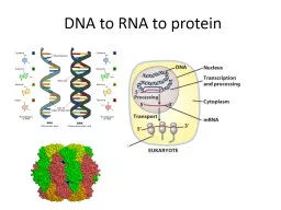
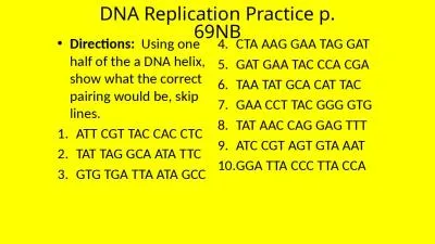
Chapters 16 and 17 Before the end of the semester we will be covering Historical DNA experiments Structure of DNARNA DNA Replication Protein Synthesis Transcription and Translation Mutations ID: 461583
Download Presentation The PPT/PDF document "DNA, RNA, and Protein Synthesis" is the property of its rightful owner. Permission is granted to download and print the materials on this web site for personal, non-commercial use only, and to display it on your personal computer provided you do not modify the materials and that you retain all copyright notices contained in the materials. By downloading content from our website, you accept the terms of this agreement.
Slide1
DNA, RNA, and Protein Synthesis
Chapters 16 and 17Slide2
Before the end of the semester we will be covering…
Historical DNA experiments
Structure of DNA/RNA
DNA Replication
Protein Synthesis (Transcription and Translation)
Mutations
Gene Expression (if time)
No more labs this semester!
Your final will be comprehensive
Both multiple choice and short answer…more info to come!Slide3
Important Historical Experiments
In
the 1940s
little was understood about inheritance and how it worked.
It
was believed the genetic material was either DNA or protein
.
It was understood that chromosomes are made of both DNA and protein
Initial experiments suggested it was
protein…little was understood about DNA’s structure or function (proteins were identified as being more complex)Slide4
Avery, McLeod,
McCarty
1944
goal was to identify if inherited substance was either DNA, RNA, or protein
used chemicals and bacteria that only allowed one of the above to be active at a time
Results determined that the transforming agent was DNA
Scientific community still skepticalSlide5
Hershey and Chase 1952
used bacteriophages to confirm DNA is the genetic material
bacteriophages were tagged with radioactive isotopes (DNA--P; protein--S)
it was shown that the DNA was able to “infect” the bacteriophages, not the protein by tracking the radioactivitySlide6
Watson and
Crick 1953
discovered
the shape of
DNA molecule
was a double helix
using pictures of the molecule, they built a model
sugar/phosphate backbone
nitrogen bases in the interior
strands are
antiparallel
helix uniform in diameter (base-pairing rules)Slide7
Wilkins and
Franklin 1953
Franklin
used x-ray diffraction to photograph DNA (her pictures were used by Watson and Crick)
Wilkins was working closely with Franklin in her lab and allegedly showed Watson and Crick the
photograph that helped them build their model
Watson,
Wilkins,
and Crick were awarded the Nobel Prize for science in 1962.
Rosalind
Franklin died
in 1958 of cancer and was never given a Nobel Prize.Slide8
Using the original papers published in
Nature
in 1953, identify the characteristics of a DNA molecule.
What important characteristics did they (Watson, Wilkins, Crick, and Franklin) discover? List/highlight as many as you can.Slide9
Deoxyribonucleic Acid (DNA)
Classified as a
nucleic acid
(biological molecule)
Genetic material
organisms inherit from their parents
Copied prior to cell division (mitosis/meiosis)
Shape of a
double helix
Made up of
nucleotides
(building blocks)
5-carbon sugar (deoxyribose), phosphate, nitrogen baseSlide10
Base-Pairing Rules:
A
T
C
G
Directionality
Complementary strands
Antiparallel
:
each strand runs in an opposite direction
Designated 5’ and 3’ ends (carbon on sugar)
Two types of basesPurines: two carbon ringsGuanine, AdeninePyrimidines: one carbon ringCytosine, Thymine, UracilSlide11
Ribonucleic Acid (RNA)
mRNA (messenger RNA)—
instructions (from DNA) for making protein
tRNA (transfer RNA)–
carries amino acids
rRNA(ribosomal RNA)—
makes up ribosomes
5-carbon sugar (ribose)
Single stranded (one gene)
Uracil instead of thymine
A
USlide12
Both RNA and DNA…
Have adenine, cytosine, and guanine
Made of nucleotides
Sugar and phosphate backbone
Nitrogen bases perpendicular to backbone
Held together by hydrogen bondsSlide13
Biochemical Gymnastics: DNA Replication
Occurs during S-phase of
Interphase
in the cell cycle
Semi-conservative process
. Each new strand of DNA produced is made of
one parental
and
one new strand
(described by Watson and Crick)
Each strand serves as a template for the new strand
In prokaryotes DNA is circularIn eukaryotes DNA is linear Slide14
DNA Replication
(Overview)
Begins at the origin of replication (specific sequences of DNA nucleotides)
Proteins recognize this sequence and attach to the DNA and separate the two strands creating a “bubble”
At either end of this “bubble” is the
replication fork
Replication then proceeds in both directions from the origin until both strands are copied.
In prokaryotes replication starts in one spot, in eukaryotes multiple spotsSlide15
The Players.
Helicase
: enzyme that unwinds and unzips the helix at the replication forks.
Single-strand Binding Protein (SSBP’s)
: binds to unpaired DNA strand to keep them from re-pairing
Topoisomerase
: enzyme that relieves tension ahead of the replication fork (from untwisting of strand)
Primase
: enzyme that synthesizes the RNA primer for replication
DNA Polymerase
: several enzymes that catalyze the synthesis of new DNA (in eukaryotes there are 11 total); also checks for errors
Ligase: links new fragmented DNA segments togetherSlide16
Replication only occurs in the 5’ to 3’ direction
Nucleotides added only to 3’ end of molecule…
This is problematic for one side of the DNA molecule
Leading strand
: continuous strand
Single RNA primer
Lagging strand
: discontinuous in fragments (Okazaki fragments)
multiple RNA primersSlide17
The steps…
1. Origin of replication is located
2. Bubble forms in DNA helix by
Helicase
3.
Primase
synthesizes primer to begin replication
Need RNA primer to have something to add nucleotides to.
4. DNA Nucleotides are added by DNA polymerase to primer to begin new strand
5. Replication proceeds in the 5’ to 3’ direction on both sides of the molecule
Leading and lagging strands
6. Replication continues until the entire molecule is copied
http://highered.mheducation.com/sites/0035456775/student_view0/chapter12/dna_replication.html http://www.dnalc.org/resources/3d/04-mechanism-of-replication-advanced.html Slide18
Ending Replication…
After every round of replication some of the DNA molecule is lost due to polymerase not being able to replicate it.
To avoid excess loss of DNA, the ends of eukaryotic chromosomes have
telomeres
(long repeating sequences)
Excess DNA nucleotides (no genetic info)
Acts as a buffer to actual genes (does shorten over time—thought to be evidence of aging)Slide19
Protein Synthesis (gene expression)
Chapter 18Slide20
Gene Expression
: process by which DNA directs the synthesis of proteins
occurs in two parts:
Transcription and Translation
this process dictates the presence of specific traits (genotype/phenotype)
occurs in all organisms
Watson and Crick describes this as the “central dogma” (
DNA
RNA
Protein)Slide21
Transcription
synthesis of RNA (mRNA) using DNA as a
template (protein instructions)
occurs in nucleus (eukaryotes) or cytoplasm (prokaryotes)
Prokaryotes can begin translation before transcription is finished
Eukaryotes have an extra step during transcription before translation can beginSlide22
DNA is a template strand, which is used to produce mRNA instructions (for protein)
mRNA is complementary to DNA
uses different nucleotides (
uracil
)
RNA
polymerase
unzips DNA and joins complementary RNA nucleotides to copy instructions
reads 5’ to
3’ , no
primer needed
promoter: DNA sequence where RNA polymerase attaches and initiates transcriptionhttp://www.dnalc.org/resources/3d/13- transcription-advanced.html Slide23
3 Stages
Initiation
RNA polymerase joins to the promoter and begins to unwind helix
helped by transcription factors (proteins), creates a “transcription-initiation complex”
Elongation
RNA polymerase unwinds/untwist 10-20 nucleotides at a time
nucleotides added to 3’ end
as mRNA is built, the molecule peels away from DNA and the double helix reforms
Termination
in prokaryotes there is a terminator sequence (stop signal)
eukaryotes transcribe a specific sequence to stop transcription (creates pre-mRNA)Slide24
RNA
Modifications
(eukaryotes only)
RNA processing
: enzymes in the nucleus modify pre-mRNA
To help protect from
degradation
5’ cap (modified G sequence)
3’ (poly-A tail)
RNA splicing
: removal of large portions of the RNA molecule (cut and paste)
eukaryotes have long stretches of non-coding DNA interspersed with coding segmentsintrons: non-coding segmentsexons: coding segments eventually expressedSlide25
RNA Splicing…
introns
are removed and
exons
are joined
together
snRNPs
: join together to form a spliceosome
Once finished, completed mRNA leaves nucleus to begin translationSlide26
Translation
synthesis of
a polypeptide
using
mRNA as instructions
occurs on
ribosomes
(
rRNA
) in cytoplasm
tRNA
: transfers amino acids to growing proteineach associated with a particular amino acidanticodon: complementary RNA sequence to mRNA Slide27
instructions for producing a protein uses three letters on mRNA (triplet code)
codon
: mRNA triplet (3 nucleotides)
methionine
is “start”
“stop”
codons
UAA, UAG, UGASlide28
3 stages
Initiation
mRNA,
tRNA
, and ribosome start with amino acid
methionine
(initiator
tRNA
)
“translation initiation complex”
Elongation
amino acid added to chain via sites on ribosome (A-P-E)“elongation factors” help process; reads 5-3’requires energyTerminationstop codons
end synthesis, codes for “release factor”release factors bind and protein is released via hydrolysisProtein is then folded into appropriate shape with help of chaperonin proteinshttp://www.dnalc.org/resources/3d/16-translation-advanced.html Slide29
Oops!
Mutation
: change to genetic information
Ultimate source of new genes
May be spontaneous or result from
mutagens
Point mutations
: change in single
nucleotide (substitution)
silent
: change doesn’t alter amino acid sequence
missense: changes one amino acid into another (minor changes)nonsense: change codon for amino acid into a stop codon (premature end to translation)Frameshift
mutation: add/lose nucleotides resulting in change to reading frame of codons (not multiples of 3)Insertion or Deletionhttp://www.bozemanscience.com/mutations/