/
Nanoscale probing of image dipole interactions Nanoscale probing of image dipole interactions

Nanoscale probing of image dipole interactions - PowerPoint Presentation

tatyana-admore
tatyana-admore . @tatyana-admore
Follow
359 views
Uploaded On 2018-03-18

Nanoscale probing of image dipole interactions - PPT Presentation

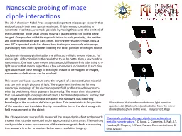
Nanoscale probing of image dipole interactions in a metallic nanostructure C Ropp Z Cummins S Nah JT Fourkas B Shapiro E Waks Nature Communications 6 6558 2015 ID: 655305

image light object quantum light image quantum object dipole resolution electromagnetic position resulting diffraction tiny nanoscale microscopy nanowire ropp imaging problem imaged

Share:

Link:

Embed:

Download Presentation from below link

Download Presentation The PPT/PDF document "Nanoscale probing of image dipole intera..." is the property of its rightful owner. Permission is granted to download and print the materials on this web site for personal, non-commercial use only, and to display it on your personal computer provided you do not modify the materials and that you retain all copyright notices contained in the materials. By downloading content from our website, you accept the terms of this agreement.


Presentation Transcript

Slide1

Nanoscale probing of image dipole interactions

“Nanoscale probing of image dipole interactions in a metallic nanostructure," C. Ropp, Z. Cummins, S. Nah, J.T. Fourkas, B. Shapiro, E. Waks, Nature Communications, 6, 6558 (2015)

The 2014 chemistry Nobel Prize recognized important microscopy research that enabled greatly improved spatial resolution. This innovation, resulting in nanometer resolution, was made possible by making the source (the emitter) of the illumination  quite small and by moving it quite close to the object being imaged. One problem with this approach is that in such proximity, the emitter and object can interact with each other, blurring the resulting image. Now, a new PFC-supported study has shown how to sharpen nanoscale microscopy (nanoscopy) even more by better locating the exact position of the light source. Traditional microscopy is limited by the diffraction of light around objects. For visible light, diffraction limits the resolution to no be better than a few hundred nanometers. One way to surmount the standard diffraction limit is by using tiny light sources that are no larger than a few nanometers in diameter. If such tiny light sources are close enough to the object meant to be mapped or imaged, nanometer-scale features can be resolved. This recent work uses quantum dots, tiny crystals of a semiconductor material that can emit single photons of light. The experiment involves performing nanoscopic mappings of the electromagnetic field profile around silver nano-wires by positioning these quantum dots nearby. The researchers discovered that sub-wavelength imaging suffered from a fundamental problem, namely that an “image dipole” induced in the surface of the nanowire was distorting knowledge of the quantum dot’s true position. This uncertainty in the position of the quantum dot translates directly into a distortion of the electromagnetic field measurement of the object. The JQI experiment successfully measured the image-dipole effect and properly showed that it can be corrected under appropriate circumstances. The resulting work provides a more accurate map of the electromagnetic fields surrounding the nanowire in order to produce better super-resolution imaging.

Illustration of the interference between light from the quantum dot (black sphere) and radiation from the mirror dipole (black sphere on the wire).

Image credit,

Ropp