/
Common Medical Myths & Misconceptions Common Medical Myths & Misconceptions

Common Medical Myths & Misconceptions - PowerPoint Presentation

tawny-fly
tawny-fly . @tawny-fly
Follow
481 views
Uploaded On 2016-06-08

Common Medical Myths & Misconceptions - PPT Presentation

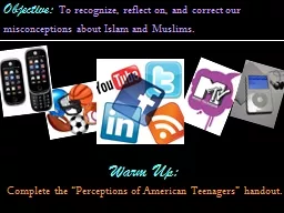
Misconceptions vs Myths For the purpose of this Discussion Misconception Common mistake but generally not perpetuated by schools and licensure exams This presentation will cover 5 common medical misconceptions ID: 353154

Share:

Link:

Embed:

Download Presentation from below link

Download Presentation The PPT/PDF document "Common Medical Myths & Misconception..." is the property of its rightful owner. Permission is granted to download and print the materials on this web site for personal, non-commercial use only, and to display it on your personal computer provided you do not modify the materials and that you retain all copyright notices contained in the materials. By downloading content from our website, you accept the terms of this agreement.


Presentation Transcript

Slide1

Common Medical Myths & MisconceptionsSlide2

Misconceptions vs. Myths

For the purpose of this Discussion:

Misconception = Common mistake, but generally not perpetuated by schools and licensure exams. This presentation will cover 5 common medical misconceptions.

Myth = More than just a common mistake; currently standard practice and propagated by schools and licensure exams. This presentation will cover 3 medical myths.Slide3

Misconception #1:

Cyclothymia

Cyclothymia

is:

An endocrine disorder that results in an underproduction of

thyroxine

An immunological disorder related to a mutation in the MHC gene

A mood disorder related to dysfunction of the thymus in the brain

None of the above

Don’t KnowSlide4

Cyclothymia

Cyclothymia

is:

An endocrine disorder that results in an underproduction of

thyroxine

An immunological disorder related to a mutation in the MHC gene

A mood disorder related to dysfunction of the thymus in the brain

None of the aboveDon’t KnowSlide5

Cyclothymia

An endocrine disorder that results in an underproduction of

thyroxine

An immunological disorder related to a mutation in the MHC gene

A mood disorder related to dysfunction of the

thymus

in the brain

None of the above (11.1%)

Don’t Know

Cyclothymia

has nothing to do with the thymusSlide6

What is the Thymus?

Cyclothymia

and

dysthymia

are named after the

greek

word “

thumos” or “thymos,” meaning spiritedness or emotion (Thumos, 2014). The thymus is also named after “thymos” though it has nothing to do with emotions, but the Greeks didn’t know that when they named it, resulting in etymological confusion that has lasted to this day.To add further confusion, the

thy

mus is right next to the

thy

roid on the trachea, and nobody ever talks about the thymus because it’s not a vital organ in children and adults, so most people don’t know what it does and the two are frequently confused.Slide7

What Does The Thymus Do?

During embryonic development lymphocytes go to the thymus to mature. Lymphocytes that fail to interact with MHCs are killed, because they wouldn’t be able to do their job if they can’t interact with MHCs

on other cells.

Lymphocytes that have strong affinity for self proteins are also killed here because they would cause autoimmunity. Failure of the thymus to kill cells that have affinity for self proteins is thought to be a possible cause of many autoimmune disorders including Diabetes Mellitus Type I (

Geenen

et al., 2005). The thymus prunes your lymphocyte population so only potentially useful lymphocytes ever make it into circulation. By your early teens your thymus starts to atrophy and turn into fat tissue and can be removed without any immune system compromise because its work has already been done (Thymus, 2015).Slide8

Misconception #2: Blood

Is blood always red?

Yes

No

Don’t KnowSlide9

Blood

Is blood always red?

Yes

No

Don’t KnowSlide10

Blood

Is blood always red?

Yes (

66.7%

)

No

Don’t KnowSlide11

Blood is Always Red.

This is a metaphor

Blood is bright red when oxygenated, and dark red when deoxygenated.Slide12

Blood is Always Red

So why are veins blue then?

This is a metaphor

And why does cyanosis happen?Slide13

Blood is Always Red

This isn’t a popular fact to share at the water cooler, because it’s a little bit complicated.

The fats and proteins in skin don’t absorb much light; they reflect it back out. Red light travels deeper than blue light before getting reflected back out (it’s better at getting through those proteins and fats).

Blood, unlike skin, absorbs light of all wavelengths, but it absorbs blue light more than red, so it appears red.

Deoxygenated blood in veins, like oxygenated blood in capillaries, reflects red light more than blue light, but compared with oxygenated blood it reflects more blue light (but still less blue than red, so it still looks red).

Kienle

et al., 1996Slide14

Even though veins absorb more blue light than red light if exposed to equal amounts of both, they don’t get exposed to equal amounts of both; they absorb a disproportionately large amount of red light under your skin because much of the blue light gets reflected back out by the proteins and fats in your skin before it can reach the vein.

Blood is Always RedSlide15

Even so, the light that comes out over top of a vein still winds up being more red than blue, but the effects discussed in the previous slide make it proportionately more blue than the surrounding skin, in a ratio of about 3 parts red to 2 parts blue (40% blue [if we ignore all the colors except red and blue for the purpose of comparison]), while the surrounding skin is 5 parts red to 3 parts blue (37.5% blue) (

Kienle

et al., 1996).

So the light from skin is mostly red, and the light from veins is mostly red, but there’s more blue in the light being reflected above veins because the veins removed more of the red light than gets removed in the surrounding skin because the red light is better at getting all the way down to the veins, and deoxygenated blood reflects more blue than oxygenated blood in surrounding capillaries.Slide16

Blood is Always Red

Color perception is skewed by our brains. The light coming out above veins is more of a reddish maroon than a blue, but it looks blue because it’s surrounded by a redder area. If you put something purple next to something red, it sometimes looks blue. This phenomenon is called relative perception.

Other examples of relative perception illusions: Different people perceive different colors in this dress.

The red squares in the top diamonds are actually the same color. The green squares in the bottom two diamonds are also the same color. Slide17

But back to the blood color responses for a second….

Is blood always red?

Yes (

66.7%

)

No

Don’t Know

But this is probably not reflective of the actual proportion of people that think that deoxygenated blood is blue, due to a type of testing bias caused by the quiz itself: the “Hawthorne effect”

Most people got it rightSlide18

The Hawthorne Effect

It is likely that people that see the question: “Is blood always red?” on a suspicious quiz will draw the conclusion that the question is only being asked because the answer is counter-intuitive, which makes the process of elimination very easy for a yes or no question, artificially inflating the proportion of correct responses.

So the blue blood misconception is likely more prevalent than these results suggest.Slide19

Misconception #3: Bipolar Disorder

Which of the following scenarios fit the diagnostic criteria for either type I or type II bipolar disorder?

No history of manic or depressive episodes. Currently experiencing a major depressive episode

No history of manic or depressive episodes. Currently experiencing a manic episode

Has experienced more than 2 years of alternating

hypomanic

and minor depressive episodes

None of the aboveDon’t knowSlide20

Bipolar Disorder

Which of the following scenarios fit the diagnostic criteria for either type I or type II bipolar disorder?

No history of manic or depressive episodes. Currently experiencing a major depressive episode

No history of manic or depressive episodes. Currently experiencing a manic episode

Has experienced more than 2 years of alternating

hypomanic

and minor depressive episodes

None of the aboveDon’t knowSlide21

Bipolar Disorder

Which of the following scenarios fit the diagnostic criteria for either type I or type II bipolar disorder?

No history of manic or depressive episodes. Currently experiencing a major depressive episode

No history of manic or depressive episodes. Currently experiencing a manic episode

(

0%

)

Has experienced more than 2 years of alternating hypomanic and minor depressive episodesNone of the above

Don’t know

All you need for a type I bipolar disorder diagnosis is mania. You almost always see depression as well, but it is not necessary for diagnosis, and it is not part of the DSM5 diagnostic criteria. Slide22

Bipolar Disorder

While Bipolar II does require at least one episode of MDD, Bipolar I does not. It doesn’t even require mild depression. It is possible for an individual to be diagnosed with Bipolar I disorder based solely on having had one or more full manic episodes (Bipolar I Disorder, 2015). These asymmetrical criteria are a product of the fact that there is a name for having depression with no elevated mood distinct from "bipolar disorder" (namely depression or MDD), but there is no special name for having elevated mood or mania with no depression distinct from bipolar disorder. This leads to a little bit of confusing

asymmetry

in the diagnostic criteria of bipolar I and bipolar II disorders.

The wording in the textbook is misleading as well. On page 429 our text says "

A bipolar disorder is characterized by mood swings from profound depression to extreme euphoria

," which makes it sound like depression is a necessary symptom for any bipolar disorder diagnosis, though it is not (Lewis et al., 2011).Slide23

Mood Disorder Chart

Depressed sometimes but never manic?

DepressionSlide24

Mood Disorder Chart

Hypo-manic sometimes & majorly depressed other times?

Type II Bipolar

Depressed sometimes but never manic?

DepressionSlide25

Mood Disorder Chart

Hypo-manic sometimes & majorly depressed other times?

Type II Bipolar

Hypomanic

sometimes &

minorly

depressed other times?

Cyclothymia

Depressed sometimes but never manic?

DepressionSlide26

Mood Disorder Chart

Manic sometimes & depressed other times?

Hypo-manic sometimes & majorly depressed other times?

Type II Bipolar

Type I Bipolar

Hypomanic

sometimes &

minorly

depressed other times?

Cyclothymia

Depressed sometimes but never manic?

DepressionSlide27

Mood Disorder Chart

Manic sometimes but never depressed?

Manic sometimes & depressed other times?

?

Hypo-manic sometimes & majorly depressed other times?

Type II Bipolar

Type I Bipolar

Hypomanic

sometimes &

minorly

depressed other times?

Cyclothymia

Depressed sometimes but never manic?

DepressionSlide28

Mood Disorder Chart

Manic sometimes but never depressed?

Manic sometimes & depressed other times?

?

We don’t really have a name for that

Hypo-manic sometimes & majorly depressed other times?

Type II Bipolar

Type I Bipolar

Hypomanic

sometimes &

minorly

depressed other times?

Cyclothymia

Depressed sometimes but never manic?

DepressionSlide29

Mood Disorder Chart

Manic sometimes but never depressed?

Manic sometimes & depressed other times?

Depressed sometimes but never manic?

?

We don’t really have a name for that

Depression

Hypo-manic sometimes & majorly depressed other times?

Type II Bipolar

Type I Bipolar

Hypomanic

sometimes &

minorly

depressed other times?

Cyclothymia

Also Type I BipolarSlide30

Our

Powerpoint

Slide on Bipolar Disorder from Psychiatric Nursing:

Not trueSlide31

Misconception #4: Osmotic Diuretics

Osmotic diuretics work because:

They are made of small molecules that pass more easily through the fenestrated membranes in the kidney

They are made of small molecules that exert a stronger colloidal osmotic pressure than large molecules

They are made of large molecules that exert a stronger colloidal osmotic pressure than small molecules

None of the above

Don’t Know Slide32

Osmotic Diuretics

Osmotic diuretics work because:

They are made of small molecules that pass more easily through the fenestrated membranes in the kidney

They are made of small molecules that exert a stronger colloidal osmotic pressure than large molecules

They are made of large molecules that exert a stronger colloidal osmotic pressure than small molecules

None of the above

Don’t Know Slide33

Osmotic Diuretics

Osmotic diuretics work because:

They are made of small molecules that pass more easily through the fenestrated membranes in the kidney

They are made of small molecules that exert a stronger colloidal osmotic pressure than large molecules

They are made of large molecules that exert a stronger colloidal osmotic pressure than small molecules

None of the above (

22.2%

)

Don’t Know Slide34

Misconception: Large molecules exert more osmotic pressure than small molecules

Blood Vessels

Same Osmotic Pressure.

Size of molecules does not affect osmotic (colloidal osmotic) pressure.

Only the number of molecules.

So the question is… Why do we specifically use large molecules for osmotic diuretics?Slide35

So the question is… Why do we specifically use large molecules for osmotic diuretics?

Because they can’t cross capillary membranes, so they never enter the interstitial fluid and they keep the relative osmotic pressure in the capillaries higher than the surrounding fluids.

As an added bonus, when they cross the fenestrated membranes in the kidney (which they can cross because the fenestrated membranes are super-permeable) they can’t get back into the blood at the semi-permeable membranes in the loop of

Henle

. Trapped in the filtrate, they draw fluid out of the vasculature with them, straight into the bladder (Pharmacological interventions for cardiopulmonary emergencies,

n.d

.).Slide36

Misconception #5: Heparin & Coumadin

What is the normal therapeutic reduction in platelet count for Coumadin and Heparin? (UVA’s normal platelet range = 150,000 – 450,000 platelets per

microliter

)

45,000 – 135,000 (about 30%)

75,000 – 225,000 (about 50%)

The goal for platelet count reduction depends on the patient’s clinical scenario and health history

None of the above

Don’t knowSlide37

Heparin & Coumadin

What is the normal therapeutic reduction in platelet count for Coumadin and Heparin? (UVA’s normal platelet range = 150,000 – 450,000 platelets per

microliter

)

45,000 – 135,000 (about 30%)

75,000 – 225,000 (about 50%)

The goal for platelet count reduction depends on the patient’s clinical scenario and health history

None of the aboveDon’t knowSlide38

Heparin & Coumadin

What is the normal therapeutic reduction in platelet count for Coumadin and Heparin? (UVA’s normal platelet range = 150,000 – 450,000 platelets per

microliter

)

45,000 – 135,000 (about 30%)

75,000 – 225,000 (about 50%)

The goal for platelet count reduction depends on the patient’s clinical scenario and health history

None of the above (22.2%

)

Don’t knowSlide39

Anticoagulants DO NOT REDUCE PLATELET COUNT

Anticoagulants reduce

coagulability

of platelets, not the number of platelets

When heparin and

coumadin

do reduce platelet count, this is a dangerous adverse reaction called heparin-induced thrombocytopenia (HIT), which means that the anticoagulants need to be discontinued immediately.

Expecting a reduction in platelet count is a dangerous misconception that can lead to a failure to recognize HIT and thrombocytopenia caused by other problemsSlide40

Bonus Slides: Physiologic Jaundice in Newborns

Physiologic Jaundice in Newborns is caused by:

Anti-

Rh

factor antibodies cross the placenta and destroy blood cells in the fetus

Breakdown of fetal hemoglobin and an immature liver & gut flora

An increase in circulating red blood cells from the mother

A, B, & CB & C

Don’t KnowSlide41

Physiologic Jaundice in Newborns

Physiologic Jaundice in Newborns is caused by:

Anti-

Rh

factor antibodies cross the placenta and destroy blood cells in the fetus

Breakdown of fetal hemoglobin and an immature liver & gut flora

An increase in circulating red blood cells from the mother

A, B, & CB & C

Don’t Know

This is a new misconception that I added after distributing the quizzes, so I have no data for prevalence of this misconception.Slide42

Blood Cells Do Not Cross the Placenta

In the Thursday OB nursing lecture in “Care of the Newborn part II” at 109:35, the lecturer states: “Increased

bilirubin

production is from an

increase in circulating red blood cells from mom

, and the fetal red blood cells also have a shorter lifespan, so between that and what’s left over from mom they have increased

hemolysis

that they have to deal with.”In “Care of the Newborn part III”, at 5:40, a guest speaker, Katie Kinsey, a pediatric nurse practitioner states: “For normal newborns this is frequently from an increase of circulating red blood cells from the mom and the fetal red blood cells have a shorter lifespan so there’s an increase in

hemolysis

.”Slide43

Blood Cells Do Not Cross the Placenta

Blood cells generally do not cross the placenta. If they could, you wouldn’t need a placenta in the first place; the umbilical cord could just plug directly into the mother’s vasculature. On occasion a few blood cells can get through, but not in any amount that would have a measurable effect on

bilirubin

levels in a fetus.Slide44

Blood Cells Do Not Cross the Placenta

Note: sometimes the fetus can swallow some of the mother’s blood during birth. In this way it is theoretically possible for maternal blood cells to contribute to physiologic jaundice in newborns by being broken down in the GI tract at which point that

bilirubin

can be absorbed into the bloodstream.Slide45

Busting Myths:

Evidence > Expert OpinionSlide46

Myth #1: Diverticulosis

Diverticulosis

is associated with:

A) Low Fiber Diets

B) High Fiber Diets

C) Neither

D) Don’t KnowSlide47

Diverticulosis

Diverticulosis

is associated with:

A) Low Fiber Diets

B) High Fiber Diets

C) Neither

D) Don’t KnowSlide48

Diverticulosis

Diverticulosis

is associated with:

A) Low Fiber Diets

B) High Fiber Diets (

16.6%

)

C) NeitherD) Don’t Know (0%)

Best current evidence suggests high fiber diets are more likely to cause

diverticulosis

, but with the general scarcity of good evidence right now, “don’t know” is a perfectly reasonable answer as well. Slide49

Diverticulosis

& Low Fiber Diet: Correlation

vs

Causation

In Fundamentals of Nursing we were told during our GI lecture that

diverticulosis

(a

blebbing-out of weak points in the colon) is associated with low fiber diets. On page 1046 our medical-surgical nursing text says: “diverticula in the sigmoid colon are thought to be associated with high luminal pressures from a deficiency in dietary fiber intake.” It also points out that, “

The disease is more prevalent in Western, industrialized populations that consume diets low in fiber […] and is uncommon in vegetarians. The disease is virtually unknown in areas of world such as rural Africa where high-fiber diets are consumed

” (Lewis et al., 2011, p. 1046)

O

ur text also cites a 2009 study in the journal of family practice that admitted: “

no direct evidence establishes a role for fiber in preventing diverticulitis” (Weisberger & Jamieson, 2009, abstract).Slide50

Diverticulosis

& Low Fiber Diet: Correlation

vs

Causation

In 2012 a study in the journal of gastroenterology with 2104 participants (as opposed to a few dozen as in most previous studies), using outpatient colonoscopy to determine incidence of

diverticulosis

(as opposed to asking patients if they felt fewer symptoms after eating more fiber as in most previous studies) found that

high fiber diets, not low fiber diets are associated with diverticulosis. (Peery et al., 2012).

This is just one study however, and history has demonstrated that no single study should be used as the basis for policy changes. The thing is though, no previous or subsequent study on the relationship between fiber and

diverticulosis

comes close to the level of evidence provided in this study. More research is badly needed.Slide51

Diverticulosis

Remember that the NCLEX still expects you to regurgitate the idea that low fiber diets cause

diverticulosis

.Slide52

Myth # 2: Contrast Media Allergies

Adverse reactions to contrast media are associated most strongly with:

Grapefruit allergies

Iodine and shellfish allergies

Nut allergies

None of the above

Don’t knowSlide53

Contrast Media Allergies

Adverse reactions to contrast media are associated most strongly with:

Grapefruit allergies

Iodine and shellfish allergies

Nut allergies

None of the above

Don’t knowSlide54

Contrast Media Allergies

Adverse reactions to contrast media are associated most strongly with:

Grapefruit allergies

Iodine and shellfish allergies

Nut allergies

None of the above (

5.6%

)Don’t knowSlide55

The Iodine & Shellfish Allergy Myth

Iodine is used in contrast media because it absorbs X-rays. Iodine is a component of proteins all over your body. It is not an allergen (

Schabelman

& Witting, 2010). If you had an allergy to iodine, you’d be dead (

Baig

et al., 2014). Even just iodine deficiency from lack of dietary intake causes hypothyroidism (Iodine Deficiency, 2015). T4 and T3 are made largely of iodine, and “70% of the body’s iodine is distributed in other tissues, including mammary glands, eyes, gastric mucosa, arterial walls, the cervix, and salivary glands. In the cells of these tissues, iodide enters directly by the sodium-iodide

symporter

(Iodine Deficiency, 2015). When an element has its own transportation mechanism in your cells, that’s a good sign that it’s important.Slide56

The Iodine & Shellfish Allergy Myth

There are no documented cases of any real allergies to iodine. As an important component of proteins used by organs all over your body it's highly

anergized

by your immune system (because it’s present in your body during embryonic development). The way our immune systems work, developing an allergy to iodine is no more possible than developing an allergy to iron. People that have an allergy to

povidone

iodine prep do not have an allergy to iodine; they have an allergy to the additives in the preparation. Slide57

The Iodine & Shellfish Allergy Myth

Moving on to the shellfish part, shellfish allergy is no more strongly associated with an adverse reaction to contrast media than any other allergy. This erroneous assumption stems from the idea that these allergies are due to iodine content in the shellfish, which is also false (shellfish allergies are due to proteins in shellfish that do not contain iodine). It is

atopy

(having a lot of allergies) in general that indicates an increased risk of an adverse reaction to contrast media, with no one allergy standing above the rest as a risk indicator (

Baig

et al., 2014).Slide58

Contrast media allergies aren’t even allergies

“Allergies” to contrast media are not even an allergy at all. Rather, people who have lots of allergies tend to have hyperactive inflammatory systems, and the

hyperosmolality

of contrast media can irritate the vasculature (

Sicherer

, 2004), causing a “direct stimulation of mast cells and

basophils

to release mediators leading to “anaphylactoid” reactions (pseudo-allergy). This may lead to urticaria, bronchospasm, hypotension, and even cardiac arrest (

Baig

et al., 2014). There are two basic types of contrast media: non-ionic low

osmolality

and ionic high osmolality

, both of which have iodine in them. The non-ionic kind generates less dissociation, which means fewer particles floating around in the blood. Recall medical misconception #4, where we discussed how osmolality is determined by the number of molecules suspended in a solvent. “These [low osmolality] compounds are usually about one-half to one-third as osmotically active as the ionic forms, and associated with fourfold or greater reduction in all adverse reaction and fivefold decrease in severe adverse reactions” (Baig et al., 2014). The iodine and shellfish assessment question we all memorized for the NCLEX is BS (bad science)Slide59

So What Should Radiologists Be Assessing For?

We discussed how people with allergies tend to have more active inflammatory systems. This means that people with any type of allergy at all are at a higher risk for an adverse reaction to contrast media. Obviously, people with a previous history of a reaction to contrast media are at the highest risk.

Radiologists should be assessing first for previous reaction to contrast media, followed by any history of any allergies, and the type and severity of reactions to those allergens.Slide60

Iodine & Shellfish Allergy Myth

Remember that the NCLEX still expects you to regurgitate the idea that iodine allergies exist and they, along with shellfish allergies indicate an increased risk of adverse reactions to contrast media.

On Tests the truth isn’t important. It’s what the test-makers think is true that’s important.Slide61

Myth # 3?: Salt Intake

Speaking of which, you had better watch out for

all of you iodine allergy people.Slide62

Salt

Reducing sodium intake to below 2300mg in the general population:

Reduces all-cause mortality

Increases all-cause mortality

Has no significant effect on all-cause mortality

Don’t know

“Salt: It’s the thing that makes food taste bad when it’s not in it.”

- unknownSlide63

Salt

Reducing sodium intake to below 2300mg in the general population:

Reduces all-cause mortality

Increases all-cause mortality

Has no significant effect on all-cause mortality

Don’t knowSlide64

Salt

Reducing sodium intake to below 2300mg in the general population:

Reduces all-cause mortality

Increases all-cause mortality

Has no significant effect on all-cause mortality

Don’t know

A new meta-analysis suggests that the safe intake range for sodium is 2645-4945mg, well above the AHA-recommended 2300mg maximum limit, which may be unhealthily low. Given the scarcity of strong evidence, “don’t know” is currently the only reasonable answer here.Slide65

How much does sodium intake reduction reduce blood pressure?

I tried asking nurses how much they thought a reduction from the national average sodium intake of 3400mg to the AHA-recommended 2300mg limit would reduce average blood pressure.

Guesses ranged from 5 to 15 points systolic and diastolic, with the average guess being 10 points systolic and 9 points diastolic.Slide66

How much does sodium intake reduction reduce blood pressure?

A Large Cochrane meta-analysis found that reducing sodium intake by 1729mg reduced BP by 4.18/2.06. A 2358mg reduction in daily sodium intake yielded 5.39/2.82 in hypertensive people and 2.42/1.00 in

normotensive

people. Note that if a person with average sodium intake (3400 mg) reduces their intake by 2358mg they would be at 1042mg/day. (He, Li, & Macgregor, 2013). Assuming a linear relationship between sodium intake reduction and BP reduction for the purpose of roughly guessing what the reduction in BP would be when reducing sodium intake from 3400mg to 2300 mg, one would expect a drop of

3.43/1.79

mmHg for hypertensive people and

1.54/0.636

mmHg for normotensive people.

So sodium intake reduction does reduce blood pressure. Just not as much as you’d think.Slide67

Salt

Until the past few years, nobody ever checked for an association between sodium intake reduction from the average amount to 2300mg with incidence of heart attacks or strokes. The logic for the 2300mg sodium restriction has always been a three-step model as illustrated below:

Sodium intake is positively correlated with blood pressure

High blood pressure is correlated with adverse cardiovascular events

Reducing sodium intake in the general population will save livesSlide68

Salt

Let’s generate a model for this logic:

A is associated with B

B is correlated with increased mortality

Reducing A will save livesSlide69

Salt

Now let’s apply that logic to automobile accidents:

Most motor vehicle crashes occur at traffic lights

Motor vehicle crashes are positively correlated with death

Removing traffic lights from cities will save lives

This logic doesn’t work. Blood pressure cannot be used as a stand-alone surrogate for the risk of adverse cardiovascular events.Slide70

Salt

The AHA recommends (ideally) reducing sodium intake to

1500 mg/day

The FDA, CDC, WHO, USDA (US department of agriculture), AND (academy of nutrition and dietetics), ADA (

american

diabetes association), and many other reputable organizations currently recommend a maximum sodium intake of

2300mg/day

for the general population.

A recent meta-analysis published in March of 2014 observed sodium intake via 24-hour urine collection and all-cause-mortality rates and found that mortality followed a J-shaped curve with an actual safe range of sodium intake to be between 2645mg and 4945mg, for both

normotensive

and hypertensive people, suggesting that the 2300mg limit is unhealthily low. Intake below 2645mg was associated with no change in the incidence of adverse cardiovascular events, but it

was

associated with an increase in all-cause-mortality, especially with hypertensive patients that suffered from

comorbidities

(Alderman et al., 2014).

In other words, according to this study, whether you’re hypertensive or not, reducing your sodium intake to 1500mg or 2300mg/day

makes you more likely to die

.Slide71

The Problem(s) With Scholarly Articles

They are hard to read.

Scholarly articles are full of esoteric, field-specific jargon and statistical expressions that researchers may be familiar with, but most health care professionals are not. Unless you’re well-educated in the specific systems being discussed in a given article, and you’re a seasoned professional at reading scholarly articles or you’ve recently taken a course in statistics, you’re lucky to get through two sentences in the methods or results section without having to spend five minutes looking something up.

Sometimes scholarly articles are wrong.

And whether an article is based on strong evidence or not, odds are you didn’t read and understand the entire article.

People like to cite them without understanding them.

Because they’re hard to read, people don’t take the time to judge an article’s research methods before they cite it.Slide72

For Example:

A recent study published in 2014 demonstrates that salt causes hypertension and kidney damage in rats!

“Salt-induced epithelial-to-

mesenchymal

transition in Dahl salt-sensitive rats is dependent on elevated blood pressure” (Wang et al., 2014)

=

Rats on a high sodium diet suffered from hypertension and kidney damage. Rats on the normal sodium diet were perfectly healthy by comparison.

Looks bad for salt. But a study is only as good as its methods.Slide73

The rats in this study received a

0.3%

NaCl

diet as a “normal” sodium diet. The rats on the “high sodium” diet received an

8%

NaCl diet. How Much salt is that? https://www.youtube.com/watch?v=gENVB6tjq_M Let’s do a few back of the envelope calculations to see how much salt that is.Americans eat an average of 3400 mg of sodium per day. Americans eat on average 4 pounds of total food (1814.37G) per day. This translates to a 0.18739287% Na diet, or 0.4572386028% NaCl diet (multiply %Na by 2.44 to get %

NaCl

).

Now that we know that Americans eat a

0.46%

NaCl diet we have a basis for comparison.The 0.3% “normal” NaCl would equate to 5.44311G NaCl = 2230mg Na/day for humans (right around the AHA maximum limit, and only a couple hundred below my meta-analysis’ minimum healthy limit, so a reasonably safe-looking number). How much salt did the “high salt” diet rats get?Deliberate Obscurantism vs. Curiosity & Free TimeSlide74

Deliberate Obscurantism vs. Curiosity & Free Time

The rats on the “high sodium” diet received an 8%

NaCl

diet, the human equivalent of 145.2G

NaCl

, or 59.49G =

59,488mg

of sodium/day. That’s 25.86 times the AHA’s recommended maximum, and 11.9 times the maximum recommended by the meta analysis that I provided, and 17.5 times the current average American intake (One of my frozen TV dinners has 1500mg of sodium. That’s like eating 40 TV dinners every day).Something worth noting: Even at this ridiculously high salt intake, only the modified salt-sensitive rats experienced a markedly higher blood pressure. The Brown Norway rats they used “do not develop elevated blood pressure on a high-salt diet as do SS rats” (Note: the term “salt resistant” they use to describe the brown Norway rats is misleading; they are not bred for special tolerance to salt. They are just normal rats, just as the terms “salt sensitive” and “salt resistant” when referring to people refer to individuals that are especially sensitive to increased salt intake and normal people, respectively). In the United States, about 25% of the population is salt-sensitive.

X 40Slide75

Deliberate Obscurantism vs. Curiosity & Free Time

Not only does this study not trump the meta-analysis, it doesn’t even disagree with it. The meta-analysis asserts that the safe sodium intake range is 2645 to 4945mg, with higher intakes resulting in hypertension and increased mortality. This study asserts that if you feed a rat the human equivalent of 60,000mg of salt it will have really bad hypertension (provided it’s a salt-sensitive rat and not a normal rat). Slide76

If you cite scholarly articles without taking the time to read and understand them

You’re going to have a bad time.Slide77

Many Studies on Sodium Intake are Deceptive

Many studies by researchers that support a reduction in sodium intake suffer from the same problems:

When using an animal model they drown the subjects in ridiculous amounts of salt that no human would be capable of eating the equivalent of

They use increased blood pressure as a proxy for an increased incidence of adverse cardiovascular events, which fails to take into account the other side effects of reducing sodium intake.

They do not examine the effects of low salt intake. They only compare a healthy salt intake with a ridiculously high salt intake, leading agencies like the AHA, FDA, and CDC to suggest restricting sodium below a maximum amount and say nothing about a lower healthy limit.Slide78

Salt

A lot of recent research and position statements by prominent national and international organizations suggest lowering sodium intake

“A reduction in salt intake lowers blood pressure and, thereby, reduces cardiovascular risk”

- “Effect of longer-term modest salt reduction on blood pressure”

(He, Li, & Macgregor, 2013)

“Reduced sodium intake results in reduced blood pressure levels. High blood pressure increases your risk for heart disease and stroke”

- Heart & Stroke Foundation

(Dietary sodium, heart disease & stroke, 2014).

“Sodium increases blood pressure,” “eating less sodium now will help curb that rise and reduce your risk of developing other conditions associated with too much sodium, such as stroke, heart failure, osteoporosis, stomach cancer, and kidney disease”

- American Heart Association

(About Sodium, 2014).

To name just a fewSlide79

Salt

There is also a lot of recent research that criticizes the 1500 and 2300mg sodium intake limits

“Fatal and nonfatal outcomes, incidence of hypertension, and blood pressure changes in relation to urinary sodium excretion”

http://www.ncbi.nlm.nih.gov/pubmed/21540421

“Low-salt intake during mating or gestation in rats is associated with low birth and survival rates of babies”

http://www.ncbi.nlm.nih.gov/pubmed/25197564

“The science upon which to base dietary sodium policy”

http://advances.nutrition.org/content/5/6/764.full

“Reducing dietary sodium the case for caution”

http://jama.jamanetwork.com/article.aspx?articleid=185293

“Data rather than opinion dictates that a definitive clinical trial must determine if the US government’s sodium guideline is safe and effective

http://ajh.oxfordjournals.org/content/24/8/859.extractSlide80

This Wouldn’t Be the First Time

Peanuts

(Huang, F., & Nowak-

Wegrzyn

, 2008)

Cholesterol

Saturated Fats

MSG (FDA & WHO say it’s fine)

Gluten

(

Biesiekierski

, 2013)Thankfully Health & Nutrition Authorities didn’t jump on the MSG or anti-gluten bandwagonshttps://www.youtube.com/watch?v=exi7O1li_wA&list=PL723E862403291665 Tom

Naughton’s

“Big Fat Fiasco”Slide81

Subjective Validation

Subjective validation is where a person believes two unrelated events are related because their hypothesis demands that they be related.

A significant amount of research is done not for the purpose of discovering causal relationships, but for the purpose of proving the researchers’ hypotheses correct.Slide82

Clarification:

Sodium restrictions are very often necessary and useful for individuals with liver and kidney dysfunction (because dysfunctional kidneys can’t dispose of excess sodium). The question is whether sodium intake reduction would be beneficial for the general population and for individuals with hypertension and otherwise healthy kidneys and livers.Slide83

The Controversy

The American Heart Association (AHA) stands by its advisory that everyone should restrict sodium intake to under 2,300 mg, or 1,500 in certain groups, warning consumers to be WARY of this new study. President elect of AHA and cardiologist at Brigham and Women’s Hospital, Dr. Elliot Altman comments,

“Given the abundance we have about excess sodium in diet and its relationship to hypertension and the ability of us as health professionals to recommend to patients that if they lower their blood pressure they will reduce their risk of heart disease and stroke, we are not distracted by these analyses [by the IOM] that have significant methodological flaws.”

Further, AHA CEO Nancy Brown cautions,

“In short, this new analysis of these studies should not be used as rationale to reverse public health policy recommendations.”

The authors of the study insist, “The findings here lend support to those who have questioned the scientific basis for sodium reduction [recommendations], which are based primarily on the assumed blood pressure effect obtained in selected intervention studies and a selected meta-analysis of intervention studies.” Gradual’s team references previous results, published by Cochrane review, supporting modest reductions in salt intake to improve blood pressure.

The American Council on Science and Health (ACSH)’s Dr. Gil Ross had this comment:

“The AHA persists in its ivory-tower warning, which is both scientifically unsound and impractical to say the least, among humans who actually eat. Again and again we’ve seen studies showing that population-wide sodium restriction is fraught with potential consequences: while it may help some groups with severe high blood pressure or fluid retention, it may harm others. Further, the AHA’s response is mere blather, as Dr. Altman immediately shifted the discussion from sodium restriction to the benefits of reducing elevated blood pressure. While the latter is indisputable, the former is at least a matter of academic discussion”

(Our sodium intake is just right, a new study finds, 2014).Slide84

The Controversy

In 2013 the Institute of Medicine (IOM) convened a committee to review the effects of sodium intake on health outcomes other than blood pressure, focusing on intake from 1500 to 2300 mg/day. The final report supported population-wide efforts to lower sodium, but concluded that there were insufficient data to support a lowering of sodium to less than 1500 mg/day as recommended by the AHA, or less than 2000 mg/day as recommended by the WHO (Cook,

Appel

, &

Whelton

, 2014).

Which leads me to a question that I find myself asking frequently: Slide85

Wikipedia, what should I believe?

“The effect of a low salt diet on mortality or cardiovascular disease is unclear with any benefit in either hypertensive or normal

tensive

people being small if present” (Low sodium diet, 2014).

WIKIPEDIA HAS SPOKENSlide86

The only thing that’s clear right now is that more research is needed as to the health effects of sodium intake reduction.

For the NCLEX, remember that the 2300mg limit is still recommended in the US. Just know that the evidence is shaky.

SaltSlide87

References

About Sodium (Salt). (2014, April 29). Retrieved March 7, 2015, from http://www.heart.org/HEARTORG/GettingHealthy/NutritionCenter/HealthyEating/About-Sodium-Salt_UCM_463416_Article.jsp

Alderman, M.,

Baslund

, B.,

Jurgens

, G., &

Graudal, N. (2014). Compared with usual sodium intake, low- and excessive-sodium diets are associated with increased mortality: A meta-analysis. American Journal of Hypertension, 27(9), 1129-1137.Baig, M.,

Faraq

, A.,

Sajid

, J., Potluri, R., Irwin, R., & Khalid, H. (2014). Shellfish allergy and relation to iodinated contrast media: United Kingdom survey.

World Journal of Cardiology, 6(3), 107-107. Biesiekierski, J., Peters, S., Newnham, E., Rosella, O., Muir, J., & Gibson, P. (2013). No effects of gluten in patients with self-reported non-celiac gluten sensitivity after dietary reduction of fermentable, poorly

a

bsorbed

,

short-chain

c

arbohydrates

.

Gastroenterology,

145

(2), 320-328.e3.

Bipolar I Disorder. (2015, January 15). Retrieved March 5, 2015, from http://en.wikipedia.org/wiki/Bipolar_I_disorder

Cook, N.,

Appel

, L., &

Whelton

, P. (2014). Lower Levels of Sodium Intake and Reduced Cardiovascular Risk.

Epidemiology and Prevention,

981-989.

Dietary Sodium, Heart Disease and Stroke. (2014, September 17). Retrieved March 7, 2015, from http://www.heartandstroke.com/site/c.ikIQLcMWJtE/b.5263133/k.696/Dietary_sodium_heart_disease_and_stroke.htm

Geenen

, V.,

Brilot

, F.,

Hansenne

, I.,

Renard

, C., & Martens, H. (2005). Importance of a thymus dysfunction in the

pathophysiology

of type 1 diabetes.

Revue

Medicale

De Liege,

60

(5), 291-296.

He, F., Li, J., & Macgregor, G. (2013). Effect of longer term modest salt reduction on blood pressure: Cochrane systematic review and meta-analysis of

randomised

trials.

BMJ,

4

, F1325-F1325.

Huang, F., & Nowak-

Wegrzyn

, A. (2008). Early Consumption Of Peanuts In Infancy Is Associated With A Low Prevalence Of Peanut Allergy.

Pediatrics,

122

(5), S118-S119.

Iodine deficiency. (2015, February 22). Retrieved March 7, 2015, from http://en.wikipedia.org/wiki/Iodine_deficiency

Kienle

, A.,

Lilge

, L.,

Vitkin

, I., Patterson, M., Wilson, B.,

Hibst

, R., & Steiner, R. (1996). Why do veins appear blue? A new look at an old question.

Applied Optics, 35

(7), 1151,1160.

Lewis, S., Dirksen,

Heitkemper

, Bucher, & Camera. (2011).

Medical-surgical nursing: Assessment and management of clinical problems

(8th ed.). St. Louis, Mo.: Elsevier/Mosby.

Low sodium diet. (2014, September 26). Retrieved March 8, 2015, from http://en.wikipedia.org/wiki/Low_sodium_diet

Our sodium intake is just right, a new study finds. (2014, April 23). Retrieved March 7, 2015, from http://acsh.org/2014/04/sodium-intake-just-right-new-study-finds/

Peery

, A., Barrett, P., Park, D., Rogers, A.,

Galanko

, J., Martin, C., & Sandler, R. (2012). A High-Fiber Diet Does Not Protect Against Asymptomatic

Diverticulosis

.

Gastroenterology,

142

(2), 266-272.

Pharmacological Interventions for Cardiopulmonary Emergencies (Clinical Essentials) (Paramedic Care) Part 7. (

n.d

.). Retrieved March 7, 2015, from http://what-when-how.com/paramedic-care/pharmacological-interventions-for-cardiopulmonary-emergencies-clinical-essentials-paramedic-care-part-7/

Pillitteri

, A. (2014).

Maternal & child health nursing: Care of the childbearing & childrearing family

(7th ed.). Philadelphia, PA: Lippincott Williams & Wilkins.

Rosenberg, K., &

Trevathan

, W. (2007). An anthropological perspective on the evolutionary context of preeclampsia in humans.

Journal of Reproductive Immunology,

76

(1-2), 91-97.

Schabelman

, E., & Witting, M. (2010). The Relationship of

Radiocontrast

, Iodine, and Seafood Allergies: A Medical Myth Exposed.

The Journal Of Emergency Medicine,

39

(5), 701-707.

Sicherer

, S. (2004). Risk of severe allergic reactions from the use of potassium iodide for radiation emergencies.

Journal of Allergy and Clinical Immunology,

114

(6), 1395-1397.

Thumos

. (2014, December 22). Retrieved March 5, 2015, from http://en.wikipedia.org/wiki/Thumos

Thymus. (2015, February 27). Retrieved March 5, 2015, from http://en.wikipedia.org/wiki/Thymus

Wang, Y., Mu, J., Liu, F.,

Ren

, K., Xiao, H., Yang, Z., & Yuan, Z. (2014). Salt-induced epithelial-to-

mesenchymal

transition in Dahl salt-sensitive rats is dependent on elevated blood pressure.

Brazilian Journal of Medical and Biological Research,

47

(3), 223-230.

Weisberger

, L., & Jamieson, B. (2009). Clinical inquiries: How can you help prevent a recurrence of diverticulitis?

Journal of Family Medicine,

58

(7), 381-382.