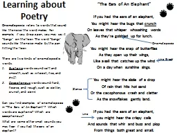
Subjective Data Concerning symptoms of the ear are Hearing loss Earache otalgia Discharge otorrhea Tinnitus Vertigo During data collection the examiner should be alert to signs of hearing ID: 757238
Download Presentation The PPT/PDF document "The Ears Dr. Zyad Saleh" is the property of its rightful owner. Permission is granted to download and print the materials on this web site for personal, non-commercial use only, and to display it on your personal computer provided you do not modify the materials and that you retain all copyright notices contained in the materials. By downloading content from our website, you accept the terms of this agreement.
Slide1
The Ears
Dr. Zyad Saleh Slide2
Subjective Data
Concerning symptoms of the ear are:
Hearing loss
Earache (otalgia)
Discharge (
otorrhea
)
Tinnitus
Vertigo
During
data
collection, the
examiner should be alert to signs of hearing
loss such
as inappropriate answers, frequent requests for
repetition, etc
.Slide3
EaracheOtitis
media: medical term for middle ear infectionSlide4
Otitis externa: inflammation of the outer ear and ear canal. Slide5
Subjective Data
Drainage (otorrhea
)
infection.
Purulent, bloody drainage infection of the external ear (external otitis). Purulent drainage associated with pain and a popping sensation otitis media with perforation of the tympanic membrane.Slide6
Subjective Data
Earache (otalgia)
ear
infections, cerumen blockage, sinus infections, or teeth
and gum
problems.Slide7
Tinnitus:
ringing, roaring or crackling no external stimulus
excessive
earwax buildup, high blood pressure,
or certain
ototoxic medicationsSlide8
Vertigo: refers to perception of rotating or spinning. (an
inner-ear problem.) It is termed subjective vertigo when clients feel that they are spinning around
objective
vertigo when clients
feel that
the room is spinning around them
.. It is important to distinguish vertigo from dizzinessSlide9
Past history:Congenital hearing loss
Removal of cerumenEar surgeryTraumaInfectionExposed to hazardous noise levelsDescribe any past treatmentsSlide10
Family history:Hearing loss
Lifestyle habits:live in an area with frequent or continuous loud noisetime
spent in swimming or in water
Otitis
externa
hearing loss affected
ability to carehearing loss affected socializinglast hearing examinationwear a hearing aidcare for earsSlide11
Objective Data
Inspect and palpate the external ear:Slide12
Objective Data
Inspect and palpate the external ear:Size and shape and ear alignment: ears are equal size bilaterally with no swelling or thickening
Skin Condition: skin color is consistent with facial skin color, skin intact, with no lump or lesions.
Enlarged
preauricular
and
postauricular lymph nodes—infectionRedness, swelling, or itching—otitis externaSlide13
Objective Data
Palpate the auricle and mastoid process
A painful auricle or tragus
otitis
externa
Tenderness over the mastoid process mastoiditis.Tenderness behind the ear otitis media.Slide14
Objective Data
Inspect the external auditory canal. (Note any discharge along with the color
and consistency of
cerumen)Slide15
Objective Data
Inspect the external auditory canal.
External
auditory meatus:
A
small amount of
odorless cerumen appearance-color varies from yellow to brown or black; moist and waxy to dry. Any other discharge? Inflammation?Slide16
Objective Data
Inspect the external auditory canal.
Observe the color and consistency
of the
ear canal walls and inspect
the character
of any nodules.The canal walls should be pink and smooth, without nodules.Slide17
Objective Data
Inspect the external auditory canal.
(abnormal)
Reddened, swollen
canals
Foul-smelling
, sticky, yellow discharge— otitis externa Bloody, purulent discharge—otitis mediaBlood or watery drainage (cerebrospinal fluid)—skull traumaSlide18
Inspect the eardrum (tympanic membrane):
Color: shiny and translucent with a pearl-gray color.no bulging or retraction.
slightly
concave, smooth, and
intact.
Cone-shaped light reflex.Slide19
Inspect the eardrum:
Red, bulging eardrum and distorted, diminished, or absent light reflex—acute otitis media
Yellowish
, bulging membrane
with bubbles
behind—serous otitis
mediaBluish or dark red color—blood behind the eardrum from skull trauma Perforations—trauma from infectionSlide20
Hearing and Equilibrium Tests:
Whisper-Voice test.ask pt to close her other ear, stand behind the pt, whisper softly toward unoccluded ear, increase intensity of sound if necessary.
Able
to correctly repeat the two-syllable word
as whispered.
hearing lossSlide21
#testing of ear and bone conduction is done if the patient has problems in hearing… Weber test:
evaluate the conduction of sound waves through bone to help distinguish between conductive hearing (
sound waves
transmitted by the external
and middle
ear) and sensorineural
hearing (sound waves transmitted by the inner ear).Slide22
#testing of ear and bone conduction is done if the patient has problems in hearing… Weber test:
Vibrations are heard equally well in both ears. No lateralization of sound to either ear.Slide23
Weber test: With conductive hearing loss, the
client reports lateralization of sound to the poor ear the poor ear receives most of the sound conducted
by bone vibration.
With sensorineural hearing loss, the
client reports
lateralization of sound to the
good ear nerve damage in the bad earSlide24
Rinne’ test: compare air conduction (AC) with bone conduction (BC). Place vibrating tuning fork on the mastoid bone, when the pt can no longer hear the sound, quickly place the fork close to the ear, and ask if the sound can be heard again.
Air conduction sound is normally heard longer than bone conduction sound (AC > BC).Slide25
Conductive hearing loss: BC=AC, or BC>AC. Sensorineural hearing loss: AC>BC. Slide26
Romberg test: Test-assesses the ability of the vestibular apparatus in the inner ear to help maintain standing balance. Slide27
Romberg test:.Ask patient to stand up with feet together. When stable have pt close eyes wait about 20 seconds; slight swaying may occur.Positive Romberg’s –loss of balance that occurs when closing eyes.