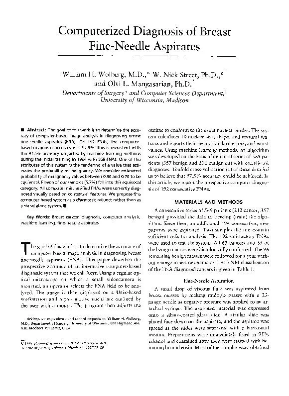
/
Computerized Diagnosis of Breast Fine-Needle Aspirates William H. Wolb Computerized Diagnosis of Breast Fine-Needle Aspirates William H. Wolb

Computerized Diagnosis of Breast Fine-Needle Aspirates William H. Wolb - PDF document

test
test . @test
Follow
411 views
Uploaded On 2016-10-09

Computerized Diagnosis of Breast Fine-Needle Aspirates William H. Wolb - PPT Presentation

78 of the confirmation was obtained by One patient had distant several recent solid masses were computeranalyzed The area sually selected operator for minimal nuclear over color videocamera was c ID: 473423

the confirmation was

Share:

Link:

Embed:

Download Presentation from below link

Download Pdf The PPT/PDF document "Computerized Diagnosis of Breast Fine-Ne..." is the property of its rightful owner. Permission is granted to download and print the materials on this web site for personal, non-commercial use only, and to display it on your personal computer provided you do not modify the materials and that you retain all copyright notices contained in the materials. By downloading content from our website, you accept the terms of this agreement.


Presentation Transcript

Computerized Diagnosis of Breast Fine-Needle Aspirates William H. Wolberg, M.D.," W. Nick Street, Ph.D.,"t and Olvi L. Mangasarian, Departments of Surgery * and Computer Sciences University of Wisconsin, Madison Abstract: The goal of this work is to determine the accu- racy of computer-based image analysis in diagnosing breast 78 of the confirmation was obtained by *One patient had distant several recent solid masses were computer-analyzed. The area sually selected operator for minimal nuclear over- color videocamera was captured ComputerEyes/RT color framegrabber board Vision, Inc., gray-scale image image analysis, are not in successfully digital im- cell nucleus. computer program called Xcyt was the approximate provide a representative rough outline computer monitor. From rough outline, actual boundary Once the analyzed have have been enclosed clear features each nucleus These features higher values the radius Nuclear shape concavity, compactness, concave points, dimension features. perimeter feature. verified using idealized mathematical programming the data based has image processing point for sample. We a surface separates the points from this 30-dimen- This method iteratively place first plane between them. are not linearly separable, MSM-T constructs a plane average distance points. Depending the separation the procedure is recursively plane until final regions but has more accurate instances, simpler perform better data than more complex ones. Therefore, also the number constructing the worst area, worst mean texture three features separating plane diagnostic separation. agnostic accuracy diagnosis together a probability nancy determined Fine-Needle Aspirates Estimated Probability ‘Lobular neoplasia correctly diagnosed visually. from the Should this fall between the sample the same way termed suspicious. lists the Xcyt-generated estimated for the consecutive FNAs diagnostic sample was obtained. hundred twenty-nine proved Adequate diagnostic obtained from accuracy projected methods during Xcyt, these results also confirm the prospective accuracy. visual diagnoses the extremes Diagnostic difficulties arise equivocal samples. the rendering estimates the probability consider Xcyt-estimated malignancy val- this equivocal category. were benign two that the malignant the separating The other a lobular clearly misclassified as benign clearly misclassified Xcyt does address the inadequate sam- constitute a small percentage in malignancy rendered clinically useful classifying some FNAs. Visually, “suspicious.” This estimated malignancy as rendered Xcyt provides following up excisional biopsy, definitive surgery. Xcyt as adjunct rather a stand-alone FNAs were correctly diagnosed visually. obtained from a multiple polyposis was incorrectly diagnosed visually, but was correctly diag- Xcyt. Although Xcyt accurately nuclear features, that are visual diagnosis. particular importance contextual features clump thickness, the adherence the presence only bare probably can samples with the original retraining. However, multiple sites multiple investigators located investigator located under Linux thus obviating for a digital image analysis coupled with machine-learning techniques accurately diagnose breast FNAs, and that the most diagnosis with visual diagnosis. INRSA Fellowship Force Office Mangasarian), and Foundation grant 80 Cross-validatory choice tistical predictions. Snakes: active feature extraction learning techniques fine needle cancer diagnosis linear programming. ations Res Linear Pro- Artificial Intelligence and Cognitive Science Society Mathematical programming ral networks. and Regression Trees. Pacific Grove, Machine Learning. critical analysis cases in et al. WEI. Negative findings solid breast masses: RWM, Hermans Fine needle aspiration cytol- with immediate reporting WH, Tanner Loh W, fine needle