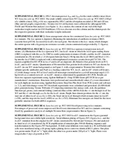
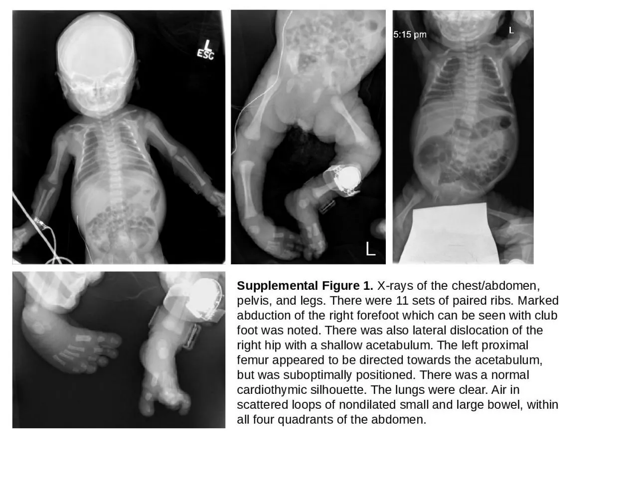
PPT-Supplemental Figure 1. X-rays of the chest/abdomen, pelvis, and legs. There were 11 sets

Author : thomas | Published Date : 2023-11-17

suboptimally positioned There was a normal cardiothymic silhouette The lungs were clear Air in scattered loops of nondilated small and large bowel within all four

Presentation Embed Code

Download Presentation

Download Presentation The PPT/PDF document "Supplemental Figure 1. X-rays of the ch..." is the property of its rightful owner. Permission is granted to download and print the materials on this website for personal, non-commercial use only, and to display it on your personal computer provided you do not modify the materials and that you retain all copyright notices contained in the materials. By downloading content from our website, you accept the terms of this agreement.

Supplemental Figure 1. X-rays of the chest/abdomen, pelvis, and legs. There were 11 sets: Transcript


suboptimally positioned There was a normal cardiothymic silhouette The lungs were clear Air in scattered loops of nondilated small and large bowel within all four quadrants of the abdomen. Explain the importance of the hip and pelvis as a support structure for the human body.. Describe the skeletal structure of the hip and pelvis. . List the primary muscles of the hip and pelvis. . Describe common injuries associated with the hip and pelvis. . SWATI VARA. Contents. How X-rays work . Radiation. Indications . DR. ABCDEFGHI. Common conditions. References. How do X-rays work?. X-rays . are.  electromagnetic . waves. Bone, fat, muscles and . tumours. Testing Dimensions. Studying attributes of items in a . cultural domain. Free listing is a way to define the contents of a . domain. . Pile . sorts and triad tests are ways to measure . similarity. . Transverse by upper margin of the pubic bone, the . ileopectal. line and . ala. of the sacrum. The normal transverse diameter in this plane is 13.5 cm . The normal AP (ant-post) diameter is 11 cm. An area bounded anterior in front by the middle of the symphysis pubis and posteriorly by the junction of the second and third sections of the sacrum. in Computed Tomography. Michael C. . Ficorelli. , RT. Lesson Description. To explain the various exams pertaining to the pelvis using computed tomography, . i. ncorporating cross sectional anatomy from images. Skeletal anatomy of the hip/pelvis. 1. Ilium – upper, lateral section of pelvis. 2. Iliac crest – upper ridge of ilium. 3. ASIS (Anterior Superior Iliac Spine) – identifies anterior end point for iliac crest. Section 6.5 Paired Difference in Means Paired Data or Separate Samples? Should data from the following situation be analyzed as paired data or separate samples? Paired Data Separate Samples To study the effect of sitting with a laptop computer on one’s lap on scrotal temperature, 29 men have their scrotal temperature tested before and then after sitting with a laptop for one hour. Figure . S1. . . (A) . Schematic shows the three KREN1, KREN2, and KREN3 ~20S editosomes, and their protein interactions identified by yeast two-hybrid and co-expression studies (dashed lines) (Schnaufer et al. 2003; Schnaufer et al. 2010; Mehta et al. 2015). (B-D) Networks show detailed editosome architecture revealed by cross-linking and mass spectrometry (CXMS) (McDermott et al. 2016). Network edge widths are proportional to the number of interlinks observed between two proteins. All previously described interactions between editosome proteins were found in our cross-linking data. . Types of Two-Sample Tests. Independent test . –. two conditions are comprised of different elements or subjects . Example: comparing children and adults on a task. Paired test . – two conditions are comprised of the same elements or subjects; same observation is measured under different testing conditions. . Supplemental Figure 1 Supplemental Figure 2 Supplemental Figure 3 Supplemental Figure 4 Supplemental Table 1 Supplemental Figure 5 fpkm. ). PD-L1 (. fpkm. ). CD8 (. fpkm. ). Iba1 (. fpkm. ). p. < 0.001. p. < 0.001. p. < 0.001. p. < 0.001. GM-CSF. GM-CSF. GM-CSF. GM-CSF. Supplemental Figure 1. Gene expression analysis in breast cancer cases from TCGA database. Gene expression levels of CD8, Iba1, PD-L1, and PD-L2 in breast cancer were compared between the cases with and without GM-CSF expression. The Mann-Whitney . A.. B.. Supplemental Figure 3. Control. α-synuclein. Vehicle. Nicotine. A.. B.. C.. D.. No RNAi. SV2L1 KD. SV2L2 KD. Scale bar: 10nm. 9.5nm. 14.7nm. Average diameter:. A. B. Supplemental figure 2. ELISA using FT and GnH-FT as antigens. A. B. * P <0.05, ** P<0.01, *** P<0.001, and **** P<0.00001. **. *. *. X Rays. Are electromagnetic radiations having wavelength of order of 1. Are highly energetic radiations which are emitted when energetic electrons are made to incident on a metallic target.. Were discovered by German Physicist W.C. Roentgen in1895..

Download Document

Here is the link to download the presentation.
"Supplemental Figure 1. X-rays of the chest/abdomen, pelvis, and legs. There were 11 sets"The content belongs to its owner. You may download and print it for personal use, without modification, and keep all copyright notices. By downloading, you agree to these terms.

Related Documents