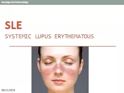
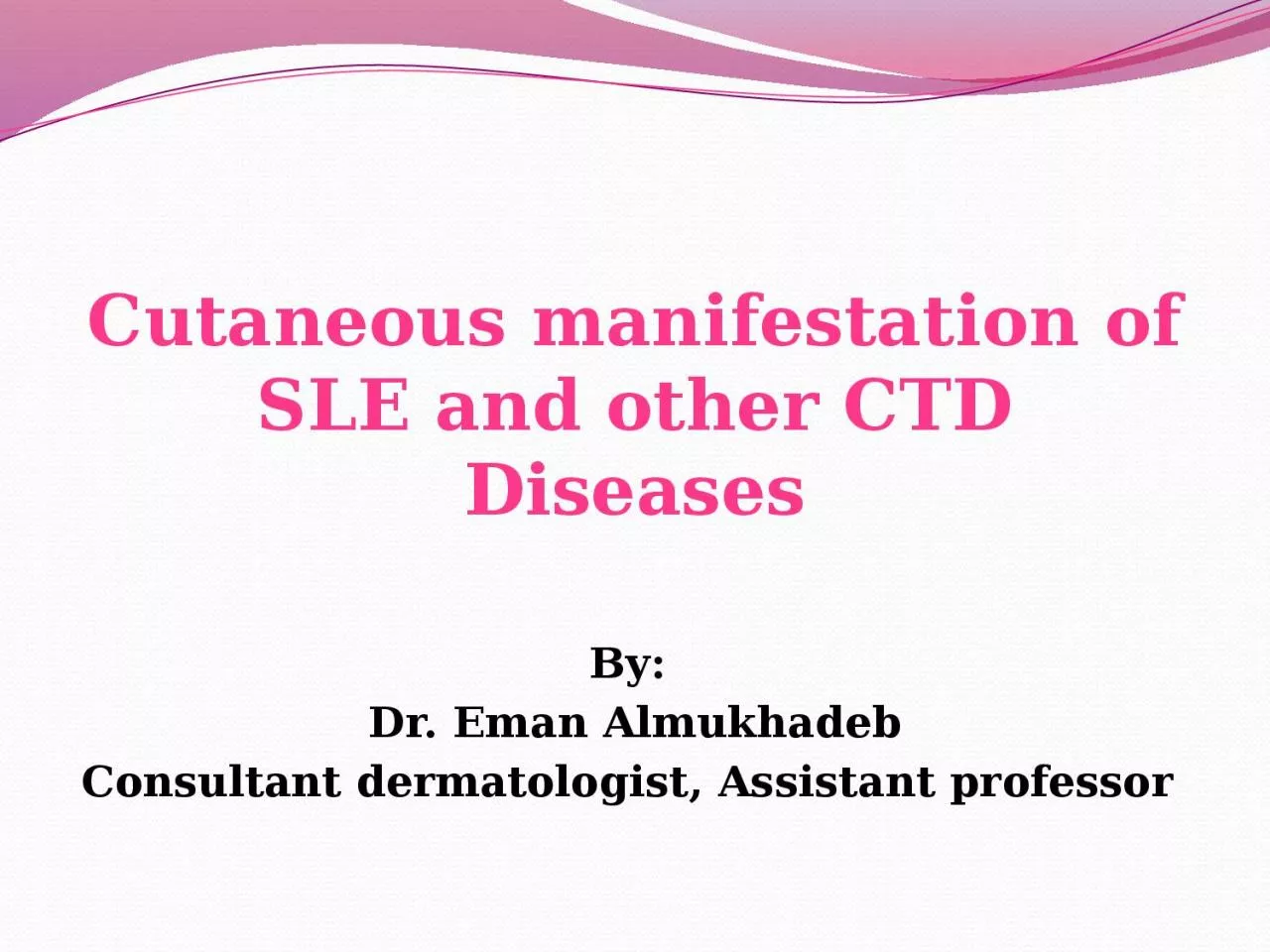
/
Cutaneous manifestation of SLE and other CTD Cutaneous manifestation of SLE and other CTD

Cutaneous manifestation of SLE and other CTD - PowerPoint Presentation

tremblay
tremblay . @tremblay
Follow
27 views
Uploaded On 2024-02-09

Cutaneous manifestation of SLE and other CTD - PPT Presentation

Diseases By Dr Eman Almukhadeb Consultant dermatologist Assistant professor Objectives At the conclusion of this lectures the student should be able to Differentiate between the various types of Lupus ID: 1045266

lupus skin cutaneous anti skin lupus anti cutaneous systemic sle rash atrophy muscle erythematosus disease sclerosis specific treatment common

Share:

Link:

Embed:

Download Presentation from below link

Download Presentation The PPT/PDF document "Cutaneous manifestation of SLE and other..." is the property of its rightful owner. Permission is granted to download and print the materials on this web site for personal, non-commercial use only, and to display it on your personal computer provided you do not modify the materials and that you retain all copyright notices contained in the materials. By downloading content from our website, you accept the terms of this agreement.


Presentation Transcript

1. Cutaneous manifestation of SLE and other CTD DiseasesBy: Dr. Eman AlmukhadebConsultant dermatologist, Assistant professor

2. Objectives At the conclusion of this lectures the student should be able to:Differentiate between the various types of LupusRecognize how Lupus affects the various systems of the bodyIdentify all of the current treatment options available for Lupus

3. Lupus Erythematosus (LE)LE is an autoimmune diseases associated with antibodies directed against components of cell nuclei. Is a multisystem disorder that may affect any tissue, kidneys, CNS, lungs and others. It might affects only skin without systemic involvement It is an immune complex disease that target collagen or ground substance .

4. Lupus Erythematosus( LE)Classification : LE classified as Cutaneous Systemic

5. Classification of cutaneous lupus erythematous AcuteSubacuteChronic

6. Cutaneous classification1- Acute LE : Specific : - Malar eruption or ‘butterfly rash’ - Erythematous papular rash on arms- Photosensitivity - Cheilitis and mouth ulcers- Blisters (bullous LE) Non-specific : as hair fall , raynaud’s phenomena, periungual telangiectasia , vasculitis

7. Cutaneous classification2- Subacute cutaneous LE ( SCLE ):Annular Papulosquamous Associated with syndromes :- neonatal LE - complement deficiency syndrome - drug induced SCLE

8. Cutaneous classification3- chronic : Discoid LE Verrucous ( hypertrophic ) LE Lupus erythematous –Lichen planus overlap Chilblain LETumid LE Lupus panniculitis

9. Chronic LE 1 – Discoid LE (DLE)Commonest form of cutaneous LECommon in young females Started as red indurated plaques and evolve with atrophy , scarring and pigment changes ( mostly Hypopigmentation or depigmentation )Hyperkeratosis characterized by follicular plugging (scarring alopecia).Common above head and neck on the sun exposed areas .localized form associated with 5% with SLE.Generalized form associated with 20% with SLE.

10. Discoid LE (DLE)may involve M.MAggressive SCC may arise from long standing DLE . Generalized form associated more with SLE , +ve ssDNA , +ve ANA , leukopenia, high ESR.Treated by superpotent topical steriod , antimalarial , retinoid .

11. Discoid Lupus Erythematosus

12. Discoid Lupus Erythematosus

13. Other chronic cutaneous LE2- Hypertrophic LE (Verrucous ): commonly seen on the extremities . 3- LE-LP : common on the extremities , palm and soles associated with oral ulcers and nail involvement . 4- Chilblain LE ( pernio): presents as tender red or purple skin lesion that involve fingertips , rim of the ears, calves and heals , exacerbated by cold exposure5- Tumid LE : presented with edematous erythematous plaques on the trunk and respond to antimalarial . 6- LE panniculitis : presented as non-tender subcutaneous nodules on face or proximal extremities that heals with atrophy

14. Chilblain LELupus Tumidus

15. Lupus panniculitis

16. Subacute cutaneous lupus erythematosus 10 to 15% of LE .Common on young females.a non-itchy rash appears on the upper back and chest( on sun exposed regions )Associated with photosensitivity and arthralgia .Systemic involvement is not usually severe.Can be annular or psoriasiform and heals without scarring.Labs: 80% ANA and 70% anti-RO , anti-LA .Treated by antimalarial and sun protection.

17. Neonatal lupus erythematosus Newborn babies born to mothers with subacute LE may develop annular rash, known as neonatal LE that resolve spontaneously. The neonates could be at risk of complete heart block, thrombocytopenia , increased liver enzymes

18. Neonatal lupus erythematosus

19. SLEOnly a few patients with cutaneous LE also have SLE. The most common presentation is with a malar eruption or butterfly. Other skin changes in SLE are photosensitivity, mouth ulcers, and diffuse hair loss. SLE may also affect joints, kidneys, lungs, heart, liver, brain, blood vessels and blood cells

20. SLE ACR criteria for the diagnosis : 4 out of 11 need for the diagnosis . Malar rash . DLE . Photosensitivity . Oral ulcers . Arthritis Serositis ( pleuritis , pericarditis )

21. SLE7- renal : proteinuria more than 0.5 g/day8- CNS : Seizure or psychosis 9- blood : hemolytic anemia, leukopenia, thrombocytopenia 10- +VE ANA 11 - +VE anti-ds DNA and anti-smith and anti-phospholipid antibodies .

22. Drug induced Lupus :associated with ANA and anti-histone antibodies.does not usually affect the skin.hydralazine, procainamide, sulfonamide, anticonvulsants, Minocycline.

23.

24. SLESpecific : - Malar eruption or ‘butterfly rash’ - Erythematous papular rash on arms- Photosensitivity - Cheilitis and mouth ulcers- Blisters (bullous LE) Non-specific : as hair fall , raynaud’s phenomena, periungual telangiectasia , vasculitis

25. Lab findings CBC : anemia , Leucopenia , thrombocytopenia High ESRCoombs test : hemolytic anemia Urinalysis and 24 hr urine collection : for proteinuria U &E . Serology : ANA (+ve in 95% of SLE patients, screening test ) , anti-ds DNA (specific ), anti-ss DNA , anti smith (specific ) , anti-RO , anti-LA , lupus anticoagulant and anti cardiolipin , anti-n RNP. low C2 and C4.Skin biopsy may be diagnostic especially in DLE. Direct IF may show positive antibody deposition along the basement membrane (lupus band test).

26. Treatment of cutaneous lupusThe aim of treatment for cutaneous LE is to alleviate symptoms and to prevent scarring. Local therapy :Sun protection.Topical or intralesional steroid Topical calcineurin inhibitor

27. Systemic antimalarial therapy : hydroxychloroquine 200 mg OD or BD in adults up to 6.5mg/kg . chloroquine ( 125-250 OD in adults up to 3.5-4 mg/kg. quinacrine 100 mg OD . Combination Side effects of hydroxychloroquine : eye toxicity . skin eruption . leukopenia and thrombocytopenia Hemolytic anemia in G6PD def .

28. Other Systemic therapy Retinoid.Thalidomide.Dapsone of bullous lupus Systemic steroidsAzathioprine ,Methotrexate, Mycophenolate mofetil,cyclosporine, cyclophosphamide, IVIG, and Rituximab.

29.

30. ObjectivesTo learn how to diagnose , investigate and manage dermatomyositis.To learn the presentation of morphea and systemic sclerosis and ways to manage them.

31. DermatomyositisAn uncommon inflammatory disease affects adults between 40-60 (females mainly) and children 5-15.Skin changes.Muscle weakness

32. ClassificationClassic DM : -Adult type -Juvenile type Amyopathic DM Paraneoplastic DMOverlap with CTD Drug induced DM : D-penicillamine, hydroxyurea ,NSAID,STATIN and Phenytoin

33. CLINICAL FEATURES Skin :Heliotrope rash : scaly edematous pinkish plaques over both eyelids .Gottron’s sign : scaly erythematous plaques over elbows and knees Gottron’s papules : pink to purple flat topped papules over knucklesErythema of upper chest (V) pattern or over shoulders ( shawl sign )

34. CLINICAL FEATURES Skin:Periungual telangectasia Mechanics Hand :hyperkeratosis , fissuring , scaling with hyperpigmentation over finger tips, sides of fingers and palmsholster sign : scaling and erythema over hips and thigh.Poikiloderma : atrophy , Mottled hyper and hypopigmentaion , telangiectasia

35. CLINICAL FEATURES Other cutaneous signs and symptoms include: Ulceration Photosensitivity Raynaud's phenomenon Calcinosis cutis especially in children

36. Heliotrope rash

37. Gottron’s papules

38. Gottron’s papules

39. Gottron’s sign

40. V-shape erythema of neck Shawl sign

41. Periungual telangiectasia and cuticle hypertrophy

42. Clinical features :Muscle Weakness, progressive proximal muscle weakness involves the hips, thighs, shoulders, upper arms and neck. The weakness is symmetrical and more in the extensor muscles.Lungs : interstitial lung disease

43. Associated disease It can be associated with: Other CTD:Such as lupus, rheumatoid arthritis, scleroderma and Sjogren's syndrome.Cancer:Especially in older patients.common : ovarian CA Cancer of the cervix, lungs, pancreas, breasts, lymphoma and gastrointestinal tract. Malignancy could precede, coincide or follow the diagnosis of DM.

44. Investigations and Diagnosis CRITERIA : 1- Typical skin rash . 2- Muscle weakness ( proximal upper or lower extremity)3- +ve ms enzymes ( CK and aldolase )4- +ve EMG ( short duration , polyphasic)5- +ve muscle biopsy Definite : rash with 3 muscle findings.OTHER LABS:Serology: Anti- Jo-1 ( histidyl- tRNA synthetase): lung disease & mechanical hands. Mi-2 : specific for DM with skin findingsSkin BxMagnetic resonance imaging (MRI).

45. Treatment SKIN :Sunscreen Topical steroid , Topical TacrolimusHydoxychloroquine Oral steroids are the mainstay treatment.Systemic therapy: Steroid sparing agents are:Methotrexate, azathioprine, mycophenolate mofetil, cyclosporine, cyclophosphamide, IVIG, and Rituximab.Physiotherapy to improve strength and flexibility of the muscles.Surgical excision or Co2 laser could be utilized to remove tender calcium deposits .

46. Systemic sclerosis(Scleroderma) A group of diseases that involve the hardening and tightening of the skin and connective tissuesaffects women more often than men . most commonly occurs between the ages of 30 and 50.

47.

48. Classification of SSCutaneous Sclerosis : morphia and linear SS.Limited SS : CREST syndrome Diffuse SS : -progressive SS - overlap with CTD.

49. MORPHIA more common in Female oval reddish or purplish patches and plaques on the skin that progress to smooth , hard , sclerotic , depressed plaque surrounded by violaceous zone It subsides on its own over time leaving dyspigmentation and atrophy Can be localized or generalized without visceral involvement

50.

51.

52. Linear scleroderma parasagittally on the forehead ( encoup de sabre ) parry-Romberg syndrome manifested as progressive hemifacial atrophy, epilepsy , exopthalmos, and alopecia. may extend on the extremities associated with muscle atrophy and flexion contracture..

53.

54.

55. Limited Systemic Sclerosis CREST syndrome : C CALCINOSIS CUTIS R RAYNAUDS PHENOMENA E EOSOPHAGEAL DISMOTILITY S SCLERODACTALY T TELANGECTASIA Good prognosis.Anticentromere antibodies ( specific)

56.

57.

58.

59.

60. Systemic Sclerosis (Diffuse SS )An autoimmune multisystem disease that results in fibrosis and vascular abnormalities in association with autoimmune changes.Usually starts between 30-40 years. More in womenPathophysiology may involve some injury to the endothelial cells and this results in excessive activation of the dermal connective tissue cells, the fibroblasts.Usually presents with Raynaud's phenomena, Thickening of the skin of the fingers, then atrophy and sclerosis. The fingers become spindle-shaped (sclerodactyly) from resorption of the fingertips

61. Skin findingsRaynaud phenomena. Face : expressionless, constricted mouth, peaked nose. Thin lips with radially furrowed. Claw like hands Neck sign : rigdging and tightening of neck on extension. Finger tip atrophy, ulceration and gangrene. Nail fold capillary hemorrhage Telangiectasia appear on the fingers, palms, face, lips, and chest Hyper or depigmented spots (salt and pepper skin )

62. Internal involvement Esophageal dysmotility( distal ) Esophageal reflux and dysphagia: Pulmonary fibrosis Pericarditis , HTN Retinopathy polyarthritisProgressive kidney disease resulting in proteinuria, high blood pressure and eventually renal failure

63.

64. Investigations and DiagnosisDiagnosis is made based on clinical features and presentation.ANA is usually positive. Anti topoisomerase I (Scl 70) is characteristic for it especially in severe cases.High ESR , thrombocytopenia, hemolytic anemiaHigh CK with muscle involvement CXR : diffuse ground glass and honeycomb lung patternCT :lung fibrosis ECG : arrythemia Skin biopsy will show skin atrophy with thickened collagen bundles

65. Treatment of SSIs symptomatic.Raynaud's phenomena: Stop smoking, keep hands warm and decrease trauma. calcium channel blockers, aspirin and vasodilating drugs including nifedipine and iloprost infusions. Calcinosis cutis: nifedipine, surgical or laser excision.Skin sclerosis: physiotherapy, phototherapy.GI: proton pump inhibitor, surgery for strictures.Kidney: ACE inhibitors.In severe cases: immunosuppressant , D-Penicillamine might be used

66. Thank you