/
Chromosomal Chromosomal

Chromosomal "Fingerprints" of Prior Exposure to Densely Ionizing Radia - PDF document

trish-goza
trish-goza . @trish-goza
Follow
385 views
Uploaded On 2016-03-12

Chromosomal "Fingerprints" of Prior Exposure to Densely Ionizing Radia - PPT Presentation

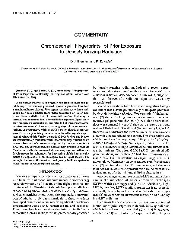
COMMENTARY D J Brenner and R K sachsi Center for Radiological Research Columbia University New York New York 10032 and eartments of Mathematics and Physics University of California B ID: 253019

COMMENTARY Brenner* and

Share:

Link:

Embed:

Download Presentation from below link

Download Pdf The PPT/PDF document "Chromosomal "Fingerprints" of Prior Expo..." is the property of its rightful owner. Permission is granted to download and print the materials on this web site for personal, non-commercial use only, and to display it on your personal computer provided you do not modify the materials and that you retain all copyright notices contained in the materials. By downloading content from our website, you accept the terms of this agreement.


Presentation Transcript

COMMENTARY Chromosomal "Fingerprints" of Prior Exposure to Densely Ionizing Radiation D. J. Brenner* and R. K. sachsi *Center for Radiological Research, Columbia University, New York, New York 10032; and +~e~artments of Mathematics and Physics, University of California, Berkeley, California 94720 Brenner, D. J. and Sachs, R. K. Chromosomal "Fingerprints" of Prior Exposure to Densely Ionizing Radiation. Radiat. Res. 140,134-142 (1994). A INTRODUCTION Various groups of people, such as inhabitants of areas with high levels of radon, uranium miners, nuclear energy research workers, well loggers, airline flight personnel and survivors of the Hiroshima A-bomb, have potentially been exposed to significant doses of densely ionizing radiation, such as a particles or neutrons. Consequently, determina- tion of whether individuals, particularly those who later developed cancer, did in fact receive significant densely ion- izing radiation doses is an important societal and legal issue. Thus there has been by densely ionizing radiation. Indeed, a recent expert report on laboratory-based methods to arrive at risk esti- mates for radiation-induced cancer in humans (I) suggested that identification of a radiation "signature" was a key research need. Several observations have been made suggesting biologi- cal lesions that may be preferentially or uniquely produced by densely ionizing radiation: For example, Vahakangas et al. (2) studied 19 lung tumors from uranium miners and Another suggested marker of high-LET radiation dam- age is the induction of sister chromatid exchanges (SCEs), which were suggested (4) to be inducible by high- LET but not low-LET radiation. Again this is not a mech- anistically driven In contrast to these reports, we discuss here a potential biomarker of prior exposure to densely ionizing radiation which is based on a preferential effect that is expected mechanistically and is observed experimentally. The pro- 0033-7587194 $5.00 01994 by Radiation Research Society. All rights of reproduction in any form reserved. COMMENTARY posed biomarker relates to yields of inter- vs intrachromo- soma1 exchange-type aberrations. The primary mechanism for the production of exchange-type chromosomal aberra- tions is the painvise interaction of two double-strand breaks (DSBS),' in which ends from different breaks meet and join in an illegitimate recombination (6). The likelihood that two DSBs become sufficiently close to undergo such an exchange depends on their relative proximity, which in turn depends on whether the DSBs were in the same or different chromosomes. Consequently, comparing yields of intra- chromosomal aberrations (from two DSBs on the same chromosome) with interchromosomal aberrations (from two DSBs on different chromosomes) can be seen as a probe of radiation track structure; it is this observation which forms the basis for the proposed marker of the pas- sage of a densely ionizing radiation track. In this Commentary, experimental data regarding the magnitude of this effect are reviewed, and it is concluded that published data are in agreement with mechanistically based expectations. Finally, the potential implications of a detectable and stable biomarker for densely ionizing radia- tion are discussed. THE NATURE OF THE FINGERPRINT After are produced by ionizing radiation, restitu- tion occurs in competition with illegitimate recombination between DSBs, the latter process producing exchange-type chromosomal aberrations; these can be interchromosomalif the DSBs are on different chromosomes, or intrachromoso-mal, interarm if the are on different arms of the chromosome. Figure 1shows schematically the production of these aberrations. If DSBs were produced at random in a human cellular nucleus, and all the DSBs were equally likely to interact with one another, the ratio (F) of interchromosomal to intrachromosomal, interarm aberrations would be approxi- mately 86, based on the patterns of chromosome arm lengths in humans (7),close to the value of 90 (4n -2) that would hold if all 92 chromosome arms were of equal length. However, it has long been known (8-10) that of DSBs on different chromosomes that are distant from each other within the nucleus are less likely to interact. If, as is observed experimentally (11,12), individual chromosomes are localized in domains that are smaller than the cell nucle- us, the increased interaction probability of nearby DSBs will result in a bias toward intrachromosomal aberrations, and thus a decrease in the Fvalue. In fact, decreased Fval- 'we refer here to the elementary lesions that interact pairwise to produce exchange-type aberrations as DNA double-strand breaks. How- ever, this identification is not essential for the arguments presented here. a interchromosomal aberrations dlcentrlc + dcentrlc frdgmfnt (unstable) b intrachromosomal pencentric inverslon (stable) FIG. 1.Panel a: Schematic of interchromosomal aberrations result- ing, in the case shown here, from two independent sparsely ionizing radi- ation tracks; each cross represents an ionization cluster of sufficient localization and multiplicity to potentially produce a DSB. This aberra- tion could also be produced by two DSBs from a single radiation track. Panel b: Intrachromosomal, interarm aberrations resulting, in the case shown here, from a single densely ionizing radiation track. ues, interpreted as evidence for chromosomal localization and a limited DSB interaction range, are observed after exposure to X and y rays (7-10). Densely ionizing radiations, however, exhibit a unique property that reduce the Fvalue even further. These radiations produce spatially inhomogeneous energy deposi- tions, and thus DSBs, that are much closer together than those produced by sparsely ionizing radiations such as X or y rays or by chemical clastogens. Consequently, it would be expected that yields of intrachromosomal aberrations would be increased further relative to interchromosomal aberrations; the resulting smaller Fvalue would then be a "fingerprint" of densely ionizing radiation. The proposed chromosomal fingerprint is the F, of interchromosomal to intrachromosomal, interarm aberra- tions, either for stable aberrations (translocations to peri- centric inversions) or for unstable aberrations (dicentrics to centric rings). Unstable aberrations are often lethal to cells 136 COMMENTARY in division, but stable aberrations are typically nonlethal, and can often be measured in irradiated cells and their progeny many years after exposure (e.g. 13-15). It is thus the measured F value in stable which has the potential to be a practical biomarker of past exposure to densely ionizing radiation. EXPERIMENTAL DATA There are many data in the literature on yields of inter- and intrachromosomal aberrations. Often F values are not well determined, because of the smaller number of intra- chromosomal aberrations produced. What adequate data are available support the notion that densely ionizing radia- tion produces characteristically low Fvalues. We quote data for sparsely densely ionizing radiations, for irradiation in vivo and in vitro, and for stable and unstable aberrations. Although it is the F value in stable aberrations that has the potential to act as a practical biomarker of past expo- sure to high-LET radiation, it is to be expected, on basis of the equalities between stable and unstable aberrations (e.g. 13,14,16), that F values for unstable aberrations fol- low the same pattern as those for stable aberrations. Thus, in investigating the validity of the proposed biomarker, it is reasonable to use data for unstable aberrations to augment those for stable aberrations. Experimental evidence sup- porting this suggestion is discussed below. In vivo, there are many measurements of Fvalues after accidental or radiotherapeutic exposures to sparsely ionizing radiations such as X or y rays. For inhabitants of the Cher- nobyl region, a ratio of 37 + 19 was measured (1 7);for indi- viduals exposed y rays in the Goiania radiation accident, the measured ratio was 20 + 3 (18). For radiotherapy patients exposed to X or y rays, where data are adequate for analysis, the mean Fvalue is 18 * 9 (19-21); these data were derived from both stable and unstable aberrations. For densely ionizing radiation, the most extensive in vivo data are for individuals injected with the contrast agent Thoro- trast, which emits a particles: the largest study yields F = 5.0 + 0.3 (22). Measurements for workers exposed to plutonium (emitting ci particles) yielded F = 5.6 + 3.0 for stable aberra- tions and F = 4.5 + 2.0 for unstable aberrations (23). Finally, measurements (24,25) for workers who were accidentally exposed to radiation consisting of -88% (by dose equiva- lent) densely ionizing neutrons yielded F = 5.7 + 3.5 for sta- ble aberrations and 5.0 + 2.4 for unstable aberrations. Two of the in vivo studies cited here (23,25) give F val-ues measured in stable and unstable aberrations several years after exposure. In both cases, the measured Fvalues from stable and unstable aberrations were very similar, sug- gesting that ratios are stable over long time scales. In vitro, very large-scale studies have been ducted. The largest data set for low-LET radiation (26) yielded F = 16.7 + 0.9, in agreement with the of 16 + 5 obtained from a literature survey (7). By contrast, analysis of human lymphocytes exposed to densely ionizing neu- trons (27) gave a significantly smaller value of F = 5.6 + 0.5. There are other studies in the literature, but many suffer from inadequate statistics or incomplete definitions of the different exchange-type aberrations. As far as we know, no data sets with adequate statistics and adequate definitions of the measured aberrations are inconsistent with the pat- tern discussed above, indicating significantly smaller F val-ues for densely ionizing radiation. Although chemical carcinogens often produce damage at specific sites within the genome, such damage is unlikely to be located preferentially on pairs of sites on opposite arms of one chromosome (28). Chemical carcinogens therefore would not be expected to produce anomalously low F values, although few adequate data sets are avail- able. An in vitro value of 14.6 + 0.8 was obtained for aber- rations induced by restriction enzymes which, on the scale of interest here, produce DSBs randomly within chromatin (29). A value of 30 + 5 for bleomycin-induced damage in vitro has been reported (30), though this value is not unex- pected in that bleomycin is a mimetic of sparsely ionizing radiation. In summary, Fvalues in vitro are consistent with those measured after irradiation in vivo and both show signifi- cantly smaller values for densely ionizing radiations than for X or y rays. These data suggest that an F value of around 6 is characteristic of densely ionizing radiation, in contrast to values of -15 or above for X or y rays and for other clastogens. MECHANISTIC BACKGROUND In this section, we argue that the Fvalues sum- marized in the previous section (-15 for sparsely ionizing radiation, and -6 for densely ionizing radiation) are quanti- tatively consistent with what we know about chromosomal localization and about DSB interaction probabilities. Should this be the case, it would provide a strong mechanis- tic underpinning for the existence of the densely ionizing chromosomal "fingerprint" discussed here. Any model for calculating F values requires information in three areas: (1)radiation track structure, to describe the initial spatial locations of ionization clusters which could produce DSBs; (2) the probability two DSBs initially formed a given distance apart will ultimately produce an exchange aberration; and (3) chromosome geometry. We use a simple two-parameter model designed to ana- lyze the measured Fvalues discussed above. In this model, detailed in the Appendix, the yield, Y, of exchanges in nuclei of diameter d is quantified in terms these three types of information: COMMENTARY 137 where t(x) describes the radiation track structure, g(x) is the DSB interaction probability, and s(x) describes the chromo- some geometry. The proportionality constants in this and subsequent equations will be irrelevant for our purposes, since we are interested in estimating yield ratios. We briefly discuss the three terms in Eq. (1): The first term in Eq. (I), describing the radiation track structure, is termed (31) the function, t(x). It can be interpreted (see Appendix) by considering a cluster of ionizations of sufficient localization and multiplicity to pro- duce a DSB in a chromosome. Then t(x)dx is proportional to the probability of another such cluster at a separation between x and x + dx. In general, t(x) is estimated by Monte Carlo track-structure simulation (32,33). However, at the large separations of interest here �(50 nm), the "LET approximation," in which energy loss occurs in straight lines with no radial extension, is valid (31): where L is the stopping power (dEldx) and D is the dose. At low doses of densely ionizing radiation, the first term, referring to pairs of energy-deposition clusters in a single radiation track, will dominate, while for sparsely ionizing radiation, the second term, referring to pairs in indepen- dent tracks, will dominate. Consequently, in the limit of both very low and very high LET, the Fvalue [which would have t(x) in both the numerator and the denominator] will be independent of dose. The second quantity in Eq. (1) describes the g(x), that two DSBs induced with separation x will eventu- ally undergo an exchange. A step-function form has often been employed (31) and has been shown (34) to constitute a reasonable approximation: g(x) = constant, x 5 di; g(x) = 0, x �di, (3) where di defines an effective range for DSB interactions and is our first relevant adjustable parameter. The normal- ization constant in Eq. (3) is again irrelevant for our pur- poses. Equation (3) is sometimes described as the "site" approximation (35). Earlier work has suggested that di is on the order of 1ym (34). The third function in Eq. (I), s(x), describes the geome- try of the chromatin in a cell nucleus. As detailed in the Appendix, s(x) is proportional to the probability that any two points in the chromatin are separated by a distance x. Here we use a simple model for chromosomal geometry whose main features are the following: (1) all 46 chromo- somes are considered identical [since corrections for actual arm length patterns are small (7)];(2) each chromosome consists of a cloud of points which randomly occupy a sphere of radius d,, where d, is our second (and last) adjustable parameter; and (3) different chromosomes inter- twine and overlap freely. As discussed in the Appendix, given these assumptions, we can write s(x) as follows: where s,(x) and s2(x) are derived in the Appendix (Eq. A9). The term s1(x)/2 refers to pairs of points on oppo- site arms of one chromosome, s2(x) refers to points on two different chromosomes. We ignore a term for points on the same arm of one chromosome, because we do not consider the corresponding aberrations in the proposed assay. From Eqs. (1-4),Fvalues can be calculated as a func- tion of d,ld and dild. At relevant doses of sparsely ionizing radiation, aberra- tions are primarily from interactions of DSBs produced by independent tracks. Thus the proximity function, t(x), is dominated by the second term in Eq. (2). Combining Eqs. (1-4) and integrating gives the Fvalue at low LET: F(low LET) = 90 (8u3 -9u4+ 2u6)lg(w), (5) where u = dild and w = dildc. While the parameters are di and d,, the model is scalable in the sense that the pre- dicted results are invariant to changes in di and d,, as long as the d,ld and dild, where d is the remain unchanged. We plot in Fig. 2a the predictions of Eq. (5) as a function of d,ld and dild. In Fig. 2a, if d, = d, i.e., if the domain of each chromo- some as large as the whole nucleus, the F value is the value, 90, obtained by assuming that all DSBs are pro- duced randomly throughout the genome, and all DSB pairs are equally likely to interact. Likewise, if the interac- tion cutoff is as large as the whole nucleus, i.e. di = d, a value of 90 is obtained. However, if both the interaction distance di and the chromosome localization diameter d, are less than the nuclear diameter d, proximity effects come into play because DSBs on the same chromosome have an increased probability to interact, and the F value decreases. At moderate doses of densely ionizing radiation, most aberrations will be produced by interactions between DSBs from one track. In this case, the proximity function will be dominated by the term in Eq. (2), and we obtain the F value at high LET: F(high LET) = 90 (~lw)~ (7) (24u -18u2 + 3u4)lh(w), 138 COMMENTARY ............ ............... ....... ......... ........._ -1 10 ...... ........ ........ _ .......... ..... 0.05 b FIG. 2. Panel a: Predicted F values for sparsely ionizing (low-LET) radiation. Here, d is the nuclear diameter, d, is the maximum DSB interac-tion distance, and d, is the diameter of the chromosomal domains. The arrow gives an indication of the consensus sparsely ionizing experimental Fvalue. Panel b: Predicted Fvalues for densely ionizing (high-LET) radia- tion. The arrow gives an indication of the consensus experimental Fvalue. where u and w are as in Eq. (5).The results for densely ion- izing radiation are shown in Fig. 2b. As with sparsely ioniz- ing radiation, if d, = d, the limiting value of 90 is reached. In contrast, for densely ionizing radiation, assuming di = d and d, d results in an F value less than 90. The reason is that ionizing radiation produces DSBs close to each other, and whose interaction probabilities are roughly pro- portional to the square of their number; thus, even with no spatial limitations on interactions (di = d), a localized chro- mosome will sometimes contain multiple DSBs, with a resulting quadratic yield of intrachromosomal that more than compensates for cases where the chromo- some is missed entirely. Assuming, on the basis of the data quoted above, that F(1ow LET) -15 and F(high LET) -6, Eqs. (5,7)can be solved numerically for d, and di. The result is where the uncertainty limits are based on corresponding estimated uncertainty limits for the Fvalues at low and high LET. For human lymphocytes, where d -6 ym, then d, = 2.0 + 0.3 ym, di = 2.0 + 0.3 ym. (10) These estimates are consistent with those derived from dif- ferent types of data (12,34). DISCUSSION The Fvalues for densely ionizing radiation appear to be around 6, significantly lower than those for any other clastogens including X rays, y rays or chemical car- cinogens. The observation of anomalously low F values for densely ionizing radiations has both mechanistic and prag- matic implications. Mechanistically, Fvalues provide significant constraints on (a) the spatial localization of individual chromosomes within the and (b) the of interaction of DSBs with each other to form exchange-type aberrations. The data suggest that the individual chromosomes are con- strained to a mean diameter about one-third that of the nucleus, and that interaction probabilities are small at distances larger than about 2 pm. These estimates are con- sistent with those from entirely different types of data (12, 34). This consistency provides the mechanistic underpinning for the suggestion that F values could be a useful biomarker of exposure to densely ionizing radiation. The fact that the proposed biomarker refers to ratios of yields results in it possessing considerable robustness against possible confounding effects. For example, such effects as cell turnover and clonogenic or interphase death, while affecting aberration yields, are unlikely to affect ratios of yields. Of course, the use of F values from stable aberra- tions, possibly measured long after exposure, presupposes that there is no differential loss over time of translocations relative to pericentric inversions. No data are available about the rates of loss of these two types of aberra- tions, though the structural similarities between them sug- gest that significant differential loss would be unlikely. An example of the application of this biomarker relates to the question of the exposure of A-bomb survivors at COMMENTARY 139 Hiroshima to densely ionizing neutrons. The 1986 reanaly- sis of the A-bomb dosimetry (36) had suggested that the neutron component at Hiroshima was negligible, a conclu- sion which affected estimates of the risk for low-LET radia- tion significantly, as well as effectively removing the only available source of estimates of risk for humans exposed to fission neutrons. However, recent measurements (37,38) have suggested that neutrons may still be significant and might even dominate the equivalent dose at relevant loca- tions. For Hiroshima A-bomb survivors, an Fvalue of 6.2 t 0.7 has been measured (39). This may be interpreted as evi- dence that a significant proportion of the dose to which the survivors were exposed was from neutrons, in accordance with recent measurements, but in contrast to the dose reassessment calculations. The most likely application of this potential "fingerprint" is in the field of radon. In the past decade, the relationship between low exposures to radon daughters and lung cancer risk has been seen to be of major importance. Detection in exposed populations of molecular markers and adducts associated with particular carcinogens and with particular cancers is now used to study chemical carcinogenesis risks (40). This approach, termed "molecular epidemiology," has become an accepted tool for assessing chemical risks. same approach could be considered in relation to radon- induced lung cancer. Currently, epidemiological studies to determine radon risks at low exposures are limited by the large "background" lung cancer rates produced by carcino- gens other than radon. For epidemiological radon studies of this kind, non- tumorous bronchial epithelial cells (basal cells and their progeny) would be obtained from bronchoscopy (using the bronchial brush technique, ref. 41), the disaggregated, stimulated to mitosis and then assayed for F values for sta- ble aberrations. In a recent report describing a technique for harvesting viable cells from the bronchial epithelium, Kelsen et al. (41) reported a 36 t4% viable cell yield and subse- quent primary-culture plating efficiencies of 50 to 60%. Although measurements of Fvalues can be undertaken using G banding, the large numbers required to reduce the confidence interval on this ratio will generally require the use of fluorescence in situ hybridization (FISH) techniques. Translocations can be measured using standard techniques described by Lucas et al. (14), involving a cocktail of com- posite DNA probes specific to several large chromosomes. For example, if chromosomes 1,2 and 4 are labeled, this covers about 23% of the genome. Theoretical considera- tions, confirmed by experiment (14), indicate that this scheme will sample about 35 % of all translocations. Pericen- tric inversions can be identified efficiently using a pancen- tromeric probe in one color, and another color labeling two loci of one arm of several large chromosomes, these two loci respectively being located close to the and close to the centromere. For example, if the probe was red and the other probes blue, the sequence blue-red would be characteristic of a normal chromosome, and blue red-blue characteristic of a pericentric inversion. Appropri- ate probes can be developed using the described by Meltzer et al. (42,43). Again, if chromosomes 1,2 and 4 were labeled as above, about 33% of pericentric inversions in the genome would be detectable. To estimate how many cells would need to be examined, suppose we examine N cells, and measure X interchromo-soma1 aberrations and Y intrachromosomal, interarm aber- rations. Assuming the detection efficiency is the same for both X and Y, the Fratio is simply XIY. It can be shown that the variance of this estimate is approximately given by (11~~3, [XY(N -X) + x2(N -Y)]. (11) As an example, suppose we are interested in subjects who actually had a cumulative exposure to radon progeny of -50 WLM (working level months: for comparison, the average lifetime domestic cumulative exposure in the U.S. is -15 WLM). Based on an estimated bronchio-epithelial dose1WLM (44), and a measured yield of dicentrics per unit a-particle dose (49, we might expect a yield of -0.24 translocations per cell and, assuming an F value of 6, 0.04 pericentric inversions per cell. Given a FISH detection efficiency of 33% (see above), examination of, say, 3000 cells would yield -240 translocations and -40 pericentric inversions, and an estimated F value of 6 + 1;this estimate would easily distinguish it from Fvalues of 215. Of course, smaller exposures would require measurements of larger numbers of cells and vice versa. In practice, the individuals under study might well have been exposed to a mixture of clastogens, such as a particles + tobacco (e.g. uranium miners) or neutrons + X rays (e.g. survivors at Hiroshima). In such cases an intermediate Fvalue might be anticipated, and experimental calibrations of F value vs proportion of damage induced by high-LET radiation would be important. In conclusion, we have suggested the existence of a potentially useful biomarker for prior exposure to high- LET radiations. The supporting theoretical and experimen- tal data are quite convincing, though further experiments with human cells other than peripheral blood lymphocytes would be desirable. For individuals or cohorts exposed to radon, measurement of the F value could provide a local history of a-particle exposure in the and, in individuals with lung cancer, in the vicinity of the tumor. As with all epidemiological studies, such results could not demonstrate a causal relationship between radon exposure and lung can- cer. They do, however, have the potential to demonstrate and quantify much stronger associations than are currently 140 COMMENTARY possible between low-dose exposure to radon daughters and induction of lung cancer. APPENDIX Here, we discuss the track-structure proximity function, t(x), the DSB interaction function g(x) and the chromoso- mal proximity function s(x). Finally, we derive Eq. (I), which combines these functions to predict aberration yields, and thus Fvalues. Radiation Track Structure Let ~(r) be the density of ionization clusters of sufficient multiplicity and localization to create a DSB if a chromo- some is hit (33). Formally, we take T to be a random func- tion. In that we are interested in interactions between spa- tially separated DSBs, the quantity needed in our calcula- tion is the cluster-density autocorrelation function, where r, r' are points within the nucleus and ( ) denotes an average. Assuming that the radiation is, on average, spatial- ly homogeneous throughout the region of interest implies DSB Interaction Probabilities Let g(x) denote the probability that two DSBs initially formed a distance x apart will ultimately interact in a chro- mosome exchange event. It has been shown (31,34) that an appropriate approximation to g(x) may be the cutoff form given in Eq. (3), where the cutoff interaction radius, di, is an adjustable parameter (of order of magnitude 1ym). We also assume that g(x) is independent of whether the DSBs are on different chromosomes or on different arms of the same chromosome; i.e., any bias toward higher interac- tion probabilities between DSBs on one chromosome is assumed to be due solely to their initial proximity, with no additional bias caused by the restricted motion of DSBs. Chromosomal Geometry Let u(r) be the of chromatin within a representa- tive cell nucleus centered at the origin. Then Here, and in subsequent equations, integrals with unspeci- fied limits go over all of three-dimensional space and have cutoffs supplied automatically by integrands such as u. Assuming that the distribution in a cell nucle- us is, on average, spatially we can define a chro- mosomal proximity function, s(x), which depends only on scalar distance, as follows: Here the factor 4m2 gives a conventional normalization which, from Eq. (A4), is d Ss(x)dx= (46)', s =O if x �d. (A61 o To compute s(x), and to distinguish between intra- and interchromosomal exchanges, we need a geometric model for chromatin geometry. We consider only interphase chromosomes on length scales of 50 nm or more, on time scales of seconds or more and with DNA locations separat- ed by lo4base pairs or more. We take all the chromosomes to have the same average geometry, so that for any i, j= 1,...,46withif j: We also take each centromere to be in the center of its chromosome. The corrections needed for differing chromo- somal arm lengths lead to only minor corrections (7). We assume each chromosome is dispersed randomly within a chromosome localization sphere of radius d, 5 d, and different chromosomes are independent of each other. The assumption of independence means different chromo- somes can overlap freely. We shall assume that point pairs on different chromosomes random pairs within the cell nucleus. For d, d this amounts to an additional assump- tion that each chromosome is centered at random within the nucleus. Inserting Eqs. (A3) and (A7) Eq. (A5), the chromosome proximity function can be written: =4m2 sd3r' [46(ui (r' +x)u,(rt)) (A81 where uj refers to chromosome number j and u is zero out- side the cell nucleus. For convenience we normalize u using +46 X 45(ul(r1+x)u2(r1))]  the chromosome number: =46 X X sl(x) +46 X '1, s,(x) +46 X 45 s2 (x),  COMMENTARY 141 where Here sl(x),which refers to pairs of points within one chro- mosome, comprises equal contributions from interarm and intra-arm terms. The function s2(x)refers to points on two different chromosomes. p(y)dy gives the probability that any two points chosen at random in a unit sphere are sepa- rated by a distance between y and y + dy (31): Equation (4), used in the estimates of Fvalues, is derived from Eq. (A8). Aberration Yields The number of exchanges that have occurred in a partic- ular cell is proportional to Sd3rSd3r1a(r)~(r)a(r')7(r1)g(r-rlI). (All) By averaging over cells, and assuming that a and T are uncor- related, the average yield, Y, of exchanges, can be calculated: Using Eqs. (A2, A5, All) now gives XSX x2 I' ajMgwdx, where t(x) a-sdflr(x), (A13) x D 0 and dfl is the element of solid angle. Equation (A13), first derived using a different argument in ref. (46),is our Eq. (1). The present derivation facilitates decomposition of the yield, Y, of exchanges into contributions which come from DSB pairs or the same or different chromosomes. ACKNOWLEDGMENTS Helpful discussions with Dr. Joe Lucas regarding the design of FISH probes, and with Dr. Charles Geard are gratefully acknowledged. This work was funded by grants from the National Institutes of Health (CA- 49062,OH-02931) and the Science Foundation (DMS 9025103). REFERENCES I.  Meeting Report, Report on a workshop to examine methods to arrive at risk estimates for radiation-induced cancer in the human based on laboratory data. Radiat. Res. 135,434-437 (1993). 2.  K. H. Vahakangas, J. M. Samet, R. A. Metcalf, J. A. Welsh, W. P. Bennett, D. P. Lane and C. C. Harris, Mutation of p53 and ras genes in radon-associated lung cancer from uranium miners. Lancet 339, 576-580 (1992). 3.  J. A. Taylor, M. A. Watson, T. R. Devereux, R. Y. Michels, G. Sac-camanno and M. Anderson, p53 mutation hotspot in radon-associat- ed lung cancer. Lancet 343,8687 (1994). 4.  S. Z. Aghamohammadi, D. T. Goodhead and J. R. K. Savage, Induc- tion of sister chromatid exchanges (SCE) in Golymphocytes by plu- tonium-238 alpha particles. Int. J. Radiat. Biol. 53,909-915 (1988). 5.  S. Grundy, L. Varga and M. A. Bender, Sister chromatid exchange frequency in human lymphocytes exposed to ionizing radiation in vivo and in vitro. Radiat. Res. 100,47-54 (1984). 6.  J. R. K. Savage, Mechanisms of chromosome aberrations. Prog. Clin. Biol. Res. 340B, 385-396 (1990). 7.  L. Hlatky, R. K. Sachs and P. Hahnfeldt, The ratio of dicentrics to centric rings produced in human lymphocytes by acute low-LET radiation. Radiat. Res. 129,304-308 (1992). 8.  K. Sax, An analysis of X-ray induced chromosomal aberrations in Tradescantia. Genetics 25,41-68 (1940). 9.  A. D. Conger and A. H. Johnston, Polyploidy and radiosensitivity. Nature 178,271 (1956). 10.  J. R. K. Savage and D. G. Papworth, The relationship of radiation- induced yield to chromosome arm number. Mutat. Res. 19, 139-143 (1973). 11.  H. Van Dekken, D. Pinkel, J. Mulliken, B. Trask, G. van den Engh and J. Gray, Three-dimensional analysis of the organization of human chromosome domains in human and hamster hybrid cells. J. Cell Sci. 94,299-306 (1989). 12.  T. Haaf and M. Schmid, Chromosome topology in mammalian inter- phase nuclei. Exp. Cell Res. 192,325-332 (1991). 13.  J. R. K. Savage and D. G. Papworth, Frequency and distribution studies of asymmetrical versus symmetrical chromosome aberra- tions. Mutat. Res. 95,7-18 (1982). 14.  J. N. Lucas, A. Awa, T. Straume, M. Poggensee, Y. Kodama, M. Nakano, K. Ohtaki, H. U. Weier, D. Pinkel, Gray and G. Little-field, Rapid translocation frequency analysis in humans decades after exposure to ionizing radiation. Int. J. Radiat. Biol. 62,5343 (1992). 15.  H. J. Evans, K. E. Buckton and A. Sumner, Cytological mapping of human chromosomes. Chromosoma 35,310-325 (1971). 16.  T. Straume and J. N. Lucas, A comparison of the yields of transloca- tions and dicentrics measured using fluorescence in situ hybridiza-tion. Int. J. Radiat. Biol. 64, 185-188 (1993). 17.  G. Stephan and U. Oestreicher, An increased frequency of structural chromosome aberrations in persons present in the vicinity of Cher- nobyl during and after the reactor accident. Mutat. Res. 223, 7-12 (1989). 18.  A. T. Ramalho and A. C. H. Nascimento, The fate of chromosomal aberrations in '37~s-exposed individuals in the Goiania radiation accident. Health Phys. 60,67-70 (1991). 19. R. H. Martin, A. Rademaker, K. Hildebrand, M. Barnes, K. Artheu, T. Rigrose, I. S. Brown and G. Douglas, A comparison of chromoso- mal aberrations induced by in vivo radiotherapy in human sperm and lymphocytes. Mutat. Res. 226,21-30 (1989). 20.  G. Stephan, A. Diener and T. Vogl, Chromosome aberrations in peripheral lymphocytes of patients irradiated with 15-MeV photons for Morbus Hodgkin. Strahlenther. Onkol. 166,46&465 (1990). 21.  A-L. Zaslav, J. Stamberg and A. Shende, Persistent chromosome damage induced by localized radiotherapy for lymphoma. Cancer Genet. Cytogenet. 30,245-251 (1988). 22.  M. S. Sasaki, T. Takatsuji, Y. Ejima, S. Kodama and C. Kido, Chro- mosome aberration frequency and radiation dose to lymphocytes by alpha-particles from internal deposit of Thorotrast. Radiat. Environ. Biophys. 26,227-238 (1987). 23.  E: J. Tawn, J. W. Hall and G. B. Schofield, Chromosome studies in plutonium workers. Int. J. Radiat. Biol. 47,599410 (1985). 142 COMMENTARY 24.  M. A. Bender and P. C. Gooch, Persistent chromosome aberrations in irradiated human subjects. Radiat Res. 16,44-53 (1962). 25.  M. A. Bender and P. C. Gooch, Persistent chromosome aberrations in irradiated human subjects. 11. Three and one-half year investiga- tion. Radiat. Res. 18,389-396 (1963). 26.  D. C. Lloyd, A. A. Edwards and J. S. Prosser, Chromosome aberra- tions induced in human lymphocytes by in vitro acute x and gamma radiation. Radiat. Prot. Dosim. 15,83-88 (1986). 27.  J. Pohl-Ruling, P. Fisher, D. C. Lloyd, A. A. Edwards, A. T. Natara-jan, G. Obe, K. E. Buckton, N. 0.Bianchi, P. P. W. van Buul, B. C. Das, F. Dashil, L. Fabry, M. Kucerova, A. Leonard, R. N. Mukher-jee, U. Mukherjee, R. Nowotny, P. Palitti, Z. Polivkova, T. Sharma and W. Schmidt, Chromosomal damage induced in human lympho- cytes by low doses of D-T neutrons. Mutat. Res. 173,267-272 (1986). 28.  B. A. Kihlman, Actions of Chemicals on Dividing Cells, pp. 143-157. Prentice-Hall, Englewood Cliffs, NJ, 1966. 29.  R. K. Sachs, B. L. Yates, J. Tarver and W. F. Morgan, Modeling the formation of polycentric chromosome aberrations. Int. J. Radiat. Biol. 62,449460 (1992). 30.  J. Dresp, E. Schmid and M. Bauchinger, The cytogenetic effect of bleomycin on human peripheral lymphocytes in vitro and in vivo. Mutat. Res. 56,341-353 (1978). 31.  A. M. Kellerer, Fundamentals of microdosimetry. In The Dosimetry of'lonizing Radiation (K. Kase, B. Bjarngard and F. Attix, Eds.), pp. 78-162. Academic Press, Orlando, 1985. 32.  D. J. Brenner and M. Zaider, The application of track calculations to radiobiology. 11. Calculations of microdosimetric quantities. Radiat. Res. 98, 14-25 (1984). 33.  D. J. Brenner and J. F. Ward, Constraints on energy deposition and target size of multiply-damaged sites associated with DNA double strand breaks. Int. J. Radiat. Biol. 61,737-748 (1992). 34.  D. J. Brenner, On the probability of interaction between elementary radiation-induced chromosomal injuries. Radiat. Environ. Biophys. 27,189-199 (1988). 35.  S. Wolff, Interpretation of induced chromosome breakage and rejoining. Radiat. Res. Suppl. 1,453462 (1959). 36.  W.C. Roesch, Ed., US-Japan Joint Reassessment of Atomic Bomb Radiation Dosimetry in Hiroshima and Nagasaki. Radiation Effects Research Foundation, Hiroshima, 1987. 37. M. Hoshi, K. Yokoru, S. Sawada, K. Shizuma, K. Iwatani, H. Hasai, T. Oka, H. Morishima and D. J. Brenner, Europium-152 activity induced by Hiroshima atomic-bomb neutrons. Comparison with the 32~,'OCO and 15'~u activities in Dosimetry System 1986 (DS86). Health Phys. 57,831-837 (1989). 38. T. Straume, S. D. Egbert, W. A. Woolson, R. C. Finkel, P. W. Kubik, H. E. Gove, P. Sharma and M. Hoshi, Neutron discrepancies in the new (DS86) Hiroshima dosimetry. Health Phys. 63,421426 (1992). 39.  R. K. Sachs, A. A. Awa, Y. Kodama, M. Nakano, K. Ohtaki and J. N. Lucas, Ratios of radiation-produced chromosome aberrations as indicators of large-scale DNA geometry during interphase. Radiat. Res. 133,345-350 (1993). 40.  F. P. Perera, R. M. Santella, D. Brenner, T. L. Young and I. B. Wein- stein, Applications of biological markers to the study of lung cancer causation and prevention. In Method for Detecting DNA Damaging Agents in Humans: Applications in Cancer Epidemiology (M. Bartsch, K. Hemminki and J. K. O'Neill, Eds.), pp. 451459. Scientif- ic Publication 89, Agency for Research on Cancer, Lyon, 1988. 41.  S. G. Kelsen, I. A. Mardini, S. Zhou, J. L. Benovic and N. C. Higgins, A technique to harvest viable tracheobronchial epithelial cells from living human donors. Am. J. Respir. Cell Mol. Biol. 7,6672 (1992). 42.  P. S. Meltzer, X-Y. Guan, A. Burgess and J. M. Trent, Rapid genera- tion of region specific probes by chromosome microdissection and their application. Nature Genet. 1,2428 (1992). 43.  X-Y. Guan, J. M. Trent and P. S. Meltzer, Generation of band-spe- cific painting probes from a single microdissected chromosome. Hum. Mol. Genet. 2,1117-1131 (1993). 44.  A. C. James, Lung dosimetry. In Radon and Its Decay Products in Indoor Air, p. 259. Wiley, New York, 1988. 45.  R. J. Purrot, A. A. Edwards, D. C. Lloyd and J. W. Stather, The induction of chromosome aberrations in human lymphocytes by in vitro irradiation with a-particles from plutonium-239. Int. J. Radi-at. Biol. 38,277-284 (1980). 46.  A. M. Kellerer and H. H. Rossi, A generalized formulation of dual radiation action. Radiat. Res. 75,471488 (1978).