/
MRI of Rectal Cancer Tumor Staging Imaging Techniques and Managemen MRI of Rectal Cancer Tumor Staging Imaging Techniques and Managemen

MRI of Rectal Cancer Tumor Staging Imaging Techniques and Managemen - PDF document

udeline
udeline . @udeline
Follow
342 views
Uploaded On 2022-08-20

MRI of Rectal Cancer Tumor Staging Imaging Techniques and Managemen - PPT Presentation

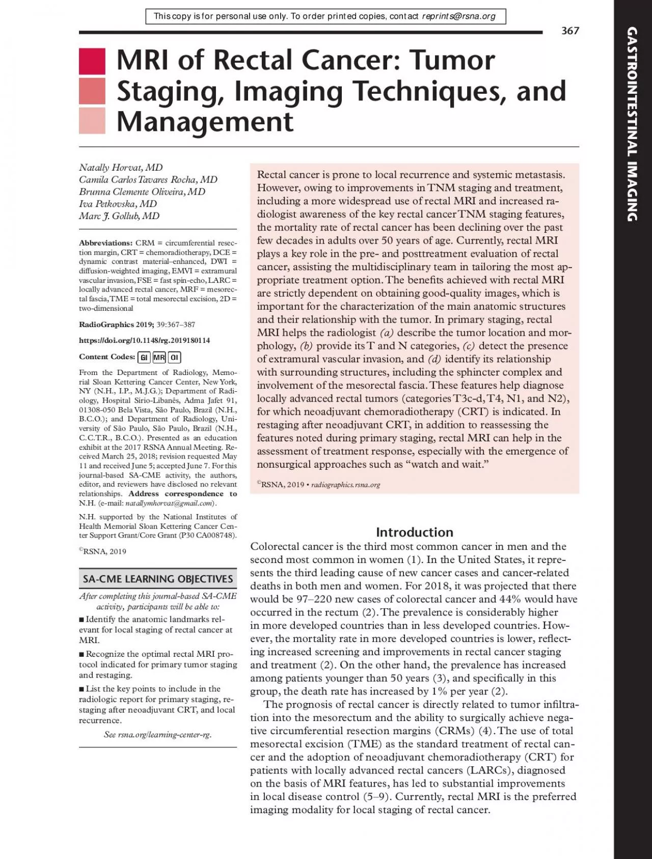
367 Rectal cancer is prone to local recurrence and systemic metastasis However owing to improvements in TNM staging and treatment diologist awareness of the key rectal cancer TNM staging features ID: 938830

cancer rectal mri tumor rectal cancer tumor mri imaging staging patients response weighted lymph nodes local crt treatment tumors

Share:

Link:

Embed:

Download Presentation from below link

Download Pdf The PPT/PDF document "MRI of Rectal Cancer Tumor Staging Imagi..." is the property of its rightful owner. Permission is granted to download and print the materials on this web site for personal, non-commercial use only, and to display it on your personal computer provided you do not modify the materials and that you retain all copyright notices contained in the materials. By downloading content from our website, you accept the terms of this agreement.


Presentation Transcript

367 MRI of Rectal Cancer: Tumor Staging, Imaging Techniques, and Management Rectal cancer is prone to local recurrence and systemic metastasis. However, owing to improvements in TNM staging and treatment, diologist awareness of the key rectal cancer TNM staging features, the mortality rate of rectal cancer has been declining over the past few decades in adults over 50 years of age. Currently, rectal MRI plays a key role in the pre- and posttreatment evaluation of rectal Natally Horvat, MDCamila Carlos Tavares Rocha, MDBrunna Clemente Oliveira, MDIva Petkovska, MDMarc J. Gollub, MDAbbreviations:tion margin, CRT = chemoradiotherapy, DCE = dynamic contrast material–enhanced, DWI = diffusion-weighted imaging, EMVI = extramural From the Department of Radiology, Memorial Sloan Kettering Cancer Center, New York, After completing this journal-based SA-CME Identify the anatomic landmarks relevant for local staging of rectal cancer at tocol indicated for primary tumor staging and restaging.radiologic report for primary staging, restaging after neoadjuvant CRT, and local recurrence. SA-CME L Colorectal cancer is the third most common cancer in men and the second most common in women (1). In the United States, it represents the third leading cause of new cancer cases and cancer-related deaths in both men and women. For 2018, it was projected that there would be 97–220 new cases of colorectal cancer and 44% would have occurred in the rectum (2). The prevalence is considerably higher in more developed countries than in less developed countries. However, the mortality rate in more developed countries is lower, reect GASTROINTESTINAL March-April 2019during follow-up for the early diagnosis of local recurrence. MRI in the recurrence setting may contribute to management by outlining disease extension within the pelvis and providing a road map to determine the resectability of lesions and the best surgical approach (21).In this article, we review rectal MRI techniques, relevant anatomic landmarks with correlating MRI ndings, and current concepts in the management of patients with rectal cancer, including the most common surgical techniques. Furthermore, we discuss the role of MRI in the assessment of local staging, restaging, and recurrence and the future directions of imaging research in rectal cancer. We also propose a schematic step-by-step approach to facilitate comprehension of the key features to address in a rectal MRI radiologic report, represented by the mnemonic RECTAL CANCER (for medical cords, valuate MRI sequences, ance, category, ocation of the tumor, RM status, nalyze prior MR images, category, sphincter omplex status, MVI status, gression).Table 1 demonstrates TNM staging of rectal cancer, where represents the tumor, sents the lymph nodes near the tumor, and (11,12). The prexes , and represent clinical, pathologic, and postneoadjuvant therapy, respectively. The management concepts of rectal cancer in the United States and Europe are summarized Surgical resection is still considered the curative treatment of rectal cancer. The surgical techniques may vary depending on the location and extent of the disease.Transanal endoscopic microsurgery (Fig 2a) is characterized by a focal endoscopic resection of the tumor and can be indicated for select patients with early rectal cancer. The selection criteria for transanal endoscopic microsurgery include well- or moderately differentiated rectal cancers and tumors that are categorized as cT1 or cN0, are less than 3 cm, are within 8 cm of the anal verge, or involve less than 30% of the wall circumference (12,23).TME is the standard transabdominal surgery indicated for the curative treatment of rectal cancer (10). After its implementation, patient outcome and quality of life improved considerably. TME involves a complete resection of the Low anterior resection (Fig 2b) is the most common transabdominal resection indicated for tumors located in the middle or upper rectum. The standard treatment in patients with MRI-staged LARC is neoadjuvant CRT followed by TME (10,11). In approximately half of patients, the disease is downstaged after CRT, and almost one-third of patients demonstrate complete pathologic response after undergoing TME (12–15). Habr-Gama et al (13) and other authors (16–18) have shown that select patients with clinical complete response to CRT can be safely followed up in a nonsurgical approach.Rectal MRI may add value in patient care in various scenarios. In primary staging (preoperative setting), MRI can assist in tients with LARC who are suitable for treatment with neoadjuvant CRT; surgical planning; and nostic factors, including extramural vascular invasion (EMVI), mucin content, and involvement of the mesorectal fascia (MRF) (19,20). In the restaging setting (after treatment with neoadjuvant CRT), rectal MRI can help in evaluating tumor regression; tailoring surgical planning; detecting a complete clinical response, along with a review of the results of digital rectal examinations and endoscopic procedures; and monitoring patients undergoing the nonsurgical treatment approach. Finally, after local treatment, performing rectal MRI is relevant The use of TME as the standard treatment of rectal cancer and the adoption of neoadjuvant CRT for patients with LARCs, diagnosed on the basis of MRI features, has led to substantial improvements in local disease control. Currently, rectal MRI is the preferred imaging modality for local staging In primary staging (preoperative setting), MRI can assist in guiding surgeons in surgical planning; and identifying poor prognostic factors, including EMVI, mucin content, and involvement of the MRF. portant MRI sequence in the evaluation of rectal cancer and anatomic structures. Standardized imaging protocols also allow for more accurate and reproducible interpretations, formed, it is important to verify the neoadjuvant treatment the patient underwent and to evaluate the results of previous treatment MRI) to understand the primary tumor’s location and morphology. The main risk factors for local recurrence are lack of preoperative radiation therapy, CRM positivity, EMVI, close proximity of the tumor to the anal verge, perforat

ion of the tumor at surgery, anastomotic leak, higher pathologic TNM stage, and lower tumor differentiation. Although 30% of patients can be asymptomatic, the majority of patients with local recurrence manifest with symptoms and an increased carcinoembryonic RG • VolumeHorvatThis technique is characterized by TME and the resection of the whole sigmoid or part of it.Ultra-low anterior resection is a sphincter-sparing surgery that can be performed in patients with low rectal cancer above the anorectal junction. The coloanal anastomosis is created 1 cm distal to the lower edge of the tumor in this procedure.Standard abdominoperineal resection (Fig 2c) with TME is indicated for tumors that inltrate the anal canal or the levator ani and/or external sphincter, located less than 1 cm from the anal verge or in cases where the resection will result in incontinence. It is characterized by the resection of the sphincter complex, resulting in a permanent colostomy.Intersphincteric abdominoperineal resection (Fig 2d) is a sphincter-sparing surgery that can be considered in cases where the intersphincteric plane is not inltrated by the tumor. Therefore, the dissection is performed within the intersphincteric plane and the external sphincter is preserved.Extralevator abdominoperineal resection (Fig 2e) is indicated for tumors that inltrate the intersphincteric plane and external sphincter and/or levator ani. The technique consists of a consequently avoids the “waist” effect that is created in a standard abdominoperineal resection, thus creating a cylindrical specimen. This surgery aims to reduce bowel and tumor perforation during the surgery and to avoid positive CRM.The potential benets achieved with rectal MRI are strictly dependent on obtaining good-quality images to allow for characterization of the main anatomic structures and their relation to the tumor. High-spatial-resolution T2-weighted imaging is the most important MRI sequence in the evaluation of rectal cancer and anatomic structures. Standardized imaging protocols also allow for more accurate and reproducible interpretations, which facilitate the widespread use of this technique (24). Figure 3 summarizes the MRI techniques frequently recommended (“Dos”), those that are not recommended (“Don’ts”), and some that are controversial practices (“Maybes”) that may be performed in select cases.Patients must be informed about the time required for imaging, and they must be positioned comfortably in the supine position in the MR imager (20). High-eld-strength MRI provides fast image acquisition, high spatial resolution, and high signal-to-noise ratio, improving the visibility of the rectal wall (25). Ideally, higher eld strengths (eg, 1.5 T or 3.0 T) are preferred, with some studies demonstrating similar accuracies for staging for both (26–28). While 1.5 T is the most widely available and used, 3.0 T may improve spatial resolution, with an increase of the signal-to-noise ratio, and may be preferable to 1.5 T. However, some experts cite greater magnetic susceptibility artifact at 3.0 T, which may occur during DWI, as a potential disadvantage (29,30). Pelvic phased-array surface coils are recommended and must cover from the aortic bifurcation to the anal verge. TableCategoryDescriptorT categoryPrimary tumor cannot be assessedNo evidence of a primary tumorCarcinoma in situ: intraepithelial or invasion of the lamina propriaMuscularis propriaSubserosa and perirectal tissueTumor penetrates to the surface of the visceral peritoneumTumor invades or is adherent to other organs or structuresN categoryRegional lymph nodes cannot be asNo regional lymph node metastasisTumor deposit(s) in the subserosa, mesentery, or nonperitonealized perirectal tissues7 or more regional lymph nodesM categoryMetastasis conned to one organ or site (eg, liver, lung, nonregional lymph or site or in the peritoneumThe subclassication of the T3 category is determined on the basis of an MRI evaluation and is used in the European guidelines for treatment recommendations (11). March-April 2019use of an endorectal coil it is also not endorsed owing to patient comfort and cost (20,28). In regard to MRI sequences, T2-weighted imaging with fat suppression is not routinely recomFor certain MRI techniques in imaging rectal cancer, there is no consensus. Administering spasmolytic agents such as glucagon (1 mg administered intravenously, intramuscularly, or subcutaneously) or hyoscine butylbromide (20 mg administered intravenously) is not mandatory but may reduce artifacts caused by peristalsis when administered immediately before the examination or just before the most motion-sensitive sequences (eg, DWI or dynamic contrast material–enhanced [DCE] sequences) are obtained (26). cause, although it may facilitate detection of small tumors with rectal distention, it may alter staging owing to compression of the mesorectal fat. This can change the distance of the tumor to the MRF, possibly leading to nonvisualization of the mesorectal nodes (35). However, there are some studies that favor endorectal lling that can be found in the literature (28,36).Another optional MRI sequence includes DWI value (), which may improve the diagnostic performance of MRI for tumor restaging after CRT. For primary staging, it may improve tumor and lymph node detection, although it is not ofcially recommended (20,25,26,31,37). Using a microenema 15 minutes before performing DWI may help remove rectal air and reduce artifacts, which can be Schematic owchart ment concepts of rectal cancer in the United States and Europe. The standard rectal MRI protocol in the evaluation of rectal cancer includes performing two-dimensional (2D) FSE T2-weighted sequences without fat suppression, using a small eld of view and a section thickness less than 3 mm (high-resolution protocol) (26). Images in this sequence should be obtained in the oblique axial plane (perpendicular to the tumor), as incorrect plane obliquity leads to blurring of the muscularis propria, which can cause incorrect T staging (31); sagittal plane, which is determined by the longitudinal tumor axis; andoblique coronal p

lane (parallel to the anal canal), which is important to depict low rectal tumors and to better evaluate their relationship with the anal sphincter. These sequences have a proven high diagnostic accuracy, between 90% and 100%, for the evaluation of tumor invasion into the MRF and adjacent organs and are recommended by the Magnetic Resonance Imaging and Rectal Cancer European Equivalence (MERCURY) group (19).FSE T2-weighted MRI with a large eld of view without fat suppression obtained in the axial plane of the entire pelvis, from the aortic bifurcation to the sphincter, allows for evaluation of distant lymph node chains (eg, inferior mesenteric, lateral, and inguinal). In the sagittal plane, from one side of the pelvic wall to the other, FSE T2-weighted MRI allows for localization of the primary tumor, enabling the measurement of its height and its relationship to the midline structures, such as the anal verge (25,28).It is not recommended to use a routine bowel preparation such as air insufation to distend the rectum with any contrast material or to use intravenous contrast material (20,25,26,31–34). The RG • VolumeHorvatparticularly helpful for assessing residual tumor at restaging MRI (38). Three-dimensional T2-weighted MRI is not routinely recommended but may be useful for evaluating response to neoadjuvant therapy. However, 2D imaging still seems preferable (39). T1-weighted imaging with a wider eld of view may help in assessment of the common iliac and lower para-aortic nodes or incidental ndings in the pelvis (with the same principle of FSE T2-weighted MRI with a large eld of view) and especially the bones. It may also be useful in cases of mucinous neoplasm when T2 signal intensities can be identical to those of fat.It is known that contrast-enhanced T1-weighted imaging does not improve the diagnostic accuracy of local staging of rectal cancer (20). Studies found no difference for distinction between T1-T2 and borderline T3 tumors for the evaluation of tumor extension into the MRF (40). However, particularly at restaging, the use of intravenous contrast material may help identify local recurrence, depicted on images as heteroge Illustrations of the anatomy of the rectum depict various surgical techniques used to treat rectal cancer. Dotted blue Illustration depicts a low anterior resection and TME and resection of the whole sigmoid or part of it, which preserves the sphincter complex. Illustration depicts an abdominoperi Illustration depicts an intersphincteric abdominoperineal al Illustration depicts an extralevator abdominoperineal resection and TME, with a broader dissection March-April 2019and some that are controversial pracwith fat saturation is rarely necessary but TableVendorsImaging ParametersAxial T2-weightedMatrixSagittal T2-weightedMatrixCoronal T2-weightedMatrixOblique axial T2-weightedMatrixNote.—Field of view and section thickness are measured in millimeters. TR/TE is measured in milliseconds. ETL = echo train length, FRFSE = fast relaxation FSE, TE = echo time, TR = repetition time, TSE = turbo spin-echo. RG • VolumeHorvatThe main MRI parameters of the most imporequipment vendors are summarized in Table 2.It is imperative for radiologists to be familiar with key anatomic landmarks of the rectum to provide an accurate local staging of rectal cancer. High-resolution T2-weighted MRI is the main sequence performed for the evaluation of relevant structures. Figure 4 summarizes the main anatomic landmarks seen at rectal MRI, provides details regarding which imaging plane is best suited for the identication of each landmark, and describes their clinical relevance.In the context of primary tumor staging, performing rectal MRI is important for the evaluation of tumor location and morphology, T category, anal sphincter complex involvement, CRM status, involvement of the pelvic sidewall, EMVI, and N category. These features should be included in the rectal MRI report. A suggested radiologic report template for primary staging It is crucial to describe the tumor location in the craniocaudal direction (lower, middle, or upper face position), as well as its length, relationship to the anterior peritoneal reection, and distance from the inferior border of the tumor to the anal verge and the anorectal junction. This information helps determine the best surgical approach. The location of the tumor is categorized as low (0–5 cm from the anal verge), middle (5.1–10 cm ing appearance. , Illustration and MR images in the oblique axial view best depict the MRF, mesorectum, rectal wall layers, , Illustration and MR image in the sagittal view best depict the retrorectal space, anorectal ring, and anal verge. Curved arrows = anterior peritoneal reection, white = mesorectum. , Illustration and MR image in the coronal view best depict the internal sphincter, external sphincter complex, and intersphincteric space. (Illustrations adapted and reprinted, under a CC BY-ND 4.0 license, from Memorial Sloan Kettering Cancer Center.) March-April 2019from the anal verge), and high (10.1–15 cm from the anal verge) (Fig 6). Tumors located above 15 cm from the anal verge are treated as colon cancer and, consequently, their staging and treatment differ from those of rectal cancer.The tumor’s morphologic pattern (polypoid, ulcerating, circumferential, or semicircumferential) and especially its appearance (nonmucinous or mucinous) should also be described (Fig 7). Mucinous tumors show high signal intensity at T2-weighted MRI and have a worse prognosis than that of nonmucinous tumors, with a higher metastatic propensity and often a higher stage at Radiologic report template lists the key imaging ndings and features that should be evaluated at primary staging RG • VolumeHorvatSometimes identifying the tumor may be challenging for radiologists who are not experienced in reading rectal MR images in daily practice. Two imaging characteristics can be helpful in tumor identication: rectal cancer usually appears with elevated borders and can accumulate mucoid material in the rectal lumen of the tumoral region, which can also be visualized in nonmuciTumor StagingFigure 9 demonstra

tes the location of rectal cancer and its corresponding T category. Diagnostic accuracy, sensitivity, and specicity of high-resolution rectal MRI in assessing T category are 85%, 87%, and 75%, respectively (43). T category is characterized by the depth of tumor penetration into the rectal wall and extramural spread into the mesorectum and adjacent structures. It is important to identify the most invasive portion of the tumor, corresponding to the area of deepest inltration, which is usually located halfway in the craniocaudal direction and at the center of the C shape depicted on oblique axial images (Fig 8). The T category is better applied to mid- and high rectal cancers and differs from that of low rectal cancer, especially owing to the narrowing of the mesorectum, which is a barrier to circumferential tumor spread (25,44), with a resultant higher risk of involvement of the MRF (45).T1 tumors inltrate the submucosa, and T2 tumors extend into the muscularis propria. Rectal MRI does not provide a reliable distinction between these two categories, except in some patients with T1 tumors when it is possible to identify a preserved submucosal layer (hyperintense signal) beneath the lesion (46,47). Therefore, patients should undergo endorectal US owing to its superior diagnostic performance in these cases (26).T3 tumors are characterized by a discontinuity of the muscularis propria, with extension of the tumor into the mesorectum without inltration of the MRF or adjacent organs (20) (Fig 10c). They are classied into four categories dependent on the distance between the outermost edge of the Figure 6.Tumor location in the craniocaudal direction. Illustration depicts the sagittal view of the rectum and provides the measurements of the tumor from the anal verge, which help categorize tumor location. Blue lines separate the low, mid-, and high rectum. (Figure 6a reprinted, under a CC BY-ND 4.0 license, from Sagittal T2-weighted MR images measurement from the rectum entrance to the tumor location. March-April 2019muscularis propria and the maximum extramural spread of the tumor (T3a, T3b, 1–5 mm; T3c, 5–15 mm; and T4d, mm;&#x;40.;က 15 mm). Differentiating T2 tumors from early T3 tumors can be difcult (48). Penetration into the muscular layers by small vessels and desmoplastic reaction are common pitfalls that can lead to overstaging a T2 tumor as a T3 tumor (20). Desmoplastic reaction is depicted as spicules with low signal intensity at T2-weighted imaging, while T3 tumors have mediate signal intensity at T2-weighted imaging (20). Lastly, T4 tumors are those that inltrate the peritoneal reection (T4a) or other pelvic organs and structures (T4b).In patients with low rectal cancer, radiologists play a pivotal role in preoperative evaluation. Accurate staging is required to determine the need for neoadjuvant CRT or more extensive surgery and to provide the surgeon with a guide for planes of excision (45,49). Conventional staging is insufcient because tumors in the lower rectum are in close proximity to the anal sphincter complex and are more likely to invade the MRF and adjacent organs, with positive surgical margins in about 30% of cases owing to the narrowing of the mesorectum in this location (49). Mucinous and nonmucinous tumors. Axial oblique T2-weighted MR images in two different patients show a mucinous tumor (arrow in ) and a nonmucinous tumor (arrow in ). Mucinous tumors ), and two 7-mm round heterogeneous mesorectal lymph nodes (solid arrows in ) are depicted, making this RG • VolumeHorvatIllustration depicts the anatomy of the rectum and the possible locations of rectal cancer, along with corresponding T categories and potential tumor sizes for each location. Rectal MR images that show distinct tumor stages obtained from three and T2-weighted MR images show a polypoid lesion (solid arrow) surrounded tached to the rectal wall and the intact muscularis propria (dashed arrow), ndings characteristic of a T1 or T2 tumor. Oblique axial T2-weighted MR image ing 7 mm beyond the muscularis propria Oblique axial T2-weighted mor invading the anterior peritoneal reTaylor et al (50) revised a specic staging system on the basis of invasion of the anal sphincter complex owing to the extension through the muscular layer for surgical planning and the risk of traditional abdominoperineal resection. The report should describe if the tumor invades the internal sphincter, intersphincteric plane, and external sphincter and/or levator ani (Fig 11). this evaluation at T2-weighted MRI.CRM is the surface of the nonperitonealized part of the rectum that is resected during surgery. MRI is the most reliable imaging modality to determine potential CRM involvement (43,51). At MRI, CRM status can be obtained by measuring the shortest distance between the outermost part of the rectal tumor and the MRF (52). The CRM status is potentially positive if this measurement is less than 1 mm, and threatened if it is between 1 and 2 mm (33). It is important to highlight that the rectum is not entirely surrounded by MRF (Fig 12), and thus CRM status is not applicable if the tumor is situated in a peritonealized aspect of the rectal wall.is a reliable predictor for negative margins after March-April 2019 semicircumferential tumor (arrowhead) in the lower rectum. (c, d) Axial oblique and coronal T2-weighted MR images show the tumor (solid arrow in ) inltrating beyond the muscularis propria tal and axial views of the peritoneal and MRF coverage of the rectum. Note that the potential CRM described in the radiologic report corresponds to the distance between the tumor and the MRF and does not include the portions of the rectum surrounded by peritoneum. (Reprinted, under a CC BY-ND 4.0 license, from Memorial TME (50). On the other hand, a positive CRM is the most important predictor of local recurrence and poor survival (53). Therefore, every report should include the CRM status and the location of potential involvement (clock-face method).In T4b tumors, it is important to describe if adjacent structures are involved, including the uterus, vagina, prostate gland, seminal vesicles, ureters, presacral fascia, sacra

l nerve roots, sacrum, iliac vessels, and pelvic muscles.VascularMRI can depict EMVI with moderate sensitivity and high specicity, which is an important prognostic factor and predictor of metastatic disease RG • VolumeHorvat(54–56). EMVI is an extension of the tumor to the vessels in the mesorectum, resulting in wall irregularity, focal enlargement, and/or signal intensity of the tumor (intermediate at T2-weighted imaging) within the vessel (56) (Fig 13).LymphCompared with the accuracy of MRI in tumor staging, the accuracy of MRI in assessing the involvement of metastatic lymph nodes in rectal cancer is less accurate, which is an important prognostic factor and indicator for the use of neoadjuvant CRT (43). The presence, number, and precise location of suspicious lymph nodes should be reported.The proximity between the suspicious lymph nodes and the MRF is also important to report for surgical planning, although it has been shown that it does not confer poor prognosis in the same manner as that of the primary tumor (12).As a large proportion of metastatic lymph nodes in rectal cancer measure less than 5 mm, size is not a reliable criterion (57,58). However, some studies have demonstrated that lymph nodes measuring greater than 8 mm in the short axis are highly specic for metastatic involvement (26,59,60). Therefore, it has been proposed for nodal assessment to include size and morphologic characteristics of malignancy, including the presence of irregular borders, heterogeneous signal intensity, and round shape (Table 3) (Fig 8) (33,57).Regional lymph nodes involved in rectal cancer include the mesorectal, superior rectal, middle rectal, inferior rectal, sigmoid mesenteric, inferior mesenteric, lateral sacral, presacral, sacral promontory, or internal iliac (61). Lymph nodes tases (M1). Extramesorectal nodes are important to describe, including those along the pelvic sidewall, as they are a negative prognostic predictor and are not routinely resected (62). Lesions that inltrate the presacral space can manifest with retroperitoneal lymph nodes; therefore, those chains are also important to evaluate. A group of tumor cells not associated with lymphoid or vascular tissues, dened as , are characterized as N1c. terized by focal enlargement of the vessel, signal intensity of the tumor replacing the ow void, and wall TableLymphNumber of malignant morphologic criteria that have manifestedTwoIrregular borders, heterogeneous signal intensity, and round shape. Measured in the largest short axis.  indicates not suspicious for malignancy, + indicates suspicious for malignancy. March-April 2019 obtained during primary staging shows an inltrative tumor (arrow) with the most invasive border between the 1-o’clock obtained after shows a scar in the tumor bed, without residual tumor. Note the wall thickening and mucosal edema within the normal rectal ). The patient was selected for a watch-and-wait protocol. Oblique axial T2-weighted MR image obtained 9 months later shows thickening in the tumor bed, with Colonoscopic image For patients with LARC, neoadjuvant CRT is considered the standard treatment. It has been shown to improve local control, inducing tumor downstaging in approximately 50% of patients, and results in pathologic complete response in 15%–38% of cases (13–15,63,64). This can allow for a sphincter-preserving surgery to be performed or may even offer a nonsurgical treatment approach in some patients. RG • VolumeHorvatDigital rectal examination and endoscopy have been used to evaluate pathologic complete response, but these assessments are limited to the luminal view, leaving residual tumors in other layers of the bowel wall undetected (65). In this context, MRI has an important role in the assessment of tumor response after neoadjuvant CRT.Before the restaging of rectal cancer after rectal MRI is performed, it is important to verify the neoadjuvant treatment the patient underwent and to evaluate the results of previous examinations (digital rectal examination, endoscopy, and pretreatment MRI) to understand the primary tumor’s location and morphology. The normal rectal wall adjacent to the tumor can manifest with post-CRT changes such as submucosal edema (thickened and intermediate to high signal intensity on T2-weighted images) that can lead to a common pitfall usually misinterpreted as residual tumor (Fig 14c). After treatment, the tumor may be similar in appearance to that of the pretreatment tumor or may appear atrophic and brotic, with low signal intensity on T2-weighted images, which As described previously, mucin within the tumor appears as an area of high signal intensity on T2-weighted images. Regarding this characteristic, tumors can manifest with one of three different mucin responses after CRT:1. Mucin (or colloid degeneration) response can occur in nonmucinous tumors that become mucinous after CRT (66). It indicates a response to treatment and better prognosis (4).2. Acellular mucin response represents a pathologic response of a mucinous tumor with no impact on recurrence-free survival (67). Until recently, there has been no reliable imaging method to differentiate cellular from acellular mucin.3. Mucinous tumor without response is characterized as a mucinous tumor at the primary staging that did not respond to CRT. It is related to an increased risk of local recurrence and poor Comparing pretreatment MR images with posttreatment MR images is important to differentiate colloid degeneration in a nonmucinous tumor from a genuinely mucinous tumor.TumorA residual tumor has intermediate signal intensity on T2-weighted images whereas brosis and/or scaring has low signal intensity. However, differentiation is still challenging as residual tumor may occur within a scar. Additional functional MRI sequences, including DWI, have demonstrated promising results in tumor restaging at MRI. Fibrosis demonstrates low signal intensity -value diffusion-weighted images, while residual tumor shows high signal intensity (Fig 15). In the tumor restaging setting, performing a microenema is suggested before performing recrectum to decrease artifacts at DWI (68,69).A tumor regression grading

system for rectal cancer has been proposed (Table 4) and demonstrates a correlation with survival outcomes (19). Regression grades range from 1 to 5, with grade 1 indicating complete radiologic response to treatment and grade 5 indicating no response.The accuracy of rectal cancer staging at MRI in post-CRT tumors is lower than that of primary staging at MRI (70). Therefore, it is important to use a multidisciplinary approach and a combination of modalities to assess response to treatment, including MRI, clinical assessment, and endoscopy. During tumor restaging, it is also important to evaluate the sphincter complex and pelvic side wall status in a similar fashion to that which is performed at primary staging.The CRM status and the smallest distance between the remaining tumor and the MRF must also be described in the radiologic report, although this evaluation is less accurate than that of the pretreatment assessment (51). The importance of reporting EMVI at rectal cancer restaging, which can disappear after treatment and be replaced by brotic tissue, is still unclear (66).must be reported at restaging. Many irradiated lymph nodes disappear, and the majority of the remaining nodes are sterilized. After CRT, evaluating nodal size in the short axis is more reliable than evaluating borders and shape to assess for residual malignancy (33). The absence of lymph nodes at DWI, the decrease in size in at least 70% of lymph nodes, and a nodal size less than 2.5 mm in the short axis have been shown to be reliable predictors of negative node status after surgery (Fig 16) (71).Due to the advent of neoadjuvant CRT and improvements in rectal surgery, the prevalence of recurrent rectal cancer began to decline in the past decade, occurring in approximately 4%–8% of patients who underwent surgery performed with a curative intent, especially in the rst 3 years after treatment (72). Early diagnosis is fundamental to avoid progression and enable surgical resection (21).The main risk factors for local recurrence are lack of preoperative radiation therapy, CRM March-April 2019 Oblique axial T2-weighted MR image obtained after neoadjuvant CRT at restaging shows areas of low signal intensity (black TableTumorComplete radiologic response: no evidence of treated tumorGood response:umor dense brosis (75%); no obvious residual tumor, signifying minimal residual disease, or no tumorModerate response:umor 50%brosis or mucinvisible intermediate signal intensitySlight response: little areas of brosis or mucin,but mostly tumorNo response: intermediate signal intensity; same appearance as that of the original tumorNote.—TRG = tumor regression grade. Data are from references 20 and 25. positivity, EMVI, close proximity of the tumor to the anal verge, perforation of the tumor at surgery, anastomotic leak, higher pathologic TNM stage, and lower tumor differentiation (73). Although 30% of patients can be asymptomatic, the majority of patients with local recurrence manifest with symptoms and an increased carcinoembryonic antigen level.Local recurrence can occur in four locations: axial, with recurrence in the anastomotic, residual mesorectum, or perirectal soft tissue in the center of the pelvis or perineum, including the pelvic oor; anterior, with recurrence in the bladder, vagina, uterus, seminal vesicles, or prostate; posterior, with recurrence in the presacral fascia, sacrum, coccyx, or sacral root sheaths; and lateral, with recurrence in the pelvic ureters, iliac vessels, lateral lymph nodes, pelvic nerves, sidewall muscles, or lateral pelvic bones.Most local recurrences are anastomotic and therefore are easily identied at clinical evaluation and/or endoscopy. However, other recurmethods. In this context, imaging is an important tool in early diagnosis, especially in asymptomatic patients. The main role of imaging is to verify the extent and precise localization of the disease, as well as the presence of metastases. RG • VolumeHorvatMRI is the most accurate imaging modality for local staging, while CT and PET/CT are more useful in detecting distant recurrence. However, posttreatment (surgery or CRT) changes may be difcult to differentiate from local recurrence, as they can share similar imaging features and may also appear uorodeoxyglucose avid at PET. An increase in size and early, heterogeneous, marked contrast enhancement, invasive behavior, and asymmetric appearance are suspicious for local recurrence (21,47,74). The contraindications for pelvic exenteration are shown in Table 5 (75,76) and should be described in the radiologic report.Figure 17 summarizes a schematic step-by-step approach to facilitate comprehension of the key features that should be addressed and described in a rectal cancer MRI report. The key features are also represented by the mnemonic RECTAL Future Directions and ResearchSome novel MRI techniques, which are not currently used in routine clinical practice, have been studied to overcome some limitations of MRI in the evaluation of rectal cancer during primary staging and restaging. The main novel techniques that have been studied in the evaluation of response after CRT are DCE MRI, magnetization transfer ratio (MTR), and textural analysis (eg, radiomics). Figure 16.No signicant change in lymph node size after CRT. Axial T2-weighted MR image obtained Axial T2-weighted MR obtained 10 months after TME without lateral pelvic lymph node TableInltration of the proximal sacrum (S2 or higher Invasion of the proximal lumbosacral plexus and sciatic nervesEncasement of the external or common iliac vessels March-April 2019In regard to lymph node assessment, novel contrast material and PET/MRI have been described as possible means of improving the evaluation of nodal status. Furthermore, some of these techniques have been assessed as imaging biomarkers to predict clinical outcomes.DCE MRI assesses tumor vascularization, which can help in tumor identication and may correlate with the degree of angiogenesis and tumor aggressiveness and may predict response to neoadjuvant CRT. Volume transfer content ) is the volume transfer coefcient that reects vascular permeability of gadolinium from b

lood into the extravascular extracellular space. Dijkhoff et al (77) published a systematic review revealing that high K values at primary stag values after CRT are signicant predictors of response (77).MTR is a technique that evaluates differences in magnetization interaction between the protons bound to macromolecules and those in free water, which corresponds to the efciency of this exchange. Tissues that are rich in macromolecules (such as those in collagen that occur in brosis) will express a high MTR. Studies have demonstrated the potential of MTR in the assessment of tumor response after CRT (78,79).tion of many quantitative features from large imaging datasets (“big data”) derived from CT, MRI, and PET, with the potential to correlate these image phenotypes that are otherwise not detectable by conventional radiologic human interpretation with disease outcomes (80). In rectal cancer, radiomics using MRI has demonstrated promising pilot results in the prediction of complete response after CRT (81–83) and using PET/CT as a predictor of survival (84).The use of lymph node MRI contrast material, such as ultrasmall superparamagnetic iron oxide (USPIO) and gadofosveset, is promising for differentiating benign from metastatic lymph nodes, but availability and safety concerns have mostly prevented their use (85,86). USPIO is an iron-based nanoparticle that is taken up by normal cells, decreasing their signal intensity at T2-weighted imaging. Malignant nodes do not take up USPIO particles and have a higher signal intensity relative to that of benign nodes and are enhanced relative to that of normal tissue. Gadofosveset is an albumin-bound gadolinium chelate that is taken up by normal and reactive lymph nodes, which enhance like vessels. Thus, malignant nodes show less enhancement.PET/MRI has demonstrated higher accuracy in T staging and at least comparable accuracy in N and M staging compared with those of PET/CT owing to the high soft-tissue contrast enhancement depicted at MRI (87). This strength may optimize local and distant staging and Chart shows a step-by-step approach to imaging and staging rectal cancer. The mnemonic RECTAL CANCER facilitates ps to RG • VolumeHorvatpotentially improve the diagnostic performance PET. However, studies with larger sample sizes are required to evaluate the importance of this modality in rectal cancer evaluation. FDG PET/CT and PET/MRI may also be helpful for restaging (67). In a meta-analysis with a total of 538 patients with locally advanced rectal cancer, Rymer et al (88) found that post-CRT PET/CT showed a statistically signicant reduction in the maximum standardized uptake value in histopathologic responders and higher response index values. The sensitivities of PET/CT in the prediction of pathologic complete response and identication of distant metastases were 75% and 97%, respectively (89).may add value in patient care, further research is still required to provide consistent results and standardized technical parameters before PET/for patients with rectal cancer.Currently, rectal MRI plays a key role in management of patients with rectal cancer in local staging, identifying risk factors for local and distant recurrence to help tailor treatment, and improving patient outcome. A systematic analysis allows for a uniform and reproducible interpretation.The authors would like to express their deepest gratitude to Joanne Chin, MFA, for her editorial support and to MSK Design and Creative Services.1.Ferlay J, Soerjomataram I, Dikshit R, et al. Cancer incidence and mortality worldwide: sources, methods and major patterns in GLOBOCAN 2012. Int J Cancer 2.American Cancer Society. Cancer facts and gures: and-figures/2018/cancer-facts-and-figures-2018.pdf. in the United States, 1975-2010. JAMA Surg 2015;150 Nagtegaal I, Gaspar C, Marijnen C, Van De Velde C, Fodde R, Van Krieken H. Morphological changes in tumour type after radiotherapy are accompanied by changes in gene expression prole but not in clinical behaviour. J Pathol Heald RJ, Ryall RD. Recurrence and survival after total mesorectal excision for rectal cancer. Lancet 1986;1(8496): Krook JE, Moertel CG, Gunderson LL, et al. Effective surgical adjuvant therapy for high-risk rectal carcinoma. N Gastrointestinal Tumor Study Group. Prolongation of the disease-free interval in surgically treated rectal carcinoma. National Institutes of Health. Adjuvant therapy for patients with colon and rectum cancer. NIH Consensus Statement Sauer R, Becker H, Hohenberger W, et al. Preoperative versus postoperative chemoradiotherapy for rectal cancer. 10.National Comprehensive Cancer Network. Clinical practice physician_gls/pdf/colon.pdf. Published 2018. Accessed Glimelius B, Tiret E, Cervantes A, Arnold D; ESMO Guidelines Working Group. Rectal cancer: ESMO Clinical Practice Guidelines for diagnosis, treatment and follow-up. org/professionals/physician_gls/pdf/rectal.pdf. Published Habr-Gama A, Perez RO, Nadalin W, et al. Operative versus nonoperative treatment for stage 0 distal rectal cancer following chemoradiation therapy: long-term results. Ann Maas M, Nelemans PJ, Valentini V, et al. Long-term outcome in patients with a pathological complete response after chemoradiation for rectal cancer: a pooled analysis of PÃ¥hlman L, Bohe M, Cedermark B, et al. The Swedish 16.Maas M, Beets-Tan RG, Lambregts DM, et al. Wait-and-see policy for clinical complete responders after chemoradiation 17.Renehan AG, Malcomson L, Emsley R, et al. Watch-and-wait approach versus surgical resection after chemoradiotherapy for patients with rectal cancer (the OnCoRe project): a propensity-score matched cohort analysis. Lancet Oncol Li J, Liu H, Yin J, et al. Wait-and-see or radical surgery for rectal cancer patients with a clinical complete response after neoadjuvant chemoradiotherapy: a cohort study. Oncotarget Patel UB, Taylor F, Blomqvist L, et al. Magnetic resonance imaging-detected tumor response for locally advanced rectal cancer predicts survival outcomes: MERCURY experience. Taylor FG, Swift RI, Blomqvist L, Brown G. A systematic approach to the interpretation of preoperative staging MRI for rectal cancer. AJR Am J Roentgenol 2008;191(6):1827

0;1835.Sinaei M, Swallow C, Milot L, Moghaddam PA, Smith A, Atri M. Patterns and signal intensity characteristics of pelvic recurrence of rectal cancer at MRI. RadioGraphics 22.Glimelius B, Oliveira J; ESMO Guidelines Working Group. nosis, treatment and follow-up. Ann Oncol 2008;19(suppl Willett CG, Compton CC, Shellito PC, Erd JT. Selection factors for local excision or abdominoperineal resection of Suzuki C, Torkzad MR, Tanaka S, et al. The importance of rectal cancer MRI protocols on interpretation accuracy. view and update on recent advances. AJR Am J Roentgenol Beets-Tan RGH, Lambregts DMJ, Maas M, et al. Magnetic resonance imaging for clinical management of rectal cancer: updated recommendations from the 2016 European Society -lished correction appears in Eur Radiol 2018;28(6):2711.]27.))) Delli Pizzi A, Basilico R, Cianci R, et al. Rectal cancer MRI: protocols, signs and future perspectives radiologists should consider in everyday clinical practice. Insights Imaging Gollub MJ, Arya S, Beets-Tan RG, et al. Use of magnetic resonance imaging in rectal cancer patients: Society of Abdominal Radiology (SAR) rectal cancer disease-focused panel (DFP) recommendations 2017. Abdom Radiol (NY) March-April 2019son of accuracy of local-regional staging with two- and three-dimensional preoperative 3T MRI. Radiology Gowdra Halappa V, Corona Villalobos CP, Bonekamp S, et al. Rectal imaging: part 1—high-resolution MRI of carcinoma of the rectum at 3 T. AJR Am J Roentgenol Hoeffel C, Mulé S, Laurent V, Bouché O, Volet J, Soyer P. Primary rectal cancer local staging. Diagn Interv Imaging Beets-Tan RG, Lambregts DM, Maas M, et al. Magnetic cer patients: recommendations from the 2012 European Society of Gastrointestinal and Abdominal Radiology (ESGAR) Brown G, Daniels IR, Richardson C, Revell P, Peppercorn D, Bourne M. Techniques and trouble-shooting in high spatial resolution thin slice MRI for rectal cancer. Br J Radiol Okizuka H, Sugimura K, Yoshizako T, Kaji Y, Wada A. Rectal carcinoma: prospective comparison of conventional and gadopentetate dimeglumine enhanced fat-suppressed Slater A, Halligan S, Taylor SA, Marshall M. Distance between the rectal wall and mesorectal fascia measured by MRI: effect of rectal distension and implications for preoperative prediction of a tumour-free circumferential Ye F, Zhang H, Liang X, Ouyang H, Zhao X, Zhou C. Journal club: preoperative MRI evaluation of primary rectal cancer: intrasubject comparison with and without rectal 37.weighted single-shot echo-planar MRI using parallel imaging value for the detection of primary induced susceptibility artefacts on diffusion-weighted MRI T: effect of applying a micro-enema to ment of 2D versus 3D T2WI and evaluation of ultra-high b-value (b=2,000 mm/s2) DWI for response assessment in Maas M, Lambregts DM, Lahaye MJ, et al. T-staging of rectal cancer: accuracy of 3.0 Tesla MRI compared with Vliegen RF, Beets GL, von Meyenfeldt MF, et al. Rectal cancer: MRI in local staging—is gadolinium-based contrast Consorti F, Lorenzotti A, Midiri G, Di Paola M. Prognostic signicance of mucinous carcinoma of colon and rectum: a prospective case-control study. J Surg Oncol Al-Sukhni E, Milot L, Fruitman M, et al. Diagnostic accuracy of MRI for assessment of T category, lymph node ment in patients with rectal cancer: a systematic review and Salerno G, Daniels I, Heald RJ, Brown G, Moran BJ. Management and imaging of low rectal carcinoma. Surg Shihab OC, Moran BJ, Heald RJ, Quirke P, Brown G. MRI 46.ing of rectal cancer. Magn Reson Imaging Clin N Am Furey E, Jhaveri KS. Magnetic resonance imaging in rectal cancer. Magn Reson Imaging Clin N Am 2014;22(2):165–Beets-Tan RG, Beets GL, Vliegen RF, et al. Accuracy free resection margin in rectal cancer surgery. Lancet Shihab OC, Brown G, Daniels IR, Heald RJ, Quirke P, Moran BJ. Patients with low rectal cancer treated by abdominoperineal excision have worse tumors and higher involved margin rates compared with patients treated by Taylor FG, Quirke P, Heald RJ, et al. One millimetre is tion of surgical margin status in rectal cancer. Br J Surg Oberholzer K, Junginger T, Heintz A, et al. Rectal cancer: MRI of the mesorectal fascia and effect of chemoradiation on assessment of tumor involvement. J Magn Reson Imaging Hermanek P, Junginger T. The circumferential resection margin in rectal carcinoma surgery. Tech Coloproctol resolution magnetic resonance imaging can identify good prognosis stage I, II, and III rectal cancer best managed by surgery alone: a prospective, multicenter, European study. Horn A, Dahl O, Morild I. Venous and neural invasion as predictors of recurrence in rectal adenocarcinoma. Dis Bugg WG, Andreou AK, Biswas D, Toms AP, Williams mural venous invasion in rectal carcinoma. Clin Radiol Smith NJ, Barbachano Y, Norman AR, Swift RI, Abula AM, Brown G. Prognostic signicance of magnetic resonance imaging-detected extramural vascular invasion in rectal Brown G, Richards CJ, Bourne MW, et al. Morphologic predictors of lymph node status in rectal cancer with use of Kotanagi H, Fukuoka T, Shibata Y, et al. The size of regional lymph nodes does not correlate with the presence or absence of metastasis in lymph nodes in rectal cancer. J Kim JH, Beets GL, Kim MJ, Kessels AGH, Beets-Tan RGH. High-resolution MRI for nodal staging in rectal cancer: are there any criteria in addition to the size? Eur J 60.Beets-Tan RG. Pretreatment MRI of lymph nodes in rectal cancer: an opinion-based review. Colorectal Dis Kaur H, Choi H, You YN, et al. MRI for preoperative evaluation of primary rectal cancer: practical considerations. Sugihara K, Kobayashi H, Kato T, et al. Indication and benet of pelvic sidewall dissection for rectal cancer. Dis Tulchinsky H, Shmueli E, Figer A, Klausner JM, Rabau  M. An interval 7 weeks between neoadjuvant therapy and free survival in patients with locally advanced rectal cancer. Garcia-Aguilar J, Marcet J, Coutsoftides T, et al. Impact of neoadjuvant chemotherapy following chemoradiation on tumor response, adverse events, and surgical complications in patients with advanced rectal cancer treated with TME. Duldulao MP, Lee W, Streja L, et al. Distribution of residual cancer cells in the bowel

wall after neoadjuvant chemoradiation in patients with rectal cancer. Dis Colon Patel UB, Blomqvist LK, Taylor F, et al. MRI after treatment of locally advanced rectal cancer: how to report tumor response—the MERCURY experience. AJR Am J Shia J, McManus M, Guillem JG, et al. Signicance of acellular mucin pools in rectal carcinoma after neoadjuvant chemoradiotherapy. Am J Surg Pathol 2011;35(1):127–134.van der Paardt MP, Zagers MB, Beets-Tan RG, Stoker J, therapy for locally advanced rectal cancer restaged by using diagnostic MRI: a systematic review and meta-analysis. RG • VolumeHorvatMaas M, Lambregts DM, Nelemans PJ, et al. Assessment of clinical complete response after chemoradiation for rectal cancer with digital rectal examination, endoscopy, and MRI: selection for organ-saving treatment. Ann Surg Chen CC, Lee RC, Lin JK, Wang LW, Yang SH. How accurate is magnetic resonance imaging in restaging rectal cancer in patients receiving preoperative combined chemo71. van Heeswijk MM, Lambregts DMJ, Palm WM, et al. DWI for assessment of rectal cancer nodes after chemoradiotherapy: is the absence of nodes at DWI proof of a negative nodal Kapiteijn E, Marijnen CA, Nagtegaal ID, et al. Preoperative radiotherapy combined with total mesorectal excision for resectable rectal cancer. N Engl J Med 2001;345(9):638–646.Jörgren F, Johansson R, Damber L, Lindmark G. Risk factors of rectal cancer local recurrence: population-based survey and validation of the Swedish rectal cancer registry. netic resonance imaging of rectal and anal cancer. Magn Moore HG, Shoup M, Riedel E, et al. Colorectal cancer pelvic recurrences: determinants of resectability. Dis Colon ing in pelvic exenteration. RadioGraphics 2015;35(4): Dijkhoff RAP, Beets-Tan RGH, Lambregts DMJ, Beets GL, Maas M. Value of DCE-MRI for staging and response evaluation in rectal cancer: a systematic review. Eur J Radiol Martens MH, Lambregts DM, Papanikolaou N, et al. Magnetization transfer imaging to assess tumour response after chemoradiotherapy in rectal cancer. Eur Radiol 79. Martens MH, Lambregts DM, Papanikolaou N, et al. Magnetization transfer ratio: a potential biomarker for the assessment of postradiation brosis in patients with rectal 80. Gillies RJ, Kinahan PE, Hricak H. Radiomics: images are more Horvat N, Veeraraghavan H, Khan M, et al. MRI of rectal cancer: radiomics analysis to assess treatment response after Cusumano D, Dinapoli N, Boldrini L, et al. Fractal-based radiomic approach to predict complete pathological response after chemo-radiotherapy in rectal cancer. Radiol Med Liu Z, Zhang XY, Shi YJ, et al. Radiomics analysis for evaluation of pathological complete response to neoadjuvant chemoradiotherapy in locally advanced rectal cancer. Clin Lovinfosse P, Polus M, Van Daele D, et al. FDG PET/CT radiomics for predicting the outcome of locally advanced rectal 85. Choi SH, Moon WK. Contrast-enhanced MRI of lymph nodes 86. hanced MRI for the assessment of rectal cancer lymph nodes: Paspulati RM, Partovi S, Herrmann KA, Krishnamurthi S, Delaney CP, Nguyen NC. Comparison of hybrid FDG PET/MRI compared with PET/CT in colorectal cancer staging and restaging: a pilot study. Abdom Imaging Rymer B, Curtis NJ, Siddiqui MR, Chand M. FDG PET/CT can assess the response of locally advanced rectal cancer to neoadjuvant chemoradiotherapy: evidence from meta-analysis Cho YB, Chun HK, Kim MJ, et al. Accuracy of MRI and rent chemoradiotherapy for rectal cancer. World J Surg This journal-based SA-CME activity has been approved for AMA PRA Category 1 Credit �2�'���s���6�O�L�U�M�E �� �� �� �.�U�M�B�E�R �� � �� �(�O�R�V�A�T �� �E�T �� �A�L �� ��� 69. Maas M, Lambregts DM, Nelemans PJ, et al. Assessment of clinical complete response after chemoradiation for rectal cancer with digital rectal examination, endoscopy, and MRI: selection for organ-saving treatment. Ann Surg Oncol 2015;22(12):3873–3880. 70. Chen CC, Lee RC, Lin JK, Wang LW, Yang SH. How accurate is magnetic resonance imaging in restaging rectal cancer in patients receiving preoperative combined chemo - radiotherapy? Dis Colon Rectum 2005;48(4):722–728. 71. van Heeswijk MM, Lambregts DMJ, Palm WM, et al. DWI for assessment of rectal cancer nodes after chemoradiotherapy: is the absence of nodes at DWI proof of a negative nodal status? AJR Am J Roentgenol 2017;208(3):W79–W84. 72. Kapiteijn E, Marijnen CA, Nagtegaal ID, et al. Preoperative radiotherapy combined with total mesorectal excision for resectable rectal cancer. N Engl J Med 2001;345(9):638–646. 73. Jörgren F, Johansson R, Damber L, Lindmark G. Risk factors of rectal cancer local recurrence: population-based survey and validation of the Swedish rectal cancer registry. Colorectal Dis 2010;12(10):977–986. 74. Torkzad MR, Kamel I, Halappa VG, Beets-Tan RG. Mag - netic resonance imaging of rectal and anal cancer. Magn Reson Imaging Clin N Am 2014;22(1):85–112. 75. Moore HG, Shoup M, Riedel E, et al. Colorectal cancer pelvic recurrences: determinants of resectability. Dis Colon Rectum 2004;47(10):1599–1606. 76. Sagebiel TL, Viswanathan C, Patnana M, Devine CE, Frumovitz M, Bhosale PR. Overview of the role of imag - ing in pelvic exenteration. RadioGraphics 2015;35(4): 1286–1294. 77. Dijkhoff RAP, Beets-Tan RGH, Lambregts DMJ, Beets GL, Maas M. Value of DCE-MRI for staging and response evaluation in rectal cancer: a systematic review. Eur J Radiol 2017;95:155–168. 78. Martens MH, Lambregts DM, Papanikolaou N, et al. Magnetization transfer imaging to assess tumour response after chemoradiotherapy in rectal cancer. Eur Radiol 2016;26(2):390–397. 79. Martens MH, Lambregts DM, Papanikolaou N, et al. Magnetization transfer ratio: a potential biomarker for the assessment of postradiation brosis in patients with rectal cancer. Invest Radiol 2014;49(1):29–34. 80. Gillies RJ, Kinahan PE, Hricak H. Radiomics: images are more than pictures, they are data. Radiology 2016;278(2):563–577. 81. Horvat N, Veeraraghavan H, Khan M, et al. MRI of rectal

cancer: radiomics analysis to assess treatment response after neoadjuvant therapy. Radiology 2018;287(3):833–843. 82. Cusumano D, Dinapoli N, Boldrini L, et al. Fractal-based radiomic approach to predict complete pathological response after chemo-radiotherapy in rectal cancer. Radiol Med (Torino) 2018;123(4):286–295. 83. Liu Z, Zhang XY, Shi YJ, et al. Radiomics analysis for evaluation of pathological complete response to neoadjuvant chemoradiotherapy in locally advanced rectal cancer. Clin Cancer Res 2017;23(23):7253–7262. 84. Lovinfosse P, Polus M, Van Daele D, et al. FDG PET/CT radiomics for predicting the outcome of locally advanced rectal cancer. Eur J Nucl Med Mol Imaging 2018;45(3):365–375. 85. Choi SH, Moon WK. Contrast-enhanced MRI of lymph nodes in cancer patients. Korean J Radiol 2010;11(4):383–394. 86. Lambregts DM, Heijnen LA, Maas M, et al. Gadofosveset-en - hanced MRI for the assessment of rectal cancer lymph nodes: predictive criteria. Abdom Imaging 2013;38(4):720–727. 87. Paspulati RM, Partovi S, Herrmann KA, Krishnamurthi S, Delaney CP, Nguyen NC. Comparison of hybrid FDG PET/MRI compared with PET/CT in colorectal cancer staging and restaging: a pilot study. Abdom Imaging 2015;40(6):1415–1425. 88. Rymer B, Curtis NJ, Siddiqui MR, Chand M. FDG PET/CT can assess the response of locally advanced rectal cancer to neoadjuvant chemoradiotherapy: evidence from meta-analysis and systematic review. Clin Nucl Med 2016;41(5):371–375. 89. Cho YB, Chun HK, Kim MJ, et al. Accuracy of MRI and 18F-FDG PET/CT for restaging after preoperative concur - rent chemoradiotherapy for rectal cancer. World J Surg 2009;33(12):2688–2694. This journal-based SA-CME activity has been approved for AMA PRA Category 1 Credit TM . See rsna.org/learning-center-rg. 386 March-April 2019 radiographics.rsna.org 29. Kim H, Lim JS, Choi JY, et al. Rectal cancer: compari - son of accuracy of local-regional staging with two- and three-dimensional preoperative 3T MRI. Radiology 2010;254(2):485–492. 30. Gowdra Halappa V, Corona Villalobos CP, Bonekamp S, et al. Rectal imaging: part 1—high-resolution MRI of carcinoma of the rectum at 3 T. AJR Am J Roentgenol 2012;199(1):W35–W42. 31. Hoeffel C, Mulé S, Laurent V, Bouché O, Volet J, Soyer P. Primary rectal cancer local staging. Diagn Interv Imaging 2014;95(5):485–494. 32. Beets-Tan RG, Lambregts DM, Maas M, et al. Magnetic resonance imaging for the clinical management of rectal can - cer patients: recommendations from the 2012 European So - ciety of Gastrointestinal and Abdominal Radiology (ESGAR) consensus meeting. Eur Radiol 2013;23(9):2522–2531. 33. Brown G, Daniels IR, Richardson C, Revell P, Peppercorn D, Bourne M. Techniques and trouble-shooting in high spatial resolution thin slice MRI for rectal cancer. Br J Radiol 2005;78(927):245–251. 34. Okizuka H, Sugimura K, Yoshizako T, Kaji Y, Wada A. Rectal carcinoma: prospective comparison of conventional and gadopentetate dimeglumine enhanced fat-suppressed MRI. J Magn Reson Imaging 1996;6(3):465–471. 35. Slater A, Halligan S, Taylor SA, Marshall M. Distance between the rectal wall and mesorectal fascia measured by MRI: effect of rectal distension and implications for preoperative prediction of a tumour-free circumferential resection margin. Clin Radiol 2006;61(1):65–70. 36. Ye F, Zhang H, Liang X, Ouyang H, Zhao X, Zhou C. Journal club: preoperative MRI evaluation of primary rectal cancer: intrasubject comparison with and without rectal distention. AJR Am J Roentgenol 2016;207(1):32–39. 37. Soyer P, Lagadec M, Sirol M, et al. Free-breathing diffusion- weighted single-shot echo-planar MRI using parallel imaging (GRAPPA 2) and high b value for the detection of primary rectal adenocarcinoma. Cancer Imaging 2010;10(1):32–39. 38. van Griethuysen JJM, Bus EM, Hauptmann M, et al. Gas- induced susceptibility artefacts on diffusion-weighted MRI of the rectum at 1.5 T: effect of applying a micro-enema to improve image quality. Eur J Radiol 2018;99:131–137. 39. Hausmann D, Liu J, Budjan J, et al. Image quality assess - ment of 2D versus 3D T2WI and evaluation of ultra-high b-value (b=2,000 mm/s2) DWI for response assessment in rectal cancer. Anticancer Res 2018;38(2):969–978. 40. Maas M, Lambregts DM, Lahaye MJ, et al. T-staging of rectal cancer: accuracy of 3.0 Tesla MRI compared with 1.5 Tesla. Abdom Imaging 2012;37(3):475–481. 41. Vliegen RF, Beets GL, von Meyenfeldt MF, et al. Rectal cancer: MRI in local staging—is gadolinium-based contrast material helpful? Radiology 2005;234(1):179–188. 42. Consorti F, Lorenzotti A, Midiri G, Di Paola M. Prog - nostic signicance of mucinous carcinoma of colon and rectum: a prospective case-control study. J Surg Oncol 2000;73(2):70–74. 43. Al-Sukhni E, Milot L, Fruitman M, et al. Diagnostic ac - curacy of MRI for assessment of T category, lymph node metastases, and circumferential resection margin involve - ment in patients with rectal cancer: a systematic review and meta-analysis. Ann Surg Oncol 2012;19(7):2212–2223. 44. Salerno G, Daniels I, Heald RJ, Brown G, Moran BJ. Management and imaging of low rectal carcinoma. Surg Oncol 2004;13(2-3):55–61. 45. Shihab OC, Moran BJ, Heald RJ, Quirke P, Brown G. MRI staging of low rectal cancer. Eur Radiol 2009;19(3):643–650. 46. Costa-Silva L, Brown G. Magnetic resonance imag - ing of rectal cancer. Magn Reson Imaging Clin N Am 2013;21(2):385–408. 47. Furey E, Jhaveri KS. Magnetic resonance imaging in rectal cancer. Magn Reson Imaging Clin N Am 2014;22(2):165– 190, v–vi. 48. Beets-Tan RG, Beets GL, Vliegen RF, et al. Accuracy of magnetic resonance imaging in prediction of tumour- free resection margin in rectal cancer surgery. Lancet 2001;357(9255):497–504. 49. Shihab OC, Brown G, Daniels IR, Heald RJ, Quirke P, Moran BJ. Patients with low rectal cancer treated by abdominoperineal excision have worse tumors and higher involved margin rates compared with patients treated by anterior resection. Dis Colon Rectum 2010;53(1):53–56. 50. Taylor FG, Quirke P, Heald RJ, et al. One millimetre is the safe cut-off for magnetic resonance imaging predic - tion of surgical margin status

in rectal cancer. Br J Surg 2011;98(6):872–879. 51. Oberholzer K, Junginger T, Heintz A, et al. Rectal cancer: MRI of the mesorectal fascia and effect of chemoradiation on assessment of tumor involvement. J Magn Reson Imaging 2012;36(3):658–663. 52. Hermanek P, Junginger T. The circumferential resection margin in rectal carcinoma surgery. Tech Coloproctol 2005;9(3):193–199; discussion 199–200. 53. Taylor FG, Quirke P, Heald RJ, et al. Preoperative high- resolution magnetic resonance imaging can identify good prognosis stage I, II, and III rectal cancer best managed by surgery alone: a prospective, multicenter, European study. Ann Surg 2011;253(4):711–719. 54. Horn A, Dahl O, Morild I. Venous and neural invasion as predictors of recurrence in rectal adenocarcinoma. Dis Colon Rectum 1991;34(9):798–804. 55. Bugg WG, Andreou AK, Biswas D, Toms AP, Williams SM. The prognostic signicance of MRI-detected extra - mural venous invasion in rectal carcinoma. Clin Radiol 2014;69(6):619–623. 56. Smith NJ, Barbachano Y, Norman AR, Swift RI, Abula AM, Brown G. Prognostic signicance of magnetic resonance imaging-detected extramural vascular invasion in rectal cancer. Br J Surg 2008;95(2):229–236. 57. Brown G, Richards CJ, Bourne MW, et al. Morphologic predictors of lymph node status in rectal cancer with use of high-spatial-resolution MRI with histopathologic compari - son. Radiology 2003;227(2):371–377. 58. Kotanagi H, Fukuoka T, Shibata Y, et al. The size of regional lymph nodes does not correlate with the presence or absence of metastasis in lymph nodes in rectal cancer. J Surg Oncol 1993;54(4):252–254. 59. Kim JH, Beets GL, Kim MJ, Kessels AGH, Beets-Tan RGH. High-resolution MRI for nodal staging in rectal cancer: are there any criteria in addition to the size? Eur J Radiol 2004;52(1):78–83. 60. Beets-Tan RG. Pretreatment MRI of lymph nodes in rectal cancer: an opinion-based review. Colorectal Dis 2013;15(7):781–784. 61. Kaur H, Choi H, You YN, et al. MRI for preoperative evaluation of primary rectal cancer: practical considerations. RadioGraphics 2012;32(2):389–409. 62. Sugihara K, Kobayashi H, Kato T, et al. Indication and benet of pelvic sidewall dissection for rectal cancer. Dis Colon Rectum 2006;49(11):1663–1672. 63. Tulchinsky H, Shmueli E, Figer A, Klausner JM, Rabau  M. An interval 7 weeks between neoadjuvant therapy and surgery improves pathologic complete response and disease- free survival in patients with locally advanced rectal cancer. Ann Surg Oncol 2008;15(10):2661–2667. 64. Garcia-Aguilar J, Marcet J, Coutsoftides T, et al. Impact of neoadjuvant chemotherapy following chemoradiation on tumor response, adverse events, and surgical complications in patients with advanced rectal cancer treated with TME. J Clin Oncol 2011;29(15 suppl):3514. 65. Duldulao MP, Lee W, Streja L, et al. Distribution of residual cancer cells in the bowel wall after neoadjuvant chemoradiation in patients with rectal cancer. Dis Colon Rectum 2013;56(2):142–149. 66. Patel UB, Blomqvist LK, Taylor F, et al. MRI after treat - ment of locally advanced rectal cancer: how to report tumor response—the MERCURY experience. AJR Am J Roentgenol 2012;199(4):W486–W495. 67. Shia J, McManus M, Guillem JG, et al. Signicance of acellular mucin pools in rectal carcinoma after neoadjuvant chemoradiotherapy. Am J Surg Pathol 2011;35(1):127–134. 68. van der Paardt MP, Zagers MB, Beets-Tan RG, Stoker J, Bipat S. Patients who undergo preoperative chemoradio - therapy for locally advanced rectal cancer restaged by using diagnostic MRI: a systematic review and meta-analysis. Radiology 2013;269(1):101–112. �2�'���s���6�O�L�U�M�E �� �� �� �.�U�M�B�E�R �� � �� �(�O�R�V�A�T �� �E�T �� �A�L �� ��� potentially improve the diagnostic performance of MRI by adding the functional capabilities of PET. However, studies with larger sample sizes are required to evaluate the importance of this modality in rectal cancer evaluation. FDG PET/CT and PET/MRI may also be helpful for restaging (67). In a meta-analysis with a total of 538 patients with locally advanced rec- tal cancer, Rymer et al (88) found that post-CRT PET/CT showed a statistically signicant reduc- tion in the maximum standardized uptake value in histopathologic responders and higher re- sponse index values. The sensitivities of PET/CT in the prediction of pathologic complete response and identication of distant metastases were 75% and 97%, respectively (89). Although these techniques are promising and may add value in patient care, further research is still required to provide consistent results and standardized technical parameters before PET/ MRI can be implemented into clinical practice for patients with rectal cancer. Conclusion Currently, rectal MRI plays a key role in manage- ment of patients with rectal cancer in local stag- ing, identifying risk factors for local and distant recurrence to help tailor treatment, and improv- ing patient outcome. A systematic analysis allows for a uniform and reproducible interpretation. Acknowledgments.— The authors would like to express their deepest gratitude to Joanne Chin, MFA, for her editorial sup - port and to MSK Design and Creative Services. References 1. Ferlay J, Soerjomataram I, Dikshit R, et al. Cancer incidence and mortality worldwide: sources, methods and major patterns in GLOBOCAN 2012. Int J Cancer 2015;136(5):e359–e386. 2. American Cancer Society. Cancer facts and gures: 2018. https://www.cancer.org/content/dam/cancer-org/ research/cancer-facts-and-statistics/annual-cancer-facts- and-figures/2018/cancer-facts-and-figures-2018.pdf. Accessed May 1, 2018. 3. Bailey CE, Hu CY, You YN, et al. Increasing disparities in the age-related incidences of colon and rectal cancers in the United States, 1975-2010. JAMA Surg 2015;150 (1):17–22. 4. Nagtegaal I, Gaspar C, Marijnen C, Van De Velde C, Fodde R, Van Krieken H. Morphological changes in tumour type after radiotherapy ar

e accompanied by changes in gene expression prole but not in clinical behaviour. J Pathol 2004;204(2):183–192. 5. Heald RJ, Ryall RD. Recurrence and survival after total mesorectal excision for rectal cancer. Lancet 1986;1(8496): 1479–1482. 6. Krook JE, Moertel CG, Gunderson LL, et al. Effective surgical adjuvant therapy for high-risk rectal carcinoma. N Engl J Med 1991;324(11):709–715. 7. Gastrointestinal Tumor Study Group. Prolongation of the disease-free interval in surgically treated rectal carcinoma. N Engl J Med 1985;312(23):1465–1472. 8. National Institutes of Health. Adjuvant therapy for patients with colon and rectum cancer. NIH Consensus Statement 1990;8(4):1–25. 9. Sauer R, Becker H, Hohenberger W, et al. Preoperative versus postoperative chemoradiotherapy for rectal cancer. N Engl J Med 2004;351(17):1731–1740. 10. National Comprehensive Cancer Network. Clinical practice guidelines in oncology (NCCN Guidelines): colon can - cer—version 1.2018. https://www.nccn.org/professionals/ physician_gls/pdf/colon.pdf. Published 2018. Accessed January 30, 2018. 11. Glimelius B, Tiret E, Cervantes A, Arnold D; ESMO Guidelines Working Group. Rectal cancer: ESMO Clinical Practice Guidelines for diagnosis, treatment and follow-up. Ann Oncol 2013;24(suppl 6):vi81–vi88. 12. National Comprehensive Cancer Network. NCCN guide - lines: version 2.2017—rectal cancer. https://www.nccn. org/professionals/physician_gls/pdf/rectal.pdf. Published September 10, 2017. Accessed May 1, 2018. 13. Habr-Gama A, Perez RO, Nadalin W, et al. Operative ver - sus nonoperative treatment for stage 0 distal rectal cancer following chemoradiation therapy: long-term results. Ann Surg 2004;240(4):711–717; discussion 717–718. 14. Maas M, Nelemans PJ, Valentini V, et al. Long-term outcome in patients with a pathological complete response after chemoradiation for rectal cancer: a pooled analysis of individual patient data. Lancet Oncol 2010;11(9):835–844. 15. Påhlman L, Bohe M, Cedermark B, et al. The Swedish rectal cancer registry. Br J Surg 2007;94(10):1285–1292. 16. Maas M, Beets-Tan RG, Lambregts DM, et al. Wait-and-see policy for clinical complete responders after chemoradiation for rectal cancer. J Clin Oncol 2011;29(35):4633–4640. 17. Renehan AG, Malcomson L, Emsley R, et al. Watch-and-wait approach versus surgical resection after chemoradiotherapy for patients with rectal cancer (the OnCoRe project): a propensity-score matched cohort analysis. Lancet Oncol 2016;17(2):174–183. 18. Li J, Liu H, Yin J, et al. Wait-and-see or radical surgery for rectal cancer patients with a clinical complete response after neoadjuvant chemoradiotherapy: a cohort study. Oncotarget 2015;6(39):42354–42361. 19. Patel UB, Taylor F, Blomqvist L, et al. Magnetic resonance imaging-detected tumor response for locally advanced rectal cancer predicts survival outcomes: MERCURY experience. J Clin Oncol 2011;29(28):3753–3760. 20. Taylor FG, Swift RI, Blomqvist L, Brown G. A systematic approach to the interpretation of preoperative staging MRI for rectal cancer. AJR Am J Roentgenol 2008;191(6):1827–1835. 21. Sinaei M, Swallow C, Milot L, Moghaddam PA, Smith A, Atri M. Patterns and signal intensity characteristics of pelvic recurrence of rectal cancer at MRI. RadioGraphics 2013;33(5):e171–e187. 22. Glimelius B, Oliveira J; ESMO Guidelines Working Group. Rectal cancer: ESMO clinical recommendations for diag - nosis, treatment and follow-up. Ann Oncol 2008;19(suppl 2):ii31–ii32. 23. Willett CG, Compton CC, Shellito PC, Erd JT. Selection factors for local excision or abdominoperineal resection of early stage rectal cancer. Cancer 1994;73(11):2716–2720. 24. Suzuki C, Torkzad MR, Tanaka S, et al. The importance of rectal cancer MRI protocols on interpretation accuracy. World J Surg Oncol 2008;6(1):89. 25. Jhaveri KS, Hosseini-Nik H. MRI of rectal cancer: an over - view and update on recent advances. AJR Am J Roentgenol 2015;205(1):W42–W55. 26. Beets-Tan RGH, Lambregts DMJ, Maas M, et al. Magnetic resonance imaging for clinical management of rectal cancer: updated recommendations from the 2016 European Society of Gastrointestinal and Abdominal Radiology (ESGAR) con - sensus meeting. Eur Radiol 2018;28(4):1465–1475. [Pub - lished correction appears in Eur Radiol 2018;28(6):2711.] 27. Delli Pizzi A, Basilico R, Cianci R, et al. Rectal cancer MRI: protocols, signs and future perspectives radiologists should consider in everyday clinical practice. Insights Imaging 2018;9(4):405–412. 28. Gollub MJ, Arya S, Beets-Tan RG, et al. Use of magnetic resonance imaging in rectal cancer patients: Society of Abdominal Radiology (SAR) rectal cancer disease-focused panel (DFP) recommendations 2017. Abdom Radiol (NY) 2018 May 21 [Epub ahead of print]. 384 March-April 2019 radiographics.rsna.org In regard to lymph node assessment, novel con - trast material and PET/MRI have been described as possible means of improving the evaluation of nodal status. Furthermore, some of these tech - niques have been assessed as imaging biomarkers to predict clinical outcomes. DCE MRI assesses tumor vascularization, which can help in tumor identication and may correlate with the degree of angiogenesis and tumor aggressiveness and may predict response to neoadjuvant CRT. Volume transfer content (K trans ) is the volume transfer coefcient that reects vascular permeability of gadolinium from blood into the extravascular extracellular space. Dijkhoff et al (77) published a systematic review revealing that high K trans values at primary stag- ing and a decrease in K trans values after CRT are signicant predictors of response (77). MTR is a technique that evaluates differences in magnetization interaction between the protons bound to macromolecules and those in free wa- ter, which corresponds to the efciency of this ex - change. Tissues that are rich in macromolecules (such as those in collagen that occur in brosis) will express a high MTR. Studies have demon- strated the potential of MTR in the assessment of tumor response after CRT (78,79). Radiomics consists of computer-aided extrac- tion of

many quantitative features from large imaging datasets (“big data”) derived from CT, MRI, and PET, with the potential to correlate these image phenotypes that are otherwise not detectable by conventional radiologic human interpretation with disease outcomes (80). In rectal cancer, radiomics using MRI has demon- strated promising pilot results in the prediction of complete response after CRT (81–83) and using PET/CT as a predictor of survival (84). The use of lymph node MRI contrast mate- rial, such as ultrasmall superparamagnetic iron oxide (USPIO) and gadofosveset, is promising for differentiating benign from metastatic lymph nodes, but availability and safety concerns have mostly prevented their use (85,86). USPIO is an iron-based nanoparticle that is taken up by normal cells, decreasing their signal intensity at T2-weighted imaging. Malignant nodes do not take up USPIO particles and have a higher signal intensity relative to that of benign nodes and are enhanced relative to that of normal tissue. Gado- fosveset is an albumin-bound gadolinium chelate that is taken up by normal and reactive lymph nodes, which enhance like vessels. Thus, malig- nant nodes show less enhancement. PET/MRI has demonstrated higher accuracy in T staging and at least comparable accuracy in N and M staging compared with those of PET/ CT owing to the high soft-tissue contrast en- hancement depicted at MRI (87). This strength may optimize local and distant staging and Figure 17. Chart shows a step-by-step approach to imaging and staging rectal cancer. The mnemonic RECTAL CANCER facilitates the comprehension of the key features that should be addressed in a rectal MRI report. Red areas = features to identify and steps to complete at restaging. �2�'���s���6�O�L�U�M�E �� �� �� �.�U�M�B�E�R �� � �� �(�O�R�V�A�T �� �E�T �� �A�L �� ��� MRI is the most accurate imaging modality for local staging, while CT and PET/CT are more useful in detecting distant recurrence. However, posttreatment (surgery or CRT) changes may be difcult to differentiate from local recurrence, as they can share similar imaging features and may also appear uorodeoxyglucose avid at PET. An increase in size and early, heterogeneous, marked contrast enhancement, invasive behavior, and asymmetric appearance are suspicious for local recurrence (21,47,74). The contraindications for pelvic exenteration are shown in Table 5 (75,76) and should be described in the radiologic report. Schematic Approach to Reporting Rectal Cancer at MRI Figure 17 summarizes a schematic step-by-step approach to facilitate comprehension of the key features that should be addressed and described in a rectal cancer MRI report. The key features are also represented by the mnemonic RECTAL CANCER. Future Directions and Research Some novel MRI techniques, which are not cur - rently used in routine clinical practice, have been studied to overcome some limitations of MRI in the evaluation of rectal cancer during primary stag - ing and restaging. The main novel techniques that have been studied in the evaluation of response after CRT are DCE MRI, magnetization transfer ratio (MTR), and textural analysis (eg, radiomics). Figure 16. No signicant change in lymph node size after CRT. (a) Axial T2-weighted MR image obtained during primary staging shows a suspicious left lateral node (arrow). ��B� Axial T2-weighted MR image ob - tained after CRT shows the lymph node (arrow), which did not reduce in size. (c, d) Axial T2-weighted MR image (c) and axial PET/CT image (d) obtained 10 months after TME without lateral pelvic lymph node dissection shows the metastatic node with marked enlargement (arrow in c ) and FDG uptake (arrow in d ). �4�A�B�L�E �� �� �� �#�O�N�T�R�A�I�N�D�I�C�A�T�I�O�N�S �� �F�O�R �� �0�E�L�V�I�C �� �%�X�E�N�T�E�R�A - tion and Features to Report in Patients with Local Recurrence Unresectable distant metastasis Inltration of the proximal sacrum (S2 or higher causes pelvic instability) Invasion of the proximal lumbosacral plexus and sciatic nerves Encasement of the external or common iliac vessels Medical comorbidities 382 March-April 2019 radiographics.rsna.org Figure 15. Partial response after neoadjuvant CRT. ��A� �� �B� Oblique axial T2-weighted MR image (a) and diffusion-weighted image ��B� obtained during primary staging shows a tumor (arrow) in the lower rectum, with intermediate signal intensity in a and restricted diffu - sion in �B . (c) Oblique axial T2-weighted MR image obtained after neoadjuvant CRT at restaging shows areas of low signal intensity (black arrow) in the tumor bed and residual tumor (white arrows) with intermediate signal intensity. (d, e) Axial diffusion-weighted image (d) shows restricted diffusion within the areas of the residual tumor (arrowhead), which was conrmed on the corresponding ADC map (e) . �4�A�B�L�E �� �� �� �4�U�M�O�R �� �2�E�G�R�E�S�S�I�O�N �� �'�R�A�D�E �� �&�E�A�T�U�R�E�S �� �A�T �� MRI TRG 1 Complete radiologic response: no evidence of treated tumor TRG 2 Good response:  dense brosis (75%); no obvious residual tumor, signifying minimal residual disease, or no tumor TRG 3 Moderate response:  50%brosis or mucin * and visible intermediate signal intensity TRG 4 Slight response: little areas of brosis or mucin, * but mostly tumor TRG 5 No response: intermediate signal intensity; same appearance as that of the original tumor Note.—TRG = tumor regression grade. * Data are from references 20 and 25. positivity, EMVI, close proximity of the tumor to the anal verge, perforation of the tumor at surgery, anastomot

ic leak, higher pathologic TNM stage, and lower tumor differentiation (73). Although 30% of patients can be asymp - tomatic, the majority of patients with local recurrence manifest with symptoms and an increased carcinoembryonic antigen level. Local recurrence can occur in four locations: (a) axial, with recurrence in the anastomotic, residual mesorectum, or perirectal soft tissue in the center of the pelvis or perineum, including the pelvic oor; (b) anterior, with recurrence in the bladder, vagina, uterus, seminal vesicles, or prostate; (c) posterior, with recurrence in the presacral fascia, sacrum, coccyx, or sacral root sheaths; and (d) lateral, with recurrence in the pelvic ureters, iliac vessels, lateral lymph nodes, pelvic nerves, sidewall muscles, or lateral pelvic bones. Most local recurrences are anastomotic and therefore are easily identied at clinical evalu- ation and/or endoscopy. However, other recur- rence sites can be difcult to diagnose by these methods. In this context, imaging is an important tool in early diagnosis, especially in asymptomatic patients. The main role of imaging is to verify the extent and precise localization of the disease, as well as the presence of metastases. �2�'���s���6�O�L�U�M�E �� �� �� �.�U�M�B�E�R �� � �� �(�O�R�V�A�T �� �E�T �� �A�L �� ��� Digital rectal examination and endoscopy have been used to evaluate pathologic complete response, but these assessments are limited to the luminal view, leaving residual tumors in other layers of the bowel wall undetected (65). In this context, MRI has an important role in the assess- ment of tumor response after neoadjuvant CRT. Before the restaging of rectal cancer after rectal MRI is performed, it is important to verify the neoadjuvant treatment the patient underwent and to evaluate the results of previous examina - tions (digital rectal examination, endoscopy, and pretreatment MRI) to understand the primary tumor’s location and morphology. The normal rectal wall adjacent to the tumor can manifest with post-CRT changes such as submucosal edema (thickened and intermediate to high signal inten - sity on T2-weighted images) that can lead to a common pitfall usually misinterpreted as residual tumor (Fig 14c). After treatment, the tumor may be similar in appearance to that of the pretreatment tumor or may appear atrophic and brotic, with low signal intensity on T2-weighted images, which is dependent on the type of response (66). Mucin Response As described previously, mucin within the tumor appears as an area of high signal intensity on T2- weighted images. Regarding this characteristic, tumors can manifest with one of three different mucin responses after CRT: 1. Mucin (or colloid degeneration) response can occur in nonmucinous tumors that become mucinous after CRT (66). It indicates a response to treatment and better prognosis (4). 2. Acellular mucin response represents a pathologic response of a mucinous tumor with no impact on recurrence-free survival (67). Until re- cently, there has been no reliable imaging method to differentiate cellular from acellular mucin. 3. Mucinous tumor without response is char- acterized as a mucinous tumor at the primary staging that did not respond to CRT. It is related to an increased risk of local recurrence and poor outcome (4,66). Comparing pretreatment MR images with post - treatment MR images is important to differentiate colloid degeneration in a nonmucinous tumor from a genuinely mucinous tumor. �2�E�S�I�D�U�A�L �� �4�U�M�O�R �� �A�N�D �� �&�I�B�R�O�S�I�S A residual tumor has intermediate signal intensity on T2-weighted images whereas brosis and/or scaring has low signal intensity. However, dif- ferentiation is still challenging as residual tumor may occur within a scar. Additional functional MRI sequences, including DWI, have demon- strated promising results in tumor restaging at MRI. Fibrosis demonstrates low signal intensity on high b -value diffusion-weighted images, while residual tumor shows high signal intensity (Fig 15). In the tumor restaging setting, performing a microenema is suggested before performing rec- tal MRI to reduce the amount of gas within the rectum to decrease artifacts at DWI (68,69). Assessing Other Posttreatment Features A tumor regression grading system for rectal cancer has been proposed (Table 4) and dem - onstrates a correlation with survival outcomes (19). Regression grades range from 1 to 5, with grade 1 indicating complete radiologic response to treatment and grade 5 indicating no response. The accuracy of rectal cancer staging at MRI in post-CRT tumors is lower than that of primary staging at MRI (70). Therefore, it is important to use a multidisciplinary approach and a combina- tion of modalities to assess response to treatment, including MRI, clinical assessment, and endos- copy. During tumor restaging, it is also important to evaluate the sphincter complex and pelvic side wall status in a similar fashion to that which is performed at primary staging. The CRM status and the smallest distance between the remaining tumor and the MRF must also be described in the radiologic report, although this evaluation is less accurate than that of the pretreatment assessment (51). The impor- tance of reporting EMVI at rectal cancer restag- ing, which can disappear after treatment and be replaced by brotic tissue, is still unclear (66). The number of remaining suspicious nodes must be reported at restaging. Many irradiated lymph nodes disappear, and the majority of the remaining nodes are sterilized. After CRT, evalu- ating nodal size in the short axis is more reliable than evaluating borders and shape to assess for residual malignancy (33). The absence of lymph nodes at DWI, the decrease in size in at least 70% of lymph nodes, and a nodal size less than 2.5 mm in the short axis have been shown to be reliable predictors of negati

ve node status after surgery (Fig 16) (71). Local Recurrence at MRI Due to the advent of neoadjuvant CRT and improvements in rectal surgery, the prevalence of recurrent rectal cancer began to decline in the past decade, occurring in approximately 4%–8% of patients who underwent surgery performed with a curative intent, especially in the rst 3 years after treatment (72). Early diagnosis is fundamental to avoid progression and enable surgical resection (21). The main risk factors for local recurrence are lack of preoperative radiation therapy, CRM 380 March-April 2019 radiographics.rsna.org Figure 14. Low rectal cancer in a 56-year-old man who underwent neoadjuvant CRT and had clinical com - plete response, with tumor regrowth 9 months later. ��A� �� �B� Oblique axial T2-weighted MR image (a) obtained during primary staging shows an inltrative tumor (arrow) with the most invasive border between the 1-o’clock and 3-o’clock positions, inltrating 1 mm beyond the muscularis propria (T3a), which corresponds to a polyp - oid lesion seen on the colonoscopic image ��B� . (c, d) Oblique axial T2-weighted MR image (c) obtained after neoadjuvant CRT shows an area with low signal intensity (arrow), and colonoscopic image (d) shows a scar in the tumor bed, without residual tumor. Note the wall thickening and mucosal edema within the normal rectal wall, which were caused by CRT (arrowhead in c ). The patient was selected for a watch-and-wait protocol. (e) Oblique axial T2-weighted MR image obtained 9 months later shows thickening in the tumor bed, with areas of intermediate signal intensity (arrow), a nding suspicious for tumor regrowth. (f) Colonoscopic image shows tumor regrowth. Restaging Rectal Cancer with MRI For patients with LARC, neoadjuvant CRT is considered the standard treatment. It has been shown to improve local control, inducing tumor downstaging in approximately 50% of patients, and results in pathologic complete response in 15%–38% of cases (13–15,63,64). This can allow for a sphincter-preserving surgery to be per- formed or may even offer a nonsurgical treatment approach in some patients. �2�'���s���6�O�L�U�M�E �� �� �� �.�U�M�B�E�R �� � �� �(�O�R�V�A�T �� �E�T �� �A�L �� ��� (54–56). EMVI is an extension of the tumor to the vessels in the mesorectum, resulting in wall irregularity, focal enlargement, and/or signal in- tensity of the tumor (intermediate at T2-weighted imaging) within the vessel (56) (Fig 13). �,�Y�M�P�H �� �.�O�D�E �� �)�N�V�O�L�V�E�M�E�N�T Compared with the accuracy of MRI in tumor staging, the accuracy of MRI in assessing the involvement of metastatic lymph nodes in rectal cancer is less accurate, which is an important prognostic factor and indicator for the use of neoadjuvant CRT (43). The presence, number, and precise location of suspicious lymph nodes should be reported. The proximity between the suspicious lymph nodes and the MRF is also important to report for surgical planning, although it has been shown that it does not confer poor prognosis in the same manner as that of the primary tumor (12). As a large proportion of metastatic lymph nodes in rectal cancer measure less than 5 mm, size is not a reliable criterion (57,58). How - ever, some studies have demonstrated that lymph nodes measuring greater than 8 mm in the short axis are highly specic for metastatic involvement (26,59,60). Therefore, it has been proposed for nodal assessment to include size and morphologic characteristics of malignancy, including the presence of irregular borders, heterogeneous signal intensity, and round shape (Table 3) (Fig 8) (33,57). Regional lymph nodes involved in rectal cancer include the mesorectal, superior rectal, middle rectal, inferior rectal, sigmoid mesenteric, inferior mesenteric, lateral sacral, presacral, sacral promontory, or internal iliac (61). Lymph nodes out of these chains are considered distant metas- tases (M1). Extramesorectal nodes are important to describe, including those along the pelvic side- wall, as they are a negative prognostic predictor and are not routinely resected (62). Lesions that inltrate the presacral space can manifest with retroperitoneal lymph nodes; therefore, those chains are also important to evaluate. A group of tumor cells not associated with lymphoid or vascular tissues, dened as tumor deposits , are characterized as N1c. Figure 13. EMVI. Sagittal T2-weighted MR images in two different patients show signs of EMVI, charac - terized by focal enlargement of the vessel, signal intensity of the tumor replacing the ow void, and wall irregularity (arrow). �4�A�B�L�E �� �� �� �-�A�L�I�G�N�A�N�T �� �-�O�R�P�H�O�L�O�G�I�C �� �#�R�I�T�E�R�I�A �� �A�N�D �� �,�Y�M�P�H �� �.�O�D�E �� �3�I�Z�E Number of malignant morpholog- ic criteria * that have manifested Lymph Node Size † 5–9 mm  9 mm None   + Two  + + Three + + + * Irregular borders, heterogeneous signal intensity, and round shape. † Measured in the largest short axis.  indicates not suspicious for malignancy, + indicates suspicious for malignancy. 378 March-April 2019 radiographics.rsna.org Figure 11. Tumor in the lower rectum. (a) Colonoscopic image shows a semicircumferential ulcer - ated tumor. Rectal MRI was performed for local staging. ��B� Sagittal T2-weighted MR image shows a semicircumferential tumor (arrowhead) in the lower rectum. (c, d) Axial oblique (c) and coronal (d) T2-weighted MR images show the tumor (solid arrow in c ) inltrating beyond the muscularis propria and invading the left external sphincter and levator ani muscle (dashed arrow), which are thickened a

nd have intermediate signal intensity. Figure 12. Illustration depicts sagit - tal and axial views of the peritoneal and MRF coverage of the rectum. Note that the potential CRM described in the radiologic report corresponds to the distance between the tumor and the MRF and does not include the portions of the rectum surrounded by peritoneum. (Reprinted, under a CC BY-ND 4.0 license, from Memorial Sloan Kettering Cancer Center.) TME (50). On the other hand, a positive CRM is the most important predictor of local recurrence and poor survival (53). Therefore, every report should include the CRM status and the location of potential involvement (clock-face method). �0�E�L�V�I�C �� �/�R�G�A�N�S �� �A�N�D �� �3�I�D�E�W�A�L�L �� �)�N�V�O�L�V�E�M�E�N�T In T4b tumors, it is important to describe if adja- cent structures are involved, including the uterus, vagina, prostate gland, seminal vesicles, ureters, presacral fascia, sacral nerve roots, sacrum, iliac vessels, and pelvic muscles. �%�X�T�R�A�M�U�R�A�L �� �6�A�S�C�U�L�A�R �� �)�N�V�A�S�I�O�N MRI can depict EMVI with moderate sensitivity and high specicity, which is an important prog- nostic factor and predictor of metastatic disease �2�'���s���6�O�L�U�M�E �� �� �� �.�U�M�B�E�R �� � �� �(�O�R�V�A�T �� �E�T �� �A�L �� ��� Figure 9. Illustration depicts the anatomy of the rectum and the possible locations of rectal cancer, along with corresponding T categories and potential tumor sizes for each location. Figure 10. Rectal MR images that show distinct tumor stages obtained from three different patients. ��A� �� �B� Sagittal (a) and axial ��B� T2-weighted MR images show a polypoid lesion (solid arrow) surrounded by mucoid material, with a thin stalk at - tached to the rectal wall and the intact muscularis propria (dashed arrow), nd - ings characteristic of a T1 or T2 tumor. (c) Oblique axial T2-weighted MR image in another patient shows a tumor inltrat - ing 7 mm beyond the muscularis propria (T3c), with positive MRF inltration (ar - rowhead). (d) Oblique axial T2-weighted MR image in a third patient shows a tu - mor invading the anterior peritoneal re - ection (arrowhead), a characteristic nd - ing of a T4a grade tumor. Taylor et al (50) revised a specic staging sys- tem on the basis of invasion of the anal sphincter complex owing to the extension through the muscular layer for surgical planning and the risk of traditional abdominoperineal resection. The report should describe if the tumor invades the internal sphincter, intersphincteric plane, and external sphincter and/or levator ani (Fig 11). The coronal oblique plane is the best plane for this evaluation at T2-weighted MRI. CRM Status CRM is the surface of the nonperitonealized part of the rectum that is resected during surgery. MRI is the most reliable imaging modality to de- termine potential CRM involvement (43,51). At MRI, CRM status can be obtained by measuring the shortest distance between the outermost part of the rectal tumor and the MRF (52). The CRM status is potentially positive if this measurement is less than 1 mm, and threatened if it is between 1 and 2 mm (33). It is important to highlight that the rectum is not entirely surrounded by MRF (Fig 12), and thus CRM status is not applicable if the tumor is situated in a peritonealized aspect of the rectal wall. A tumor–MRF distance of more than 1 mm is a reliable predictor for negative margins after 376 March-April 2019 radiographics.rsna.org muscularis propria and the maximum extramural spread of the tumor (T3a, T3b, 1–5 mm; T3c, 5–15 mm; and T4d,  15 mm). Differentiating T2 tumors from early T3 tumors can be difcult (48). Penetration into the muscular layers by small vessels and desmoplastic reaction are common pitfalls that can lead to overstaging a T2 tumor as a T3 tumor (20). Desmoplastic reac - tion is depicted as spicules with low signal inten - sity at T2-weighted imaging, while T3 tumors have a broad-based or nodular appearance with inter - mediate signal intensity at T2-weighted imaging (20). Lastly, T4 tumors are those that inltrate the peritoneal reection (T4a) or other pelvic organs and structures (T4b). Low Rectal Cancer and Anal Sphincter Complex Status In patients with low rectal cancer, radiologists play a pivotal role in preoperative evaluation. Accurate staging is required to determine the need for neoadjuvant CRT or more extensive surgery and to provide the surgeon with a guide for planes of excision (45,49). Conventional staging is insufcient because tumors in the lower rectum are in close proximity to the anal sphincter complex and are more likely to invade the MRF and adjacent organs, with positive surgical margins in about 30% of cases owing to the narrowing of the mesorectum in this loca - tion (49). Figure 7. Mucinous and nonmucinous tumors. Axial oblique T2-weighted MR images in two different patients show a mucinous tumor (arrow in a ) and a nonmucinous tumor (arrow in �B ). Mucinous tumors typically show high signal intensity, and nonmucinous tumors show intermediate signal intensity. Figure 8. Tumor within the middle rectum clinically staged as T3b, with positive mesorectal lymph nodes, accumulation of mucoid material within the rectal lumen ( * in a and �B ), and elevated borders in the superior and inferior edge of the tumor. (a) Sagittal T2-weighted MR image shows a tumor with elevated borders (arrows) in the superior and inferior edge of the tumor. Dashed line = plane of the axial oblique MR image shown in �B . ��B� �� �C� Axial oblique T2-weighted MR image ��B� and magnied area of interest (square outline in

�B ) (c) obtained perpendicular to the tumor, halfway in the craniocaudal direction, show a C-shape tumor (dashed arrows). An added gray overlay in c depicts the shape of the tumor. The most invasive portion of the tumor is fre- quently located around the center of the C shape. The tumor inltrates beyond the muscularis propria, 2 mm into the mesorectum (T3b) (arrowhead in �B ), and two 7-mm round heterogeneous mesorectal lymph nodes (solid arrows in c ) are depicted, making this tumor positive for lymph node involvement. �2�'���s���6�O�L�U�M�E �� �� �� �.�U�M�B�E�R �� � �� �(�O�R�V�A�T �� �E�T �� �A�L �� ��� Sometimes identifying the tumor may be chal- lenging for radiologists who are not experienced in reading rectal MR images in daily practice. Two imaging characteristics can be helpful in tu- mor identication: rectal cancer usually appears with elevated borders and can accumulate mu- coid material in the rectal lumen of the tumoral region, which can also be visualized in nonmuci- nous lesions (Fig 8). Mid- and H igh Rectal Cancer Tumor Staging Figure 9 demonstrates the location of rectal cancer and its corresponding T category. Diagnos - tic accuracy, sensitivity, and specicity of high- resolution rectal MRI in assessing T category are 85%, 87%, and 75%, respectively (43). T category is characterized by the depth of tumor penetra - tion into the rectal wall and extramural spread into the mesorectum and adjacent structures. It is important to identify the most invasive portion of the tumor, corresponding to the area of deepest inltration, which is usually located halfway in the craniocaudal direction and at the center of the C shape depicted on oblique axial images (Fig 8). The T category is better applied to mid- and high rectal cancers and differs from that of low rectal cancer, especially owing to the narrowing of the mesorectum, which is a barrier to circumferential tumor spread (25,44), with a resultant higher risk of involvement of the MRF (45). T1 tumors inltrate the submucosa, and T2 tumors extend into the muscularis propria. Rectal MRI does not provide a reliable distinction be - tween these two categories, except in some patients with T1 tumors when it is possible to identify a preserved submucosal layer (hyperintense signal) beneath the lesion (46,47). Therefore, patients should undergo endorectal US owing to its supe - rior diagnostic performance in these cases (26). T3 tumors are characterized by a discontinuity of the muscularis propria, with extension of the tumor into the mesorectum without inltration of the MRF or adjacent organs (20) (Fig 10c). They are classied into four categories dependent on the distance between the outermost edge of the Figure 6. Tumor location in the craniocaudal direction. (a) Illustration depicts the sagittal view of the rectum and provides the measurements of the tumor from the anal verge, which help categorize tumor location. Blue lines separate the low, mid-, and high rectum. (Figure 6a reprinted, under a CC BY-ND 4.0 license, from Memorial Sloan Kettering Cancer Center.) ��B�n�D� Sagittal T2-weighted MR images show tumors (arrow) in the high ��B� , mid- (c) , and low (d) rectum. Dotted line = measurement from the rectum entrance to the tumor location. 374 March-April 2019 radiographics.rsna.org from the anal verge), and high (10.1–15 cm from the anal verge) (Fig 6). Tumors located above 15 cm from the anal verge are treated as colon cancer and, consequently, their staging and treat- ment differ from those of rectal cancer. The tumor’s morphologic pattern (polypoid, ulcerating, circumferential, or semicircumferen - tial) and especially its appearance (nonmucinous or mucinous) should also be described (Fig 7). Mucinous tumors show high signal intensity at T2-weighted MRI and have a worse prognosis than that of nonmucinous tumors, with a higher metastatic propensity and often a higher stage at the time of diagnosis (42). Figure 5. Radiologic report template lists the key imaging ndings and features that should be evaluated at primary staging and included in the rectal MRI report. �2�'���s���6�O�L�U�M�E �� �� �� �.�U�M�B�E�R �� � �� �(�O�R�V�A�T �� �E�T �� �A�L �� ��� The main MRI parameters of the most impor- tant rectal MRI sequences among common MRI equipment vendors are summarized in Table 2. Anatomic MRI Features It is imperative for radiologists to be familiar with key anatomic landmarks of the rectum to provide an accurate local staging of rectal cancer. High-resolution T2-weighted MRI is the main sequence performed for the evaluation of rel- evant structures. Figure 4 summarizes the main anatomic landmarks seen at rectal MRI, provides details regarding which imaging plane is best suited for the identication of each landmark, and describes their clinical relevance. Staging Rectal Cancer with MRI In the context of primary tumor staging, per- forming rectal MRI is important for the evalu- ation of tumor location and morphology, T category, anal sphincter complex involvement, CRM status, involvement of the pelvic sidewall, EMVI, and N category. These features should be included in the rectal MRI report. A suggested radiologic report template for primary staging can be found in Figure 5. Location and Morphology It is crucial to describe the tumor location in the craniocaudal direction (lower, middle, or upper rectum) and in the circumferential plane (clock- face position), as well as its length, relationship to the anterior peritoneal reection, and distance from the inferior border of the tumor to the anal verge and the anorectal junction. This informa- tion helps determine the best surgical approach. The location of the tumor is categorized as low (0–5 cm from the anal verge

), middle (5.1–10 cm Figure 4. Chart shows the anatomic landmarks of the rectum, describes their clinical relevance, and summarizes their imag - ing appearance. A , Illustration and MR images in the oblique axial view best depict the MRF, mesorectum, rectal wall layers, and anterior peritoneal reection. B , Illustration and MR image in the sagittal view best depict the retrorectal space, anorectal ring, and anal verge. Curved arrows = anterior peritoneal reection, white * = mesorectum. C , Illustration and MR image in the coronal view best depict the internal sphincter, external sphincter complex, and intersphincteric space. (Illustrations adapted and reprinted, under a CC BY-ND 4.0 license, from Memorial Sloan Kettering Cancer Center.) 372 March-April 2019 radiographics.rsna.org Figure 3. Chart categorizes rectal MRI protocol according to techniques that are frequently recommended (“Dos”), those that are not recommended (“Don’ts”), and some that are controversial prac - tices (“Maybes.”) T2-weighted imaging with fat saturation is rarely necessary but may be useful for imaging mucinous tu - mors. AX = axial, COR = coronal, DWI = diffusion-weighted imaging, FOV = eld of view, FSE = fast spin-echo, OBL-AX = oblique axial, SAG = sagittal, 3D = three dimensional. �4�A�B�L�E �� �� �� �2�E�C�T�A�L �� �-�2�) �� �0�A�R�A�M�E�T�E�R�S �� �A�M�O�N�G �� �#�O�M�M�O�N �� �6�E�N�D�O�R�S Imaging Parameters GE Siemens Phillips 1.5 T 3.0 T 1.5 T 3.0 T 1.5 T 3.0 T Axial T2-weighted Sequence/ETL FRFSE/19 FRFSE/19 FRFSE/24 TSE/29 TSE/32 TSE/29 Field of view 200–240 200–240 380 200–240 200–240 200–240 Section thickness 5 5 5 5 5 5 Matrix 320  224 320  320 320  320 320  320 348  248 348  248 TR/TE 2500– 3500/120 2500– 3500/120 3500–5000/90– 150 2500– 3500/100 2500– 3500/100 2500–3500/100 Sagittal T2-weighted Sequence/ETL FRFSE/19 FRFSE/19 FRFSE/24 TSE/29 TSE/32 TSE/29 Field of view 200–240 200–240 380 200–240 250 200–240 Section thickness 4 4 4 4 4 4 Matrix 320  224 416  384 320  240 320  320 312  256 360  243 TR/TE 2500– 3500/120 2500– 3500/120 3500–5000/90– 150 2500– 3500/100 3000– 5000/100 2500–3500/100 Coronal T2-weighted Sequence/ETL FRFSE/19 FRFSE/19 FRFSE/24 TSE/29 TSE/32 TSE/29 Field of view 180 180 180 200–240 250 200–240 Section thickness 3 3 3 3 3 3 Matrix 320  224 320  320 320  240 320  320 464  288 360  243 TR/TE 2500– 3500/120 2500– 3500/120 3500–5000/90– 150 2500– 3500/100 3000– 5000/120 2500–3500/100 Oblique axial T2-weighted Sequence/ETL FRFSE/19 FRFSE/19 FRFSE TSE/29 TSE/29 TSE/29 Field of view 180 180 180 200–240 250 200–240 Section thickness 3 3 3 3 3 4 Matrix 320  224 320  320 256  256 320  320 416  266 360  243 TR/TE 4000– 6000/120 4000– 6000/120 3500–5000/90– 150 2500– 3500/100 3000– 5000/120 2500–3500/100 Note.—Field of view and section thickness are measured in millimeters. TR/TE is measured in milliseconds. ETL = echo train length, FRFSE = fast relaxation FSE, TE = echo time, TR = repetition time, TSE = turbo spin-echo. �2�'���s���6�O�L�U�M�E �� �� �� �.�U�M�B�E�R �� � �� �(�O�R�V�A�T �� �E�T �� �A�L �� ��� particularly helpful for assessing residual tumor at restaging MRI (38). Three-dimensional T2-weighted MRI is not routinely recommended but may be useful for evaluating response to neoadjuvant therapy. However, 2D imaging still seems preferable (39). T1-weighted imaging with a wider eld of view may help in assessment of the common iliac and lower para-aortic nodes or incidental ndings in the pelvis (with the same principle of FSE T2- weighted MRI with a large eld of view) and es- pecially the bones. It may also be useful in cases of mucinous neoplasm when T2 signal intensities can be identical to those of fat. It is known that contrast-enhanced T1- weighted imaging does not improve the diag- nostic accuracy of local staging of rectal cancer (20). Studies found no difference for distinction between T1-T2 and borderline T3 tumors for the evaluation of tumor extension into the MRF (40). However, particularly at restaging, the use of intravenous contrast material may help identify local recurrence, depicted on images as heteroge- neous enhancement (41). Figure 2. Illustrations of the anatomy of the rectum depict various surgical techniques used to treat rectal cancer. Dotted blue lines = anatomic structures removed during the procedure. Red area = rectal tumor. (a) Illustration shows a transanal endo - scopic microsurgery with focal endoscopic resection of a tumor. ��B� Illustration depicts a low anterior resection and TME and resection of the whole sigmoid or part of it, which preserves the sphincter complex. (c) Illustration depicts an abdominoperi - neal resection and TME, with resection of the sphincter complex. (d) Illustration depicts an intersphincteric abdominoperineal resection and TME, with dissection within the intersphincteric plane and a portion of the internal sphincter. The entire external sphincter is preserved. (e) Illustration depicts an extralevator abdominoperineal resection and TME, with a broader dissection of the sphincter complex. (Reprinted, under a CC BY-ND 4.0 license, from Memorial Sloan Kettering Cancer Center.) 370 March-April 2019 radiographics.rsna.org use of an endorectal coil it is also not endorsed owing to patient comfort and cost (20,28). In regard to MRI sequences, T2-weighted imag- ing with fat suppression is not routinely recom- mended (28,33). �-�A�Y�B�E�S For certain MRI techniques in imaging rectal cancer, there is no consensus. Administering spasmolytic agents such as glucagon (1 mg administered

intravenously, intramuscularly, or subcutaneously) or hyoscine butylbromide (20 mg administered intravenously) is not manda - tory but may reduce artifacts caused by peri - stalsis when administered immediately before the examination or just before the most motion- sensitive sequences (eg, DWI or dynamic con - trast material–enhanced [DCE] sequences) are obtained (26). Endorectal lling is not routinely used be- cause, although it may facilitate detection of small tumors with rectal distention, it may alter staging owing to compression of the mesorectal fat. This can change the distance of the tumor to the MRF, possibly leading to nonvisualization of the mesorectal nodes (35). However, there are some studies that favor endorectal lling that can be found in the literature (28,36). Another optional MRI sequence includes DWI with a high b value ( �• 800 sec/mm 2 ), which may improve the diagnostic performance of MRI for tumor restaging after CRT. For primary staging, it may improve tumor and lymph node detec- tion, although it is not ofcially recommended (20,25,26,31,37). Using a microenema 15 min- utes before performing DWI may help remove rectal air and reduce artifacts, which can be Figure 1. Schematic owchart summarizes the current manage - ment concepts of rectal cancer in the United States and Europe. The standard rectal MRI protocol in the evalu - ation of rectal cancer includes performing two-di - mensional (2D) FSE T2-weighted sequences with - out fat suppression, using a small eld of view and a section thickness less than 3 mm (high-resolution protocol) (26). Images in this sequence should be obtained in the (a) oblique axial plane (perpendicu - lar to the tumor), as incorrect plane obliquity leads to blurring of the muscularis propria, which can cause incorrect T staging (31); (b) sagittal plane, which is determined by the longitudinal tumor axis; and (c) oblique coronal plane (parallel to the anal canal), which is important to depict low rectal tumors and to better evaluate their relationship with the anal sphincter. These sequences have a proven high diagnostic accuracy, between 90% and 100%, for the evaluation of tumor invasion into the MRF and adjacent organs and are recommended by the Magnetic Resonance Imaging and Rectal Cancer European Equivalence (MERCURY) group (19). FSE T2-weighted MRI with a large eld of view without fat suppression obtained in the axial plane of the entire pelvis, from the aortic bifur- cation to the sphincter, allows for evaluation of distant lymph node chains (eg, inferior mesen- teric, lateral, and inguinal). In the sagittal plane, from one side of the pelvic wall to the other, FSE T2-weighted MRI allows for localization of the primary tumor, enabling the measurement of its height and its relationship to the midline struc- tures, such as the anal verge (25,28). Don’ts It is not recommended to use a routine bowel preparation such as air insufation to distend the rectum with any contrast material or to use intra- venous contrast material (20,25,26,31–34). The �2�'���s���6�O�L�U�M�E �� �� �� �.�U�M�B�E�R �� � �� �(�O�R�V�A�T �� �E�T �� �A�L �� ��� This technique is characterized by TME and the resection of the whole sigmoid or part of it. Ultra-low anterior resection is a sphincter-spar - ing surgery that can be performed in patients with low rectal cancer above the anorectal junction. The coloanal anastomosis is created 1 cm distal to the lower edge of the tumor in this procedure. Standard abdominoperineal resection (Fig 2c) with TME is indicated for tumors that inltrate the anal canal or the levator ani and/or external sphincter, located less than 1 cm from the anal verge or in cases where the resection will result in incontinence. It is characterized by the resection of the sphincter complex, resulting in a perma- nent colostomy. Intersphincteric abdominoperineal resection (Fig 2d) is a sphincter-sparing surgery that can be considered in cases where the intersphincteric plane is not inltrated by the tumor. Therefore, the dissection is performed within the inter- sphincteric plane and the external sphincter is preserved. Extralevator abdominoperineal resection (Fig 2e) is indicated for tumors that inltrate the intersphincteric plane and external sphincter and/or levator ani. The technique consists of a broader dissection of the sphincter complex and consequently avoids the “waist” effect that is cre- ated in a standard abdominoperineal resection, thus creating a cylindrical specimen. This surgery aims to reduce bowel and tumor perforation dur- ing the surgery and to avoid positive CRM. Rectal MRI Protocol The potential benets achieved with rectal MRI are strictly dependent on obtaining good-quality images to allow for characterization of the main anatomic structures and their relation to the tumor. High-spatial-resolution T2-weighted im- aging is the most important MRI sequence in the evaluation of rectal cancer and anatomic struc- tures. Standardized imaging protocols also allow for more accurate and reproducible interpreta- tions, which facilitate the widespread use of this technique (24). Figure 3 summarizes the MRI techniques frequently recommended (“Dos”), those that are not recommended (“Don’ts”), and some that are controversial practices (“Maybes”) that may be performed in select cases. Dos Patients must be informed about the time required for imaging, and they must be positioned comfort - ably in the supine position in the MR imager (20). High-eld-strength MRI provides fast image ac - quisition, high spatial resolution, and high signal- to-noise ratio, improving the visibility of the rectal wall (25). Ideally, higher eld strengths (eg, 1.5 T or 3.0 T) are preferred, with some studies dem - onstrating similar accuracies for staging for both (26–28). While 1.5 T is the most widely available and used, 3.0 T may improve spatial resolution, with an increase of the signal-to-noise ra

tio, and may be preferable to 1.5 T. However, some experts cite greater magnetic susceptibility artifact at 3.0 T, which may occur during DWI, as a potential disadvantage (29,30). Pelvic phased-array surface coils are recommended and must cover from the aortic bifurcation to the anal verge. �4�A�B�L�E �� �� �� �4�.�- �� �#�L�A�S�S�I�l�C�A�T�I�O�N �� �O�F �� �2�E�C�T�A�L �� �#�A�N�C�E�R Category Descriptor T category Tx Primary tumor cannot be assessed T0 No evidence of a primary tumor Tis Carcinoma in situ: intraepithelial or invasion of the lamina propria T1 Submucosa T2 Muscularis propria T3 Subserosa and perirectal tissue a * b * 1–5 mm c * 5–15 mm d *  15 mm T4 a Tumor penetrates to the surface of the visceral peritoneum b Tumor invades or is adherent to other organs or structures N category Nx Regional lymph nodes cannot be as- sessed N0 No regional lymph node metastasis N1 a 1 lymph node b 2–3 lymph nodes c Tumor deposit(s) in the subserosa, mesentery, or nonperitonealized perirectal tissues N2 a 4–6 lymph nodes b 7 or more regional lymph nodes M category M0 No distant metastasis M1 Distant metastasis a Metastasis conned to one organ or site (eg, liver, lung, nonregional lymph nodes) b Metastasis in more than one organ and/ or site or in the peritoneum * The subclassication of the T3 category is de - termined on the basis of an MRI evaluation and is used in the European guidelines for treatment recommendations (11). 368 March-April 2019 radiographics.rsna.org during follow-up for the early diagnosis of local recurrence. MRI in the recurrence setting may contribute to management by outlining disease extension within the pelvis and providing a road map to determine the resectability of lesions and the best surgical approach (21). In this article, we review rectal MRI tech- niques, relevant anatomic landmarks with cor- relating MRI ndings, and current concepts in the management of patients with rectal cancer, including the most common surgical techniques. Furthermore, we discuss the role of MRI in the assessment of local staging, restaging, and recurrence and the future directions of imag- ing research in rectal cancer. We also propose a schematic step-by-step approach to facilitate comprehension of the key features to address in a rectal MRI radiologic report, represented by the mnemonic RECTAL CANCER (for medical r e- cords, e valuate MRI sequences, C -shape appear- ance, T category, a ppearance and l ocation of the tumor, C RM status, a nalyze prior MR images, N category, sphincter c omplex status, E MVI status, and tumor re gression). Managing Rectal Cancer and Current Concepts Table 1 demonstrates TNM staging of rectal cancer, where T represents the tumor, N repre- sents the lymph nodes near the tumor, and M represents whether the tumor has metastasized (11,12). The prexes c , p , and y represent clinical, pathologic, and postneoadjuvant therapy, respec- tively. The management concepts of rectal cancer in the United States and Europe are summarized in Figure 1. Surgical resection is still consid- ered the curative treatment of rectal cancer. The surgical techniques may vary depending on the location and extent of the disease. Transanal endoscopic microsurgery (Fig 2a) is characterized by a focal endoscopic resection of the tumor and can be indicated for select patients with early rectal cancer. The selection criteria for transanal endoscopic microsurgery include well- or moderately differentiated rectal cancers and tumors that are categorized as cT1 or cN0, are less than 3 cm, are within 8 cm of the anal verge, or involve less than 30% of the wall circumference (12,23). TME is the standard transabdominal surgery indicated for the curative treatment of rectal cancer (10). After its implementation, patient outcome and quality of life improved consider- ably. TME involves a complete resection of the mesorectum along the MRF plane. Low anterior resection (Fig 2b) is the most common transabdominal resection indicated for tumors located in the middle or upper rectum. The standard treatment in patients with MRI- staged LARC is neoadjuvant CRT followed by TME (10,11). In approximately half of patients, the disease is downstaged after CRT, and almost one-third of patients demonstrate complete pathologic response after undergoing TME (12–15). Habr-Gama et al (13) and other authors (16–18) have shown that select patients with clinical complete response to CRT can be safely followed up in a nonsurgical approach. Rectal MRI may add value in patient care in various scenarios. In primary staging (preopera- tive setting), MRI can assist in (a) selecting pa- tients with LARC who are suitable for treatment with neoadjuvant CRT; (b) guiding surgeons in surgical planning; and (c) identifying poor prog- nostic factors, including extramural vascular inva- sion (EMVI), mucin content, and involvement of the mesorectal fascia (MRF) (19,20). In the restaging setting (after treatment with neoadju- vant CRT), rectal MRI can help in (a) evaluating tumor regression; (b) tailoring surgical plan- ning; (c) detecting a complete clinical response, along with a review of the results of digital rectal examinations and endoscopic procedures; and (d) monitoring patients undergoing the non - surgical treatment approach. Finally, after local treatment, performing rectal MRI is relevant T EAC H I NG P OI N TS  The use of TME as the standard treatment of rectal cancer and the adoption of neoadjuvant CRT for patients with LARCs, diagnosed on the basis of MRI features, has led to substantial improvements in local disease control. Currently, rectal MRI is the preferred imaging modality for local staging of rectal cancer.  In primary staging (preoperative setting), MRI can assist in (a) selecting patients with LARC who are suitable for neoad - juvant CRT; (b) guiding surgeons in surgical planning; and (c) ide

ntifying poor prognostic factors, including EMVI, mucin content, and involvement of the MRF.  High-spatial-resolution T2-weighted imaging is the most im - portant MRI sequence in the evaluation of rectal cancer and anatomic structures. Standardized imaging protocols also allow for more accurate and reproducible interpretations, which facilitate the widespread use of this technique.  Before the restaging of rectal cancer after rectal MRI is per - formed, it is important to verify the neoadjuvant treatment the patient underwent and to evaluate the results of previous examinations (digital rectal examination, endoscopy, and pre - treatment MRI) to understand the primary tumor’s location and morphology.  The main risk factors for local recurrence are lack of preopera - tive radiation therapy, CRM positivity, EMVI, close proximity of the tumor to the anal verge, perforation of the tumor at surgery, anastomotic leak, higher pathologic TNM stage, and lower tumor differentiation. Although 30% of patients can be asymptomatic, the majority of patients with local recurrence manifest with symptoms and an increased carcinoembryonic antigen level. 367 MRI of Rectal Cancer: Tumor Staging, Imaging Techniques, and Management Rectal cancer is prone to local recurrence and systemic metastasis. However, owing to improvements in TNM staging and treatment, including a more widespread use of rectal MRI and increased ra - diologist awareness of the key rectal cancer TNM staging features, the mortality rate of rectal cancer has been declining over the past few decades in adults over 50 years of age. Currently, rectal MRI plays a key role in the pre- and posttreatment evaluation of rectal cancer, assisting the multidisciplinary team in tailoring the most ap - propriate treatment option. The benets achieved with rectal MRI are strictly dependent on obtaining good-quality images, which is important for the characterization of the main anatomic structures and their relationship with the tumor. In primary staging, rectal MRI helps the radiologist (a) describe the tumor location and mor - phology, (b) provide its T and N categories, (c) detect the presence of extramural vascular invasion, and (d) identify its relationship with surrounding structures, including the sphincter complex and involvement of the mesorectal fascia. These features help diagnose locally advanced rectal tumors (categories T3c-d, T4, N1, and N2), for which neoadjuvant chemoradiotherapy (CRT) is indicated. In restaging after neoadjuvant CRT, in addition to reassessing the features noted during primary staging, rectal MRI can help in the assessment of treatment response, especially with the emergence of nonsurgical approaches such as “watch and wait.” © RSNA, 2019 ���s�� radiographics.rsna.org Natally Horvat, MD Camila Carlos Tavares Rocha, MD Brunna Clemente Oliveira, MD Iva Petkovska, MD Marc J. Gollub, MD Abbreviations: CRM = circumferential resec - tion margin, CRT = chemoradiotherapy, DCE = dynamic contrast material–enhanced, DWI = diffusion-weighted imaging, EMVI = extramural vascular invasion, FSE = fast spin-echo, LARC = locally advanced rectal cancer, MRF = mesorec - tal fascia, TME = total mesorectal excision, 2D = two-dimensional RadioGraphics 2019; 39:367–387 https://doi.org/10.1148/rg.2019180114 Content Codes: From the Department of Radiology, Memo - rial Sloan Kettering Cancer Center, New York, NY (N.H., I.P., M.J.G.); Department of Radi - ology, Hospital Sírio-Libanês, Adma Jafet 91, 01308-050 Bela Vista, São Paulo, Brazil (N.H., B.C.O.); and Department of Radiology, Uni - versity of São Paulo, São Paulo, Brazil (N.H., C.C.T.R., B.C.O.). Presented as an education exhibit at the 2017 RSNA Annual Meeting. Re - ceived March 25, 2018; revision requested May 11 and received June 5; accepted June 7. For this journal-based SA-CME activity, the authors, editor, and reviewers have disclosed no relevant relationships. Address correspondence to N.H. (e-mail: natallymhorvat@gmail.com ). N.H. supported by the National Institutes of Health Memorial Sloan Kettering Cancer Cen - ter Support Grant/Core Grant (P30 CA008748). © RSNA, 2019 After completing this journal-based SA-CME activity, participants will be able to:  Identify the anatomic landmarks rel- evant for local staging of rectal cancer at MRI.  Recognize the optimal rectal MRI pro- tocol indicated for primary tumor staging and restaging.  List the key points to include in the radiologic report for primary staging, re- staging after neoadjuvant CRT, and local recurrence. See rsna.org/learning-center-rg. SA-CME L EAR N I NG OBJ ECTI V ES Introduction Colorectal cancer is the third most common cancer in men and the second most common in women (1). In the United States, it repre - sents the third leading cause of new cancer cases and cancer-related deaths in both men and women. For 2018, it was projected that there would be 97–220 new cases of colorectal cancer and 44% would have occurred in the rectum (2). The prevalence is considerably higher in more developed countries than in less developed countries. How - ever, the mortality rate in more developed countries is lower, reect - ing increased screening and improvements in rectal cancer staging and treatment (2). On the other hand, the prevalence has increased among patients younger than 50 years (3), and specically in this group, the death rate has increased by 1% per year (2). The prognosis of rectal cancer is directly related to tumor inltra - tion into the mesorectum and the ability to surgically achieve nega - tive circumferential resection margins (CRMs) (4). The use of total mesorectal excision (TME) as the standard treatment of rectal can - cer and the adoption of neoadjuvant chemoradiotherapy (CRT) for patients with locally advanced rectal cancers (LARCs), diagnosed on the basis of MRI features, has led to substantial improvements in local disease control (5–9). Currently, rectal MRI is the preferred imaging modality for local staging of rectal cancer. This copy is for personal use only. To order printed copies, contact rep