/
Intracranial hemorrhage from metastatic CNS Intracranial hemorrhage from metastatic CNS

Intracranial hemorrhage from metastatic CNS - PDF document

victoria
victoria . @victoria
Follow
342 views
Uploaded On 2022-08-19

Intracranial hemorrhage from metastatic CNS - PPT Presentation

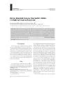
69 lymphoma A case report and literature review 1 JiQing Qiu PhD 2 Yu Cui MD 3 LiChao Sun MD 1 Bin Qi PhD 1 ZhanPeng Zhu PhD JQ Qiu and Y Cui contributed equally to this work ID: 938195

cns lymphoma brain hemorrhage lymphoma cns hemorrhage brain primary patient cell gastrointestinal hematoma x0066006c case patients nervous system central

Share:

Link:

Embed:

Download Presentation from below link

Download Pdf The PPT/PDF document "Intracranial hemorrhage from metastatic ..." is the property of its rightful owner. Permission is granted to download and print the materials on this web site for personal, non-commercial use only, and to display it on your personal computer provided you do not modify the materials and that you retain all copyright notices contained in the materials. By downloading content from our website, you accept the terms of this agreement.


Presentation Transcript

69 Intracranial hemorrhage from metastatic CNS lymphoma: A case report and literature review * 1 Ji-Qing Qiu PhD , * 2 Yu Cui MD , 3 Li-Chao Sun MD , 1 Bin Qi PhD , 1 Zhan-Peng Zhu PhD *JQ Qiu and Y Cui contributed equally to this work and are co-�rst authors Departments of 1 Neurosurgery, 2 Otolaryngology and 3 Emergency Medicine, The First Hospital of Jilin University, Changchun, Jilin, China Abstract Metastatic brain lymphomas, which belong to secondary central nervous system lymphomas, usually commonly metastasize to the lung or heart. We report here a case of brain hemorrhage due to metastasis from primary gastrointestinal diffuse large B-cell lymphoma (DLBCL). A 30-year-old male presented with headache. He was diagnosed to have gastrointestinal lymphoma 6 months earlier, and treated with gastrointestinal surgery. Pathological diagnosis was DLBCL. A PET-CT scan immediately after gastrointestinal surgery demonstrated no brain metastasis. On admission to the ward, imaging of the brain showed right temporoparietal hematoma. In the ward, the patient deteriorated with impaired consciousness. Repeat brain imaging showed enlargement of the hematoma. He underwent right temporoparietal craniotomy for the removal of a hematoma, and tumor nodules adherent to the cortex this was the �rst reported case of brain hemorrhage from metastatic lymphoma. Metastatic central nervous system lymphoma should be considered as a differential diagnosis in patients with a history of gastrointestinal lymphoma presenting with neurological symptoms. Keywords: Diffuse large B-cell lymphoma; gastrointestinal; brain metastasis; brain hemorrhage Neurology Asia 2018; 23(1) : 69 – 75 INTRODUCTION Lymphomas are hematological malignancies with extranodal manifestations in approximately 40% of cases. 1 Central nervous system (CNS) lymphoma includes primary CNS lymphoma and secondary CNS lymphoma. 2 Secondary CNS lymphoma is generally considered as CNS involvement in lymphoma that was not evident at the initiation of treatment for systemic lymphoma outside the CNS. 3 Metastatic brain lymphoma is considered a secondary CNS lymphoma. 2,3 The primary sites of metastatic CNS lymphoma include the nasal cavity or paranasal sinus , peripheral blood 4,5 , orbit, 4 bone marrow 4,6 , testis 4 , bone 4 , and breast. 7 To the authors’ knowledge, there are no previous reports of brain metastasis from gastrointestinal lymphoma. Primary gastrointestinal non-Hodgkin lymphoma (PGI NHL) is one of the most common types of extranodal lymphomas, accounting for 30-50% of all extranodal lymphomas. 8 Gastrointestinal lymphoma is known to metastasize to the lung or heart. 9,10 Intracranial hemorrhage is a neurological emergency usually caused by high blood

pressure, vessel malformation, or arterial aneurysm. Intracranial hemorrhage may also be caused by tumors such as glioma. 11 There is no previous reports of cerebral hemorrhage due to metastatic lymphoma. We report here a case of brain metastasis from primary gastrointestinal diffuse large B-cell lymphoma (DLBCL) with hemorrhage. The literature on hemorrhage in cerebral lymphoma was reviewed. CASE REPORT A 30-year-old male presented at our institution with complaints of headache for four days. His medical history revealed a diagnosis of Address correspondence to : Dr Zhan-Peng Zhu, Department of Neurosurgery, The First Hospital of Jilin University, 71 Xinmin Street, Changchun, Jilin 130021, P.R. China. E-mail: 282324491@qq.com CASE REPORTS 70 gastrointestinal lymphoma and gastrointestinal surgery six months earlier; pathological diagnosis was DLBCL (Figure 1). The initial stage of the lymphoma was stage Ⅲ B. Whole body positron emission tomography–computed tomography (PET-CT) scan immediately after surgery did not demonstrate any brain metastasis. The initial risk score of CNS metastasis was intermediate risk (NCCU guidelines: NHL 2016.3). The patient underwent six courses of rituximab, cyclophosphamide, doxorubicin, vincristine and prednisone (R-CHOP) chemotherapy after abdominal surgery. The remission status of the initial 6 cycles of chemotherapy was CR (CT- Based Response). The patient had no history of hypertension, brain trauma, congenital abnormality, and was not administered any immunosuppressive agents. Clinical examination and laboratory investigations were unremarkable, including a negative serologic test for human immunode�ciency virus (HIV). Computed tomography (CT) of the brain demonstrated a right temporoparietal hematoma of mixed density with no mass effect, edema, or midline shift (Figure 2). Magnetic resonance imaging (MRI) of the brain revealed that the hematoma was hypointense on T1-weighted imaging and mildly hyperintense on fluid- attenuated inversion recovery sequence and T2-weighted imaging (Figure 3). The patient was diagnosed with hematoma in the cortex of the right temporoparietal lobe. Dynamic contrast enhanced CT angiography (CTA) of the blood vessels in the brain showed no abnormality. The hematoma was initially managed conservatively. However, on day six of hospitalization the patient’s condition rapidly deteriorated and his Glasgow Coma Score was 8/15. CT scan showed the enlarged hematoma accompanied by edema in the right temporoparietal lobe (Figure 4). Right temporoparietal craniotomy was performed immediately, a 35-ml hematoma was removed. During surgery, it was noted that some gyri were covered with tumor nodes, the largest of which was 1.0 cm in diameter. Based on the pati

ent’s history of gastrointestinal lymphoma, the hematoma was thought to be caused by metastasis. Therefore, gross resection of the hematoma was performed, and some of the abnormal brain lesions were removed. After surgery the patient’s neurologic status became normal. Histopathologic examination of the tissues revealed that the cortex included a portion of abnormal, small round cells with prominent nuclei. Immunohistochemistry showed that cells were positive for B-cell markers (CD20 and CD79a) and negative for T-cell markers (CD3) (Figure 5). The pathologic diagnosis was NHL DLBCL with acute hemorrhage. The patient was discharged after surgery and went to another hospital in another province. Because of poverty and side effect of chemotherapy, the patient’s family refused further Figure 1. Immunohistochemistry-confirmed gastrointestinal diffuse large B-cell lymphoma (DLBCL): (a) hematoxylin and eosin staining (magni�cation, ×20); the tumor was (b) LCApositive (magni�cation, ×20), (c) CD20positive (magni�cation, ×20), and (d) CD79apositive (magni�cation, ×20). 71 chemotherapy. At four months post-surgery, the patient had tumor dissemination and died. No autopsy was performed. DISCUSSION Gastrointestinal lymphoma is a malignant tumor. 12 Our patient was diagnosed with gastrointestinal DLBCL based on histopathology following gastrointestinal surgery. The postoperative PET- CT scan revealed no metastasis to the CNS. This was considered a de�nitive diagnosis, as PET-CT was thought to be able to exclude CNS metastasis. 13 The patient presented to our hospital complaining of sudden headache. CT of the brain demonstrated a cerebral hematoma. The patient had no history of hypertension, and the cause of the hematoma was unknown. The primary sites of metastatic CNS lymphoma are usually the bone marrow 4,6 , testis 4 , or orbit. 4 As there have been no reports of gastrointestinal lymphoma metastasizing to the brain, the hematoma due to metastatic lymphoma were not suspected. This case showed that CNS metastasis in DLBCL is not solely depends on the site of the tumor. CNS involvement should be suspected when the patient with lymphoma presents with CNS symptoms. Right temporoparietal craniotomy was performed in our patient with removal of the hematoma and tumor nodules. Pathology con�rmed DLBCL and hemorrhage in the brain. The median survival time (MST) of secondary CNS lymphoma is less than 6 months. 3 Our patient died four months after undergoing right temporoparietal craniotomy. Some evidence suggests that BCL-2 overexpression confers resistance to chemotherapy, and Ki-67 overexpression is associated with poor prognosis in patients treated with R-CHOP

. 14-16 The present Ⅲcase was Ki-67 (+) 90% and BCl-2(+) 80%. Bleeding in secondary CNS lymphoma is rare. Therefore, we reviewed the literature on hemorrhage in cerebral lymphoma (Table 1). PubMed and Web of Science databases were independently searched from inception to February 1, 2017 by two reviewers using Multi-slice CT imaging showing hemorrhage in the right tempoparietal lobe Figure 3. MRI showing a hematoma that was (a) hypointense on T1-weighted imaging, and (b) mildly hyperintense on T2-weighted imaging and (c) �uid-attenuated inversion recovery sequence. 72 the following keywords and subject terms: “lymphoma”, “diffuse large B-cell lymphoma”, “non-Hodgkin lymphoma”, “brain”, “cerebral”, and “central nervous system” in combination with “hemorrhage”, “hematoma”. The search revealed nine reports with seven cases of intracranial hemorrhage in primary CNS lymphoma, one case each of relapse CNS lymphoma, and systemic lymphoma. The mean age of the patients was 60. 7 years (range: 29-96 years), suggesting that bleeding in CNS lymphoma occurs more in the elderly patients. There were six males and three females. The lymphomas of seven patients originated from B cells. Five patients were vascular endothelial growth factor (VEGF) positive. VEGF is thought to induce spontaneous hemorrhage in CNS lymphoma. 17 Only one patient was HIV positive. In six patients, the hemorrhage was located in the frontal lobe, and in one patient each, the hemorrhage was located CT scan showed the enlarged hematoma accompanied by edema in the right temporoparietal lobe. Figure 5. Immunohistochemistry con�rmed that the abnormal cortex originated from DLBCL: (a) Hematoxylin and eosin staining (magni�cation, ×20); the tumor was (b) CD20positive (magni�cation, ×20), (c) positive (magni�cation, ×20), and (d) CD3negative (magni�cation, ×20). 73 Table 1: Clinical features of previously reported patients of CNS lymphoma with hemorrhage Case Author Age/ Gender Clinical presentation Physical examination Hemorrhage location CT/MRI Diagnosis Origin Treatment after hemorrhage Treatment before hemorrhage VEGF HIV test Result 1 Fukui et al. 20 29/M Oral dyskinesia, headache, nausea Left-sided facial droop Lt. Fr T2-hypointense T1-mixed signal intensity Primary CNS lymphoma ND Only biopsy ND Not examined Positive ND 2 Rubenstein et al. 17 55/M Right arm tonic clonic seizures Right upper extremity weakness Lt. Posterior Fr ND Primary CNS lymphoma diffuse large cell non- Hodgkin’s lymphoma Left posterior frontal craniotomy Chemotherapy and radiotherapy ND Intense reactivity ND Remission discharge 3 Kim et a

l. 21 49/F Sudden deterioration of mental status Stuporous mental state, right hemiparesis grade Ⅲ Lt. Fr Mass effect of left frontal lobe, midline shifting to the right Primary CNS lymphoma B-cell Chemotherapy and radiotherapy ND High immuno- reactivity Negative Discharge 4 Kimura et al. 22 57/F Drowsy Mild right hemifacial palsy, mild right hemiparesis, and hyperre�exia in the right extremities without pathologic Lt. Fr T1-Hyperintense, T2-hypointense Primary CNS lymphoma B-cell Left frontal craniotomy i ntrathecal chemo- therapy Positive Negative Discharge 5 Kim et al. 23 75/M Sensory aphasia ND Lt. temporoparietal ND Relapsed CNS lymphoma B-cell Chemotherapy and radiotherapy ND Positive ND ND 6 Low et al. 24 62/M Mild right upper limb, facial weakness Right facial droop, grade 4+/5 power in the right upper limb Lt. parietofrontal chronic subdural hematoma T1-hypointense, T2 and FLIR- mildly hyperintense Primary dural lymphoma B-cell left frontoparietal craniotomy ND Not examined Negative Continue oncology and radiology treatment 7 Matsuyama et al. 25 67/M Comatose ND Lt. Fr Intr acerebral bleeding i n the left frontal lobe, midline shifting to the right side Primary CNS lymphoma B-cell Emergency endoscopic removal of the hematoma chemo- therapy High levels of immuno- reactivity Negative Died 8 Yang et al. 26 56/F Lower extremities Weakness, progressive cognitive decline C ognitive de�cits, positive left babinski sign Lt. T T1-h ypointense area with focal hyperintensity, T2 and FLAIR- hyperintensity Systemic Lymphoma B-cell Refuse treatment ND ND Negative Died 9 Kameda et al. 27 96/M P r ogressing gait disturbance and appetite loss ND Lt. Fr T2 - hypointense and by contrast enhancement P rimary CNS lymphoma B-cell Left frontal craniotomy ND ND Negative Died F = female; M = male; Lt. =left; Fr= frontal; T= temporal; P= parietal; CNS= central nervous system; ND=not described; FLAIR= �uid-attenuated inversion recovery; VEGF= vascular endothelial growth factor 74 in the temporal lobe, parietofrontal subdura, and temporoparietal lobe. Most bleeding was intraparenchymal, and patients’ presentation varied. In our case, the hemorrhage was located in the right temporoparietal lobe. Intracranial hemorrhage is a common emergency in patients with brain cancer, with an incidence estimated at 2.4%. 18 Intratumoral hemorrhage and coagulopathy are the main cause of intracranial hemorrhage. 11 Lung metastatic brain tumors hemorrhage is due to blood vessel invasion, neoangiogenesis, and tumor cell necrosis. 19 Hemorrhage in primary glioblastoma multiforme is caused by highly invasive tumor cells. 19 In our patient, coagulopathy was normal. The mechanisms responsible

for the hemorrhage of CNS lymphoma are unknown, as published reports are scarce. In conclusion, CNS involvement may occur in gastrointestinal lymphoma and may occur due to the inadequate initial treatment. Physicians should consider the possibility of metastatic CNS lymphoma in patients with intracerebral hemorrhage who have a history of gastrointestinal lymphoma presenting with neurological disturbances. ACKNOWLEDGEMENTS This manuscript has been edited and proofread by Medjaden Bioscience Limited. The study was approved by the ethics committee of First Hospital of Jilin University. Informed parental consent was obtained in this case. We have received a signed release form from the patient parents authorizing the publication of her material. DISCLOSURE Financial support: None Con�ict of interest: None REFERENCES Gigli S, Buonocore V, Barchetti F, et al . Primary colonic lymphoma: An incidental �nding in a patient with a gallstone attack. World J Clin Cases 2014;2:146-50. Baraniskin A, Deckert M, Schulte-Altedorneburg G, Schlegel U, Schroers R. Current strategies in the diagnosis of diffuse large B-cell lymphoma of the central nervous system. Br J Haematol 2012;156:421- 32. Tomita N, Kodama F, Kanamori H, Motomura S, Ishigatsubo Y. Secondary central nervous system lymphoma. Int J Hematol 2006;84:128-35. 4.Liang R, Chiu E, Loke SL. Secondary central nervous system involvement by non-Hodgkin’s lymphoma: the risk factors. Hematol Oncol 1990;8:141-5. Gendelman S, Rizzo F, Mones RJ. Central nervous system complications of leukemic conversion of the lymphomas. Cancer 1969;24:676-82. Bunn PA, Jr., Schein PS, Banks PM, DeVita VT, Jr. Central nervous system complications in patients with diffuse histiocytic and undifferentiated lymphoma: leukemia revisited. Blood 1976;47:3-10. Aviles A, Delgado S, Nambo MJ, Neri N, Murillo E, Cleto S. Primary breast lymphoma: results of a controlled clinical trial. Oncology 2005;69:256-60. Koch P, del Valle F, Berdel WE, et al . Primary gastrointestinal non-Hodgkin’s lymphoma: I. Anatomic and histologic distribution, clinical features, and survival data of 371 patients registered in the German Multicenter Study GIT NHL 01/92. J Clin Oncol 2001;19:3861-73. Kakkar N, Vasishta RK, Sharma Y, Bhasin DK. Intracavitary metastatic non-Hodgkin’s lymphoma simulating an atrial myxoma. Indian J Pathol Microbiol 2003;46:60-2. Dorenbeck U, Hollerbach S, Geissler A, Andus T. Pulmonary metastasis of extranodal high malignancy B-cell non-Hodgkin lymphoma of the bulbus duodeni and pylorus of the stomach. Z Gastroenterol 2000;38:173-6. (in German) 11. et al . Intracerebral and subarachnoid hemorrhage in patients with cancer. Neurology 2010;74:494-501. 12.Nakamura S, Matsumoto T.

Gastrointestinal lymphoma: recent advances in diagnosis and treatment. Digestion 2013;87:182-8. 13.Maza S, Buchert R, Brenner W, et al . Brain and whole- body FDG-PET in diagnosis, treatment monitoring and long-term follow-up of primary CNS lymphoma. Radiol Oncol 2013;47:103-10. 14.Salles G, de Jong D, Xie W, et al . Prognostic signi�cance of immunohistochemical biomarkers in diffuse large B-cell lymphoma: a study from the Lunenburg Lymphoma Biomarker Consortium. Blood 2011;117:7070-8. Zinzani PL, Magagnoli M, Frezza G, et al . Isolated central nervous system relapse in aggressive non- Hodgkin’s lymphoma: the Bologna experience. Leuk Lymphoma 1999;32:571-6. Gleissner B, Chamberlain M. Treatment of CNS dissemination in systemic lymphoma. J Neurooncol 2007;84:107-17. Rubenstein J, Fischbein N, Aldape K, Burton E, Shuman M. Hemorrhage and VEGF expression in a case of primary CNS lymphoma. J Neurooncol 2002;58:53-6. Schrader B, Barth H, Lang EW, et al . Spontaneous intracranial haematomas caused by neoplasms. Acta Neurochir (Wien) 2000;142:979-85. Velander AJ, DeAngelis LM, Navi BB. Intracranial hemorrhage in patients with cancer. Curr Atheroscler Rep 2012;14:373-81. 20.Fukui MB, Livstone BJ, Meltzer CC, Hamilton RL. Hemorrhagic presentation of untreated primary CNS lymphoma in a patient with AIDS. AJR Am J Roentgenol 1998;170:1114-5. 75 Kim IY, Jung S, Jung TY, Kang SS, Choi C. Primary central nervous system lymphoma presenting as an acute massive intracerebral hemorrhage: case report with immunohistochemical study. Surg Neurol 2008;70:308-11. Kimura N, Ishibashi M, Masuda T, et al . Primary central nervous system lymphoma with cortical laminar hemorrhage. J Neurol Sci 2009;287:281-4. Kim TK, Wajima D, Yonezawa T, Takamura Y, Miyamae S. Intra-tumoral hemorrhage of a relapsed CNS diffuse large B-cell lymphoma: a case report. No Shinkei Geka 2014;42:651-8. (in Japanese) Low YY, Lai SH, Ng WH. An unusual presentation of primary malignant B-cell-type dural lymphoma. Singapore Med J 2014;55:e187-90. Matsuyama J, Ichikawa M, Oikawa T, et al . Primary CNS lymphoma arising in the region of the optic nerve presenting as loss of vision: 2 case reports, including a patient with a massive intracerebral hemorrhage. Brain Tumor Pathol 2014;31:222-8. Yang XZ, Ni J, Cui LY. Multifocal hemosiderin depositions on T2*-weighted magnetic resonance imaging in a patient with pathology-proven systemic diffuse large B-cell lymphoma. BMC Neurol 2014;14:184. Kameda K, Shono T, Takagishi S, et al . Epstein-Barr virus-positive diffuse large B-cell primary central nervous system lymphoma associated with organized chronic subdural hematoma: a case report and review of the literature. Pathol Int 2015;65:138-43. Neurology Asia Marc