/
Multiple Intracranial Aneurysms from Syphilitic Infection in a Patient Multiple Intracranial Aneurysms from Syphilitic Infection in a Patient

Multiple Intracranial Aneurysms from Syphilitic Infection in a Patient - PDF document

vivian
vivian . @vivian
Follow
342 views
Uploaded On 2022-08-22

Multiple Intracranial Aneurysms from Syphilitic Infection in a Patient - PPT Presentation

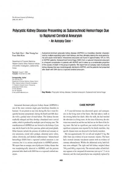
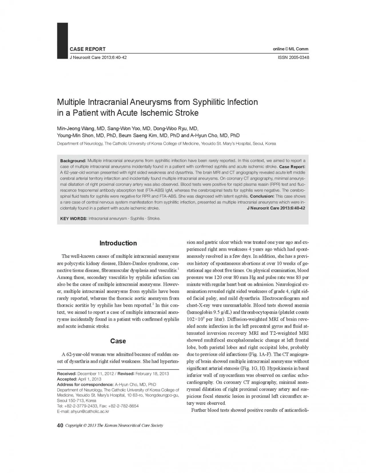
Address for correspondenceDepartment of Neurology The Catholic University of Korea College of CASE REPORTJ Neurocrit Care 201364042ISSN 20050348 MJ Wang et alpin antibody IgG antiphosopholip ID: 939698

aneurysms intracranial syphilis multiple intracranial aneurysms multiple syphilis infection tests x00660069 antibody case syphilitic acute patient left stroke negative

Share:

Link:

Embed:

Download Presentation from below link

Download Pdf The PPT/PDF document "Multiple Intracranial Aneurysms from Syp..." is the property of its rightful owner. Permission is granted to download and print the materials on this web site for personal, non-commercial use only, and to display it on your personal computer provided you do not modify the materials and that you retain all copyright notices contained in the materials. By downloading content from our website, you accept the terms of this agreement.


Presentation Transcript

Multiple Intracranial Aneurysms from Syphilitic Infection in a Patient with Acute Ischemic StrokeMin-Jeong Wang, MD, Sang-Won Yoo, MD, Dong-Woo Ryu, MD, Young-Min Shon, MD, PhD, Beum Saeng Kim, MD, PhD and A-Hyun Cho, MD, PhDDepartment of Neurology, The Catholic University of Korea College of Medicine, Yeouido St. Mary’s Hospital, Seoul, Korea Multiple intracranial aneurysms from syphilitic infection have been rarely reported. In this context, we aimed to report a case of multiple intracranial aneurysms incidentally found in a patient with conrmed syphilis and acute ischemic stroke. A 62-year-old woman presented with right sided weakness and dysarthria. The brain MRI and CT angiography revealed acute left middle cerebral arterial territory infarction and incidentally found multiple intracranial aneurysms. On coronary CT angiography, minimal aneurysmal dilatation of right proximal coronary artery was also observed. Blood tests were positive for rapid plasma reasin rescence treponemal antibody absorption test FTA-ABS Address for correspondence:Department of Neurology, The Catholic University of Korea College of CASE REPORTJ Neurocrit Care 2013;6:40-42ISSN 2005-0348 MJ Wang, et al.pin antibody IgG, antiphosopholipid antibody IgG, M, anti-β2-glycoprotein I antibody, lupus anticoagulant antibody and antinuclear antibody. Considering her previous medical history of vascular thrombosis ous abortions, she was diagnosed with antiphospholipid antibody syndrome. The current stroke might be caused by an-tiphospholipid syndrome. She was treated with hydroxychlo-roquine and steroid. Antithrombotic agents were not used be-cause of anemia and bleeding tendency at the time of dischar-ge. Her blood tests were positive for rapid plasma reasin RPRtest and �uorescent treponemal antibody absorption test FTA- IgM. The cerebrospinal �uid tests for syphilis were negative for RPR and FTA-ABS without signi�cant leu-kocytosis nor high protein level. The other autoimmune serologic tests suggesting vasculitis including rheumatoid arthritis, antinuclear cytoplasmic antibody, and anti-Ro/La antibodies showed negative results. Accordingly, we concluded that the most probable cause of intracranial aneurysms in this case might be syphilitic infection of cerebral vessels. She was treated with penicillin G Benzathin 2.4 milchemic stroke patient with seropositive syphilis. The con�rm-ation of syphilis through serologic tests, the exclusion of other causes for multiple intracranial aneurysms and the coexist-The process of aortic aneurysm from syphilitic infection begins from in�ammatory in�ltrate around the vasa vasorum of adventitia, followed by endarteritis obliterans leading to elastic tissue of the media. We assume that the mechanism of uld be similar to that. To date, a case about intracranial sy-philitic aneurysms has been rarely reported. We think it is due to their low probability of rupture, remaining as asymptomatThe serological �ndings in this case are insuf�cient to diagnose neurosyphilis e.g., meningovascular syphilis5 because the CS

F study was normal. However, we cannot exclu-de neurosyphilis only with nonreactive CSF FTA-ABS tests.Clinical diagnosis of neurosypilis can refer to an infection in-volving the central nervous system. There are two possible Diffusion-weighted MRI and apparent diffusion-coef�cient map reveal acute infarction in the left precentral gyrus and multifocal encephalomalacic change of left frontal lobe, both parietal lobes and right occipital lobe, probably due to previous old infarctions on T2-weighted and �uid attenuated inversion recovery MR image . CT angiography of brain shows an about 3 mm sized saccular aneurysm at bifurcation site of right middle cerebral artery, multiple fusiform aneurysm at A1 segment of right anterior cerebral artery , A2 segment of right anterior cerebral artery J Neurocrit Care explanations for the negative results of CSF study. First, over several decades may have passed after aneurysmal change from primary infection. Late neurosyphilis occurs 4-25 years after the initial infection. Therefore, the regressed activity of syphilitic infection may show sero-negativity.ous study, 11% of individuals with a �rst episode of primary syphilis were sero-negative with the FTA-ABS test at 1 year post-treatment, and 24% were negative by 3 years. Secondly, vessel wall not disrupting blood-brain barrier might be the rea-ence in the CSF. In summary, this report shows a rare case of CNS manifest-ation from syphilitic infection, presented as multiple intracr-anial aneurysms which were incidentally found in a patient This research was supported by Basic Science Research Program through the National Research Foundation of Korea funded by the Ministry of Education, Science and Technology No.2012R1A1B Tonn J, Hoffmann O, Hofmann E, Schlake HP, Sörensen N, Roosen K. “De novo” formation of intracranial aneurysms who is at risk? roradiologyPugh PJ, Grech ED. Images in clinical medicine. Syphilitic aortitis. Tomey MI, Murthy VL, Beckman JA. Giant syphilitic aortic aneu a case report and review of the literature. Vasc Med 2011Wardlaw JM, White PM. The detection and management of unrupChahine LM, Khoriaty RN, Tomford WJ, Hussain MS. The changing Int J Stroke 2011Jaffe HW, Larsen SA, Peters M, Jove DF, Lopez B, Schroeter AL. Tests for treponemal antibody in CSF. Arch Intern Med Romanowski B, Sutherland R, Fick GH, Mooney D, Love EJ. Serologic response to treatment of infectious syphilis. Ann Intern Med 114 Multiple Intracranial Aneurysms from Syphilitic Infection in a Patient with Acute Ischemic StrokeMin-Jeong Wang, MD, Sang-Won Yoo, MD, Dong-Woo Ryu, MD, Young-Min Shon, MD, PhD, Beum Saeng Kim, MD, PhD and A-Hyun Cho, MD, PhDDepartment of Neurology, The Catholic University of Korea College of Medicine, Yeouido St. Mary’s Hospital, Seoul, Korea Multiple intracranial aneurysms from syphilitic infection have been rarely reported. In this context, we aimed to report a case of multiple intracranial aneurysms incidentally found in a patient with conrmed syphilis and acute ischemic stroke. A 62-year-old woman presented with right sided weakness and dysarth

ria. The brain MRI and CT angiography revealed acute left middle cerebral arterial territory infarction and incidentally found multiple intracranial aneurysms. On coronary CT angiography, minimal aneurysmal dilatation of right proximal coronary artery was also observed. Blood tests were positive for rapid plasma reasin rescence treponemal antibody absorption test FTA-ABS IgM, whereas the cerebrospinal tests for syphilis were negative. The cerebrospinal uid tests for syphilis were negative for RPR and FTA-ABS. She was diagnosed with latent syphilis. Conclusion This case shows a rare case of central nervous system manifestation from syphilitic infection, presented as multiple intracranial aneurysms which were incidentally found in a patient with acute ischemic stroke. Intracranial aneurysm · Syphilis · Stroke. 2013 The Korean Neurocritical Care Society Address for correspondence:Department of Neurology, The Catholic University of Korea College of Medicine, Yeouido St. Mary’s Hospital, 10 63-ro, Yeongdeungpo-gu, Seoul 150-713, KoreaTel: +82-2-3779-2433, Fax: +82-2-782-8654are polycystic kidney disease, Ehlers-Danlos syndrome, connective tissue disease, �bromuscular dysplasia and vasculitis.Among these, secondary vasculitis by syphilis infection can also be the cause of multiple intracranial aneurysms. However, multiple intracranial aneurysms from syphilis have been rarely reported, whereas the thoracic aortic aneurysm from thoracic aortitis by syphilis has been reported.rysms incidentally found in a patient with con�rmed syphilis A 62-year-old woman was admitted because of sudden onset of dysarthria and right sided weakness. She had hypertension and gastric ulcer which was treated one year ago and exaneously resolved in a few days. In addition, she has a previ-ous history of spontaneous abortions at over 10 weeks of ge-stational age about �ve times. On physical examination, blood pressure was 120 over 80 mm Hg and pulse rate was 83 per minute with regular heart beat on admission. Neurological ex-amination revealed right sided weakness of grade 4, right sid-ed facial palsy, and mild dysarthria. Electrocardiogram and chest-X-ray were unremarkable. Blood tests showed anemia hemoglobin 9.5 g/dL and thrombocytopenia platelet counts 10210 per liter. Diffusion-weighted MRI of brain reve-aled acute infarction in the left precentral gyrus and �uid attenuated inversion recovery MRI and T2-weighted MRI showed multifocal encephalomalacic change at left frontal lobe, both parietal lobes and right occipital lobe, probably due to previous old infarctions Fig. 1A-F. The CT angiogra-phy of brain showed multiple intracranial aneurysms without signi�cant arterial stenosis Fig. 1G, H. Hypokinesia in basal inferior wall of myocardium was observed on cardiac echocardiography. On coronary CT angiography, minimal aneurysmal dilatation of right proximal coronary artery and suspicious focal stenotic lesion in proximal left circum�ex ar-Further blood tests showed positive results of anticardioli- CASE REPORTJ Neurocrit Care 2013;6:40-42ISSN 2005-0348 online