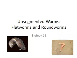
/
Parasitic  Helminths  By: Nader Parasitic  Helminths  By: Nader

Parasitic Helminths By: Nader - PowerPoint Presentation

victoria
victoria . @victoria
Follow
343 views
Uploaded On 2022-06-15

Parasitic Helminths By: Nader - PPT Presentation

Alaridah MD PhD Medical parasitology is classified into Medical helminthology Medical protozoology Deals with parasitic worms Deals with unicellular parasites 1Phylum Platyhelminthes ID: 919636

eggs worms larvae adult worms eggs adult larvae intestine intestinal host infection stage humans length infections female human larval

Share:

Link:

Embed:

Download Presentation from below link

Download Presentation The PPT/PDF document "Parasitic Helminths By: Nader" is the property of its rightful owner. Permission is granted to download and print the materials on this web site for personal, non-commercial use only, and to display it on your personal computer provided you do not modify the materials and that you retain all copyright notices contained in the materials. By downloading content from our website, you accept the terms of this agreement.


Presentation Transcript

Slide1

Parasitic Helminths

By: Nader

Alaridah

MD, PhD

Slide2

Medical

parasitology

is classified into

Medical helminthology

Medical protozoology

Deals with parasitic worms

Deals with unicellular parasites

1-Phylum : Platyhelminthes

(flat worms)

2-Phylum : Nemathelminthes

(round worms)

Class:

Trematoda

Class:

Cestoidea

Class:

Nematoda

1-Class:

Rhizopoda:

(move by pseudopodia)

2-

Class:

Ciliata

(move by cilia)

3-Class:

Zoomastigophora

(move by flagellae)

4-Class:

Sporozoa

(move by gliding movement)

Slide3

Key concepts

Most intestinal helminthic infections are fairly benign, except when worm burdens are high and numbers of adult worms in the intestine reach the hundreds.

In intestinal worm infections, the intestine usually harbors the adult stage of the parasite, except for Strongyloides, Trichinella,  and Taenia solium,  which not only reside in the intestine as adults but also have larvae capable of migrating throughout tissues.

In the case of the three most common intestinal infections (whipworm, hookworm, and ascariasis), the eggs require incubation in the soil for several days or weeks in warm, tropical climates.Most infections are acquired by ingestion of the egg or larval stage, with the exception of the hookworms, human threadworms, and schistosomes, whose larvae penetrate the skin, and the filarids, which are vectorborne

.

Slide4

The nematodes (Round worms)

They are small ,round elongated worms , Non segmented , with body cavity, have separate sexes ,usually don’t need an intermediate host and do not multiply in human host because eggs don’t hatch unless they leave the body first.

They can parasitize either an intermediate host (worms in the juvenile, larval or developmental stages ) or final / definitive host (worms occur in the adult or in the sexually reproductive stage)Infection pattern vary widely. Human intestinal nematodes infect via food borne, water borne and soil borne routs .

People infected with Ascaris often show no symptoms. If symptoms do occur they can be light and include abdominal discomfort. Heavy infections can cause intestinal blockage and impair growth in children. Other symptoms such as cough are due to migration of the worms through the body. 

Slide5

Ascaris lumbricoids

Infect by eggs ,that are strongly resistant to dissection and environmental conditions.Eggs are 75x40 with a thick mamillated brownish shell.Freshly passed eggs with stool are not infective they require 2-3 weeks to develop to be embryonated (contain larva).Ascaris lumbricoides is the largest nematode (roundworm) parasitizing the human intestine. (Adult females: 20 to 35 cm; adult male: 15 to 30 cm.)

Humans can also be infected by pig roundworm (Ascaris suum). Ascaris lumbricoides (human roundworm) and Ascaris suum (pig roundworm) are indistinguishable. It is unknown how many people worldwide are infected with Ascaris suum

.

Slide6

Pathology and pathogenesis

 If present in high numbers, adult worms may cause mechanical obstruction of the bowel and bile and pancreatic ducts.

Worms tend to migrate if drugs such as anesthetics or steroids are given, leading to bowel perforation and peritonitis, anal passage of worms, vomiting, and abdominal pain. Larvae migrating through lungs induce an inflammatory response (pneumonitis), especially after second infection, leading to bronchial spasm, mucus production, and Löffler syndrome (cough, eosinophilia, and pulmonary infiltrates).

Slide7

Slide8

Slide9

ENTEROBIUS VERMICULARIS

(Pinworm—intestinal nematode)

 Female pinworms  (about 10 mm in length) have a slender, pointed posterior end. Males are approximately 3 mm in length and have a curved posterior end. Pinworms are found worldwide but more commonly in temperate than tropical climates. They are among the most common helminthic infection and infect mostly children.

Slide10

ENTEROBIUS VERMICULARIS

Eggs are football shaped, have a thin outer shell, and are approximately 50–60

μ m in length. Infectious larvae are often visible inside the eggEggs are recovered using the “Scotch Tape” technique in the morning before a bowel movement.The main symptom associated with pinworm infections is perianal pruritus, especially at night, caused by a hypersensitivity reaction to the eggs that are laid around the perianal region by female worms, which migrate down from the colon at night.

Slide11

Slide12

TRICHURIS TRICHIURA (Whipworm—

intestinal nematode)

 Adult female whipworms  are approximately 30–50 mm in length; adult male worms are smaller. The anterior end of the worms is slender, and the posterior end is thicker, giving it a “buggy whip” appearance, hence the name whipworm. Adult whipworms inhabit the colon, where male and female worms mate. Females release eggs that are passed in the feces, and eggs become infective after about 3 weeks of incubation in moist and shady soil.

Whipworm eggs (50 μm) with distinct polar plugs.

Slide13

ANCYLOSTOMA DUODENALE AND NECATOR AMERICANUS (Human

hookworms—intestinal nematode)

Female hookworms  are approximately 10 mm in length; males are slightly smaller and have a taxonomically characteristic copulatory bursa (broadened posterior end), which is used to mate with females. Females can release more than 10,000 eggs per day into the feces, where a larva hatches from the egg within a day or two.

Eggs are oval 60x40 mm , they hatch in 48h to give the rhabiditiform larva, after 2 days it moults to filariform larva which is the infective stage that penetrate skin and mucous membranes.

Slide14

Pathology

Larvae can survive in moist soil for several weeks, waiting for an unsuspecting barefooted host to walk by. These larvae penetrate host skin and migrate throughout the host similarly to Ascaris  and end up in the small intestine where they mature into adult worms.

In the intestine, adult worms attach to intestinal villi with their buccal teeth and feed on blood and tissue with the aid of anticoagulants .A few hundred worms in the intestine can cause hookworm disease, which is characterized by severe anemia and iron deficiency. Intestinal symptoms also include abdominal discomfort and diarrhea. The initial skin infection by the larvae causes a condition known as “ground itch,” characterized by  erythema and intense pruritus. Feet and ankles are common sites of infection due to exposure from walking barefoot.

Slide15

Slide16

STRONGYLOIDES STERCORALIS (Human threadworm—intestinal and tissue nematode)

 Adult females (about 2 mm long) of

Strongyloides stercoralis  that inhabit the intestine are parthenogenic; that is, they do not need to mate with male worms to reproduce.

They lay eggs within the intestine; larvae hatch from the eggs and are passed into the feces. These larvae can either develop into parasitic forms or develop into free-living male and female worms that mate and produce several generations of worm in the soil, a great example of an evolutionary adaptation to sustain a population.

Slide17

Slide18

TRICHINELLA SPIRALIS (Intestinal

and tissue nematode)

Trichinella spiralis is acquired by eating raw or improperly cooked pork infected with the larval stage of these nematodes. In the small intestine, the larvae molt into adult worms, and, after mating with male worms, the female worms release live larvae. The larvae penetrate the intestine, circulate in the blood, and eventually encyst in muscle tissue.

Adult female worms live for several weeks and after the first week of infection may cause diarrhea, abdominal pain, and nausea. Intestinal symptoms are mild to none and often go unnoticed.

Slide19

Tissue Nematodes

Adults are parasite of the lymphatic system or connective tissue .they are filariform or thread like.

Female lay larvae but not eggs.Larvae require an intermediate host to complete development resulting in the production of the infective stage.

Family Filariidae, members are :Wucherieria

bancrofti (Mosquito)Brugia malayi (Mosquito)

Loa loa (eye worm) (fly -genus Chrysops, day-biting flies)Onchcerca volvulus (River blindness) (

black flies)

Slide20

LYMPHATIC FILARIA

Members to be found in lymphatics , body cavities and

subcatenous tissues.Progenies are embryos which are not fully developed (microfilariae), these are between eggs & larvae.Microfilariae require an intermediate host which sucks them . the infection is transmitted by mosquitoes.

Elephantiasis; true elephantiasis is the result of parasitic infection caused by  The filarid nematodes, Wuchereria bancrofti, Brugia

malayi,  and Onchocerca volvulus,  are long, slender worms  whose adult forms are found in tissues.The long thread like worms blocks the lymphatic system causing fluid to collect in tissues which lead to great swelling called ‘’lymphedema’’. Limbs can swell so enormously that they resemble an elephant’s foreleg in size texture and color.

Slide21

Slide22

Platyhelminthis

(flat worms)

Platyhelminthes are flatworms that are dorsoventrally flattened in cross section and are hermaphroditic. All medically important species belong to two classes: 

Cestoda (tapeworms)

Trematoda  leaf shaped with two muscular suckers. (flukes) Familes:

Fascilodae , Heterophyidae and schistosomatidae(Bilharzidae)

The Trematodes : fertilization occur either cross between 2 worms or self fertilization (hermaphroditic).All trematodes undergo a complex asexual reproductive phase .larval stage in a snail (their 1st intermediate host ).eggs are oval, operculated, pass to fresh water , hatch and release a ciliated snail seeking the 1st larval form –meracedium

-swims to find its snail host and develops to the final larval stage –cercariae (infective stage)-these swarm out to penetrate a 2nd intermediate host and may encyst as metacercariae (infective stage).

Slide23

Fascilodae

Large sized trematodes in which the ventral sucker is near the anterior end.

Liver flukesCLONORCHIS SINENSIS (Chinese/oriental liver fluke)FASCIOLA HEPATICA (Sheep liver fluke)Lung fluke

PARAGONIMUS WESTERMANI (lung fluke)

Slide24

SCHISTOSOMA MANSONI, S JAPONICUM,

AND S HAEMATOBIUM (Blood flukes)

The adult worms are long and slender (males are 6–12 mm in length; females are 7–17 mm in length) and can live for 10–20 years within the venous system.S mansoni

:  inferior mesenteric veins of large intestine; fresh water snails of Biomphalaria alexandrina are important hosts for this parasite. It has round eggs with lateral spine.S japonicum:  inferior and superior mesenteric veins of small intestine; fresh water snails of the

oncomelania genus are important host. Egg has a small curved rudimentary spine.S haematobium: veins of urinary bladder. fresh water snails of the bulinus trancatus are an important hosts for this parasite. Egg has a terminal spine .

Slide25

The most significant pathology is associated with the schistosome eggs, not the adult worms. Female schistosomes can lay hundreds or thousands of eggs per day within the venous system. When eggs are released, many are swept back into the circulation and lodge in the liver (S

mansoni

 and S japonicum ) or urinary bladder (S haematobium ), while other eggs are able to reach the lumen of the intestine and pass out with the feces or urine. A granulomatous reaction surrounds the eggs and leads to fibrosis of the liver with S mansoni  and japonicum.  In chronic cases, blood flow to the liver is impeded, which leads to portal hypertension, accumulation of ascites in the abdominal cavity, hepatosplenomegaly, and esophageal varices.With S haematobium  infections, there is urinary tract involvement: urethral pain, increased urinary frequency, dysuria, hematuria, and bladder obstruction leading to secondary bacterial infections.

Schistosomiasis pathology

Slide26

Slide27

Cestoda

(Tapeworms)

Flat-rippon like chain of segments with no mouth or digestive tract, adult worms are hermaphroditic ,have complex life cycle and human acquire infection by eating infected flesh.3 groups infect humans:Taenia

HymenolypisDiphylopothrium latum

Slide28

Taenia saginata

(beef tapeworm)

Worldwide , acquired by ingestion of contaminated , undercooked beef (cysticercus), a common infection but causes minimal symptoms.It is about 6-7 mm in width. The adult T.saginata usually grows to be about 4-8 m in length with about 1000 segments called proglottids.

Slide29

Taenia solium

(pork tapeworm)

Morphologically similar to T.saginata, Taenia solium is slightly shorter and have a modified scolex. the adult tapeworm grows to be about 6mm in width and 2-7 meter in length with about 800 proglottids.Cysticercosis is the presence of larval stage (

cysticercus cellulosae) in human tissue . It is a systemic disease where cysticerci encyst in muscle and in the brain, may lead to epilepsy .

Slide30

ECHINOCOCCUS GRANULOSUS (Hydatid cyst)

Echinococcus

granulosus is a small, three-segmented tapeworm found only in the intestine of dogs and other canids. But has important intermediate host such as live stock and humans. Where it causes hydatid cyst.The adult tapeworm is about 5mm .In humans, cysts containing the larvae develops after ingestion of eggs .Cysts forms primarily in the liver and the lung (hydatid cyst).

Slide31

HYMENOLEPIS NANA (Dwarf tapeworm—intestinal cestode)


Hymenolepis nana, the dwarf tapeworm of humans (and rodents), is only about 4 cm in length. It is found worldwide and is one of the most common tapeworm infections in humans owing to the fact that the eggs can short-circuit the usual developmental phase in an insect and infect humans directly from eggs passed in feces of other humans (direct life cycle).Alternatively, if the insect that harbors the larval stage is inadvertently eaten, the larvae develop into adult worms in humans (indirect life cycle). Humans can be infected in both ways

Occasionally, massive infections, mostly in children, occur as a result of internal autoreinfection when the eggs hatch in the gut without leaving the intestine. Other than these instances of extremely heavy infection, disease caused by these worms is limited to minor intestinal disturbance.

Slide32

DIPHYLLOBOTHRIUM LATUM (Broad fish tapeworm—intestinal cestode)

 Diphyllobothrium latum, the broad fish tapeworm of humans (and many other fish-eating animals), reaches enormous size, sometimes exceeding 10 m in length.

Humans acquire the infection when they eat improperly cooked or raw fish that is infected with the larvae known as plerocercoids, which look like white grains of rice in the fish flesh. In the intestine, the worm rapidly grows and develops a chain of segments capable of releasing more than 1 million eggs per day.

Slide33

The End

Slide34

Slide35