
3rd lecture in Antibiotics The Antimicrobial




Description: 3rd lecture in Antibiotics The Antimicrobial Resistance Mechanisms In the previous lecture, we took about the mechanism by which the antimicrobial agents can act against the different pathogens. in this lecture, we will demonstrate the
Related Topics
Download Presentation
"3rd lecture in Antibiotics The Antimicrobial" is the property of its rightful owner. Permission is granted to download and print the materials on this website for personal, non-commercial use only, and to display it on your personal computer provided you do not modify the materials and that you retain all copyright notices contained in the materials. By downloading content from our website, you accept the terms of this agreement.
Presentation Transcript
slide1. 3rd lecture in Antibiotics
The Antimicrobial Resistance Mechanisms<br>
slide2. In the previous lecture, we took about the mechanism by which the antimicrobial agents can act against the different pathogens. in this lecture, we will demonstrate the mechanisms of microbial resistance.
So we have:
Antibiotic action ----- against microorganism reaction
Antibiotic activity ------against microbial resistance
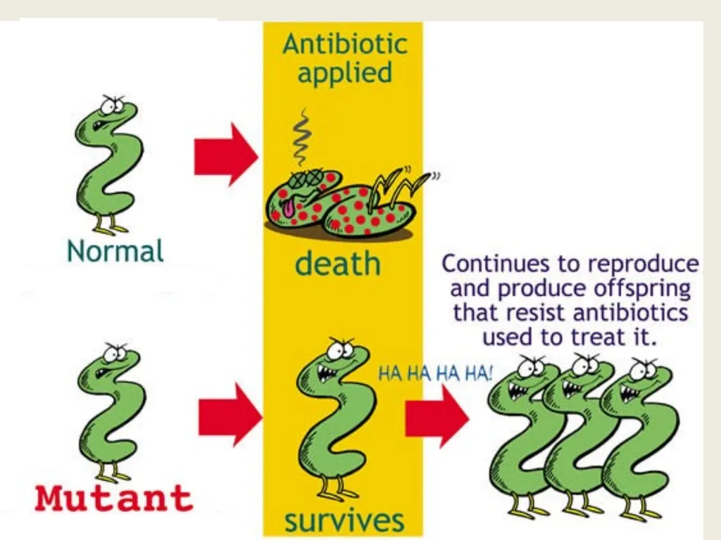
Antimicrobial resistance is the ability of microbes to grow and multiply in the presence of antimicrobial agents that would normally kill them or limit their growth. The concentration of drug at the site of infection must inhibit the organism and also remain below the level that is toxic to human cells.<br>
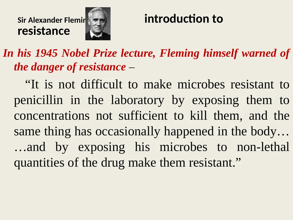
slide4. Sir Alexander Fleming introduction to resistance In his 1945 Nobel Prize lecture, Fleming himself warned of the danger of resistance –
“It is not difficult to make microbes resistant to penicillin in the laboratory by exposing them to concentrations not sufficient to kill them, and the same thing has occasionally happened in the body… …and by exposing his microbes to non-lethal quantities of the drug make them resistant.”<br>
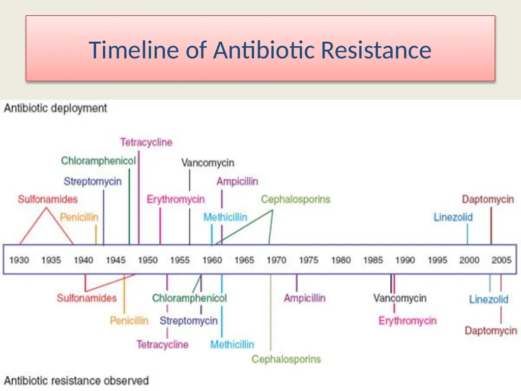
slide5. Timeline of Antibiotic Resistance<br>
slide6. Resistant organisms lead to treatment failure
Increased mortality
Resistant bacteria may spread in Community
Added burden on healthcare costs
Threatens to return to the pre-antibiotic era
Selection pressure
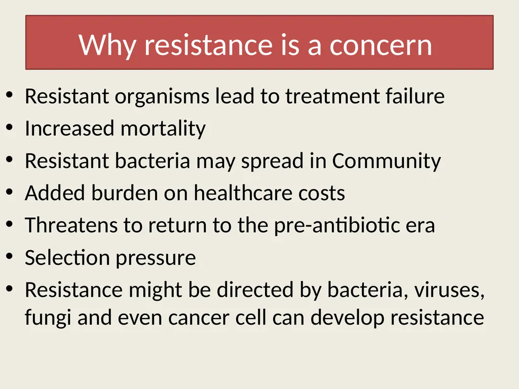
Resistance might be directed by bacteria, viruses, fungi and even cancer cell can develop resistance Why resistance is a concern<br>
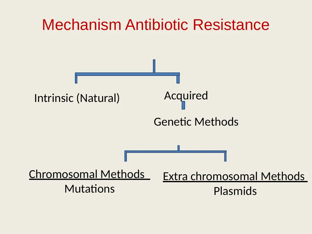
slide7. Mechanism Antibiotic Resistance Intrinsic (Natural) Acquired Genetic Methods Chromosomal Methods
Mutations Extra chromosomal Methods
Plasmids<br>
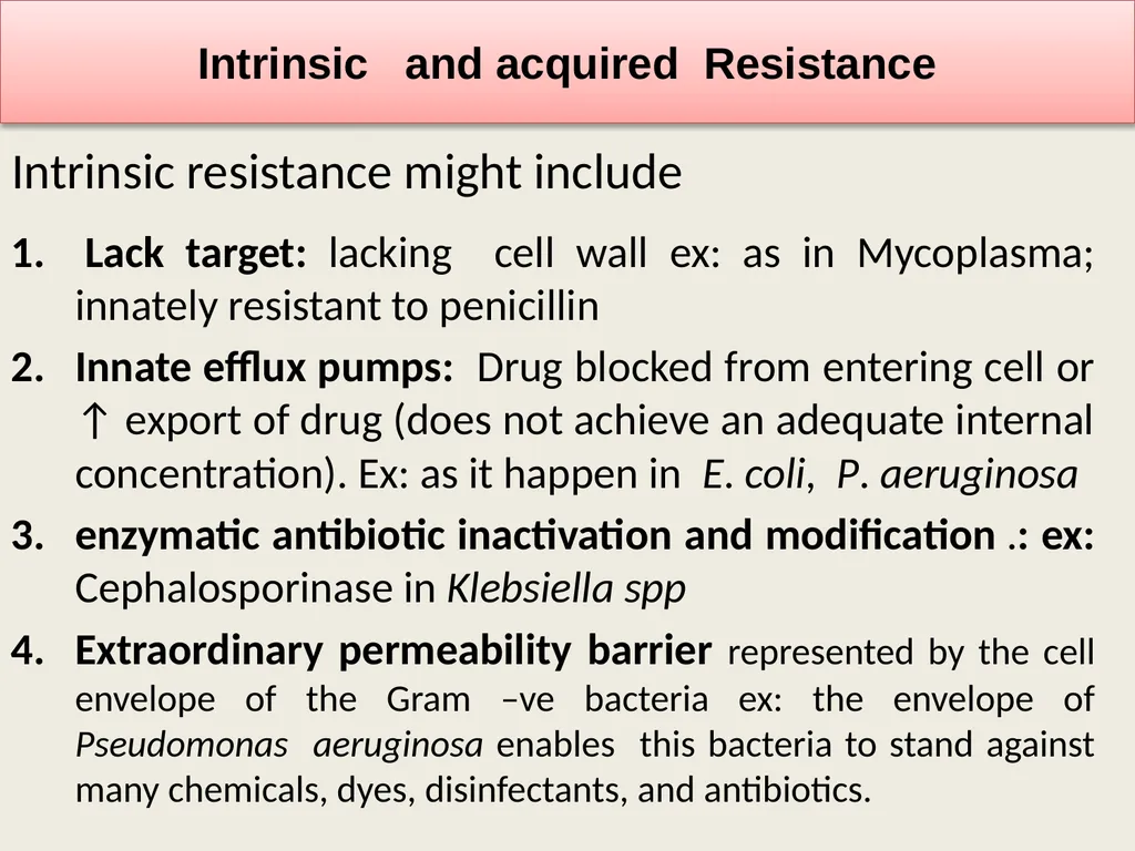
slide8. Intrinsic resistance might include Intrinsic and acquired Resistance Lack target: lacking cell wall ex: as in Mycoplasma; innately resistant to penicillin
Innate efflux pumps: Drug blocked from entering cell or ↑ export of drug (does not achieve an adequate internal concentration). Ex: as it happen in E. coli, P. aeruginosa
enzymatic antibiotic inactivation and modification .: ex: Cephalosporinase in Klebsiella spp
Extraordinary permeability barrier represented by the cell envelope of the Gram –ve bacteria ex: the envelope of Pseudomonas aeruginosa enables this bacteria to stand against many chemicals, dyes, disinfectants, and antibiotics.<br>
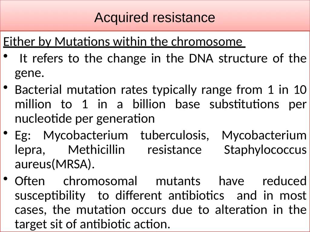
slide9. Acquired resistance Either by Mutations within the chromosome
It refers to the change in the DNA structure of the gene.
Bacterial mutation rates typically range from 1 in 10 million to 1 in a billion base substitutions per nucleotide per generation
Eg: Mycobacterium tuberculosis, Mycobacterium lepra, Methicillin resistance Staphylococcus aureus(MRSA).
Often chromosomal mutants have reduced susceptibility to different antibiotics and in most cases, the mutation occurs due to alteration in the target sit of antibiotic action.<br>
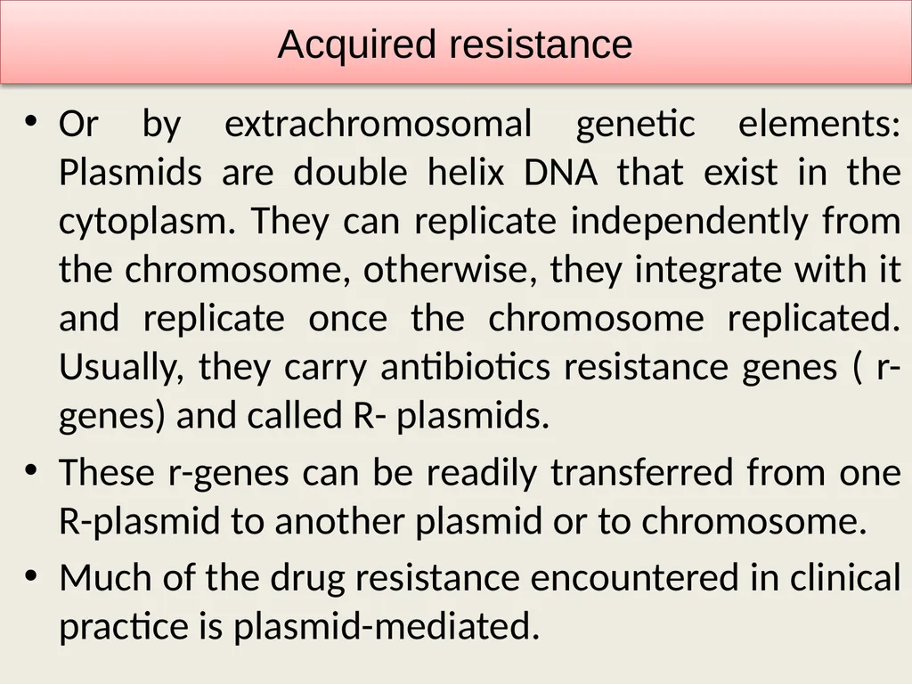
slide10. Or by extrachromosomal genetic elements: Plasmids are double helix DNA that exist in the cytoplasm. They can replicate independently from the chromosome, otherwise, they integrate with it and replicate once the chromosome replicated. Usually, they carry antibiotics resistance genes ( r-genes) and called R- plasmids.
These r-genes can be readily transferred from one R-plasmid to another plasmid or to chromosome.
Much of the drug resistance encountered in clinical practice is plasmid-mediated. Acquired resistance<br>
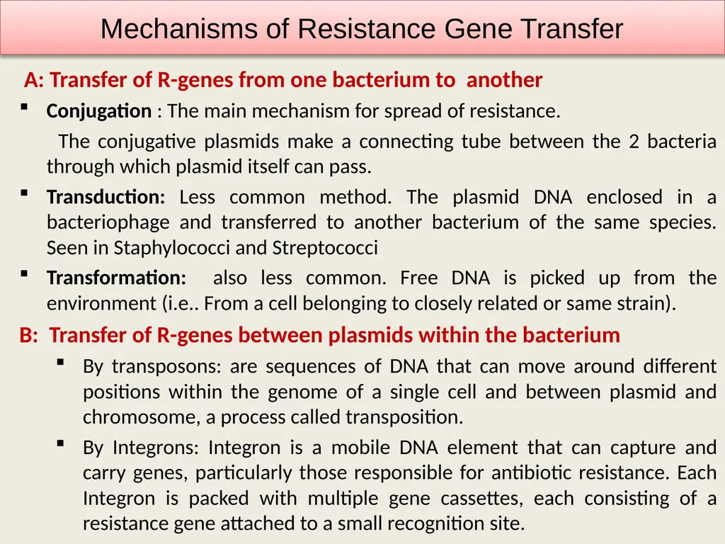
slide11. A: Transfer of R-genes from one bacterium to another
Conjugation : The main mechanism for spread of resistance.
The conjugative plasmids make a connecting tube between the 2 bacteria through which plasmid itself can pass.
Transduction: Less common method. The plasmid DNA enclosed in a bacteriophage and transferred to another bacterium of the same species. Seen in Staphylococci and Streptococci
Transformation: also less common. Free DNA is picked up from the environment (i.e.. From a cell belonging to closely related or same strain).
B: Transfer of R-genes between plasmids within the bacterium
By transposons: are sequences of DNA that can move around different positions within the genome of a single cell and between plasmid and chromosome, a process called transposition.
By Integrons: Integron is a mobile DNA element that can capture and carry genes, particularly those responsible for antibiotic resistance. Each Integron is packed with multiple gene cassettes, each consisting of a resistance gene attached to a small recognition site. Mechanisms of Resistance Gene Transfer<br>
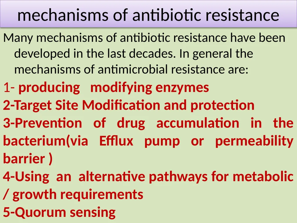
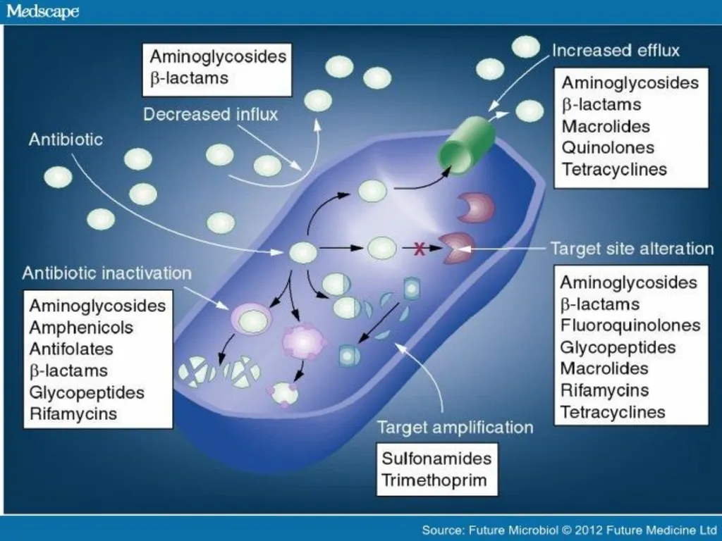
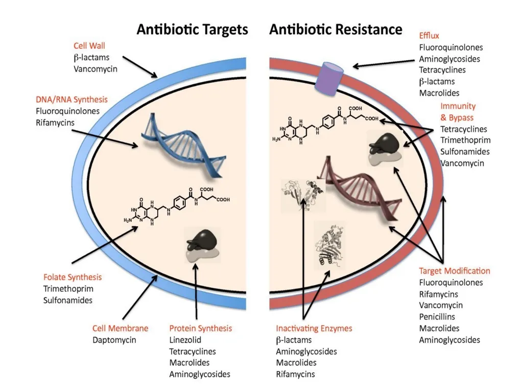
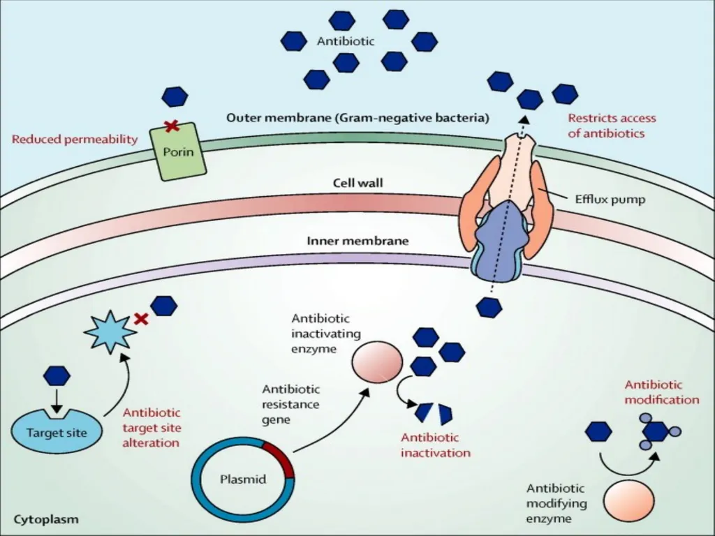
slide12. mechanisms of antibiotic resistance Many mechanisms of antibiotic resistance have been developed in the last decades. In general the mechanisms of antimicrobial resistance are: 1- producing modifying enzymes
2-Target Site Modification and protection
3-Prevention of drug accumulation in the bacterium(via Efflux pump or permeability barrier )
4-Using an alternative pathways for metabolic / growth requirements
5-Quorum sensing<br>
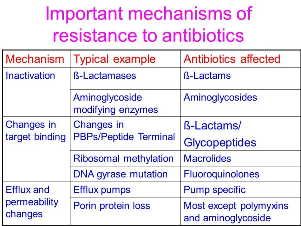
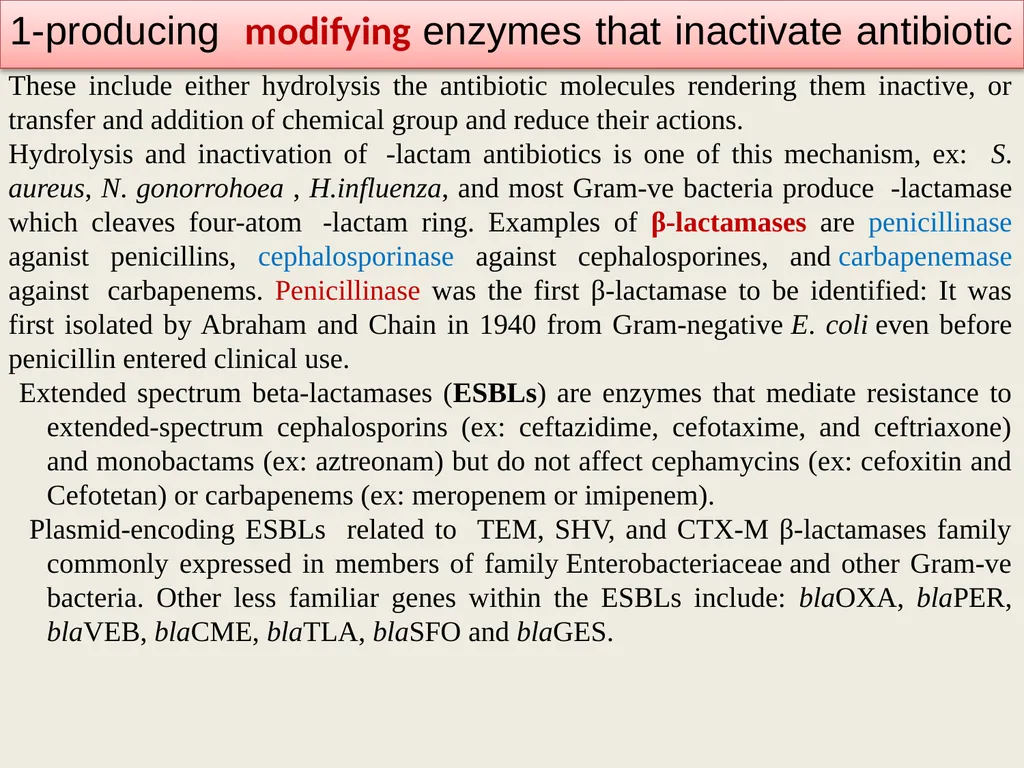
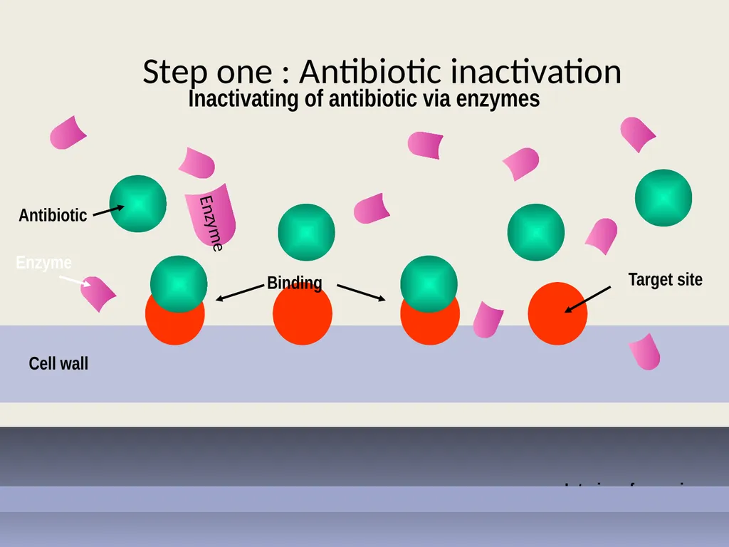
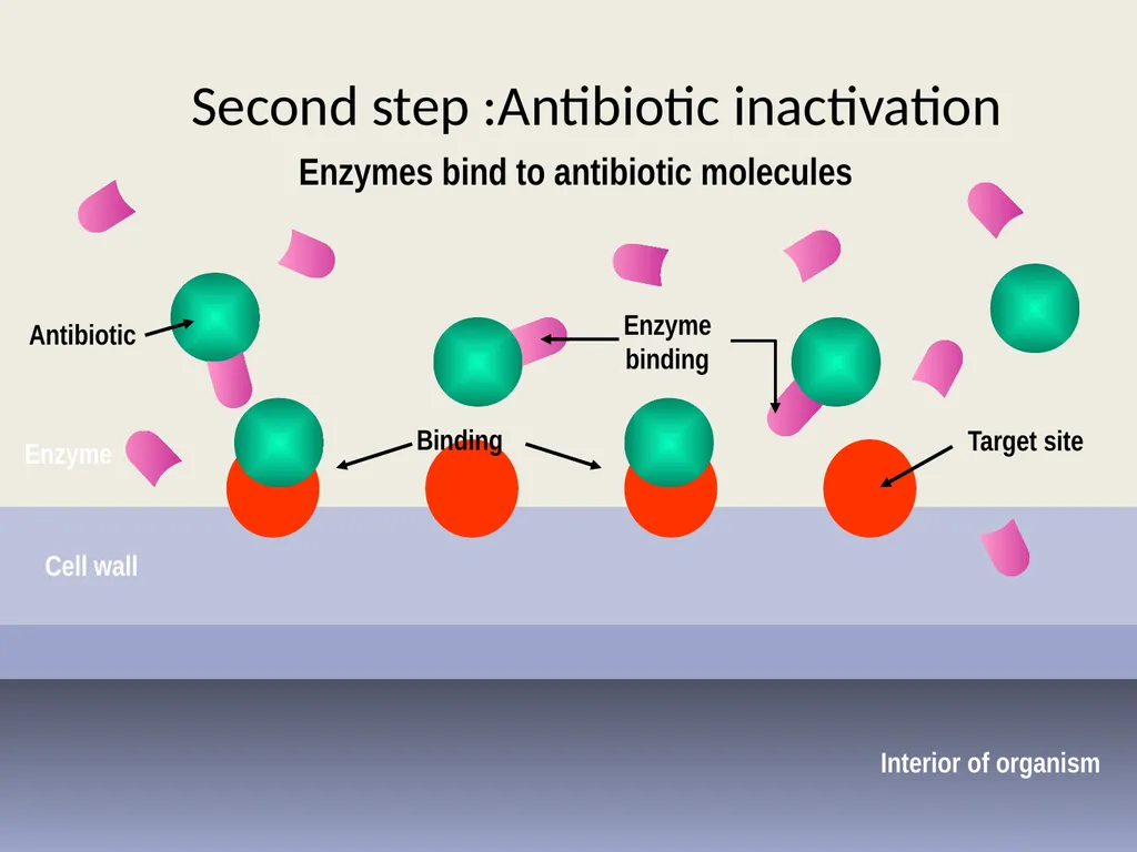
slide16. 1-producing modifying enzymes that inactivate antibiotic These include either hydrolysis the antibiotic molecules rendering them inactive, or transfer and addition of chemical group and reduce their actions.
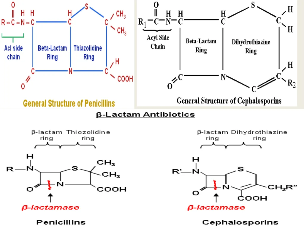
Hydrolysis and inactivation of -lactam antibiotics is one of this mechanism, ex: S. aureus, N. gonorrohoea , H.influenza, and most Gram-ve bacteria produce -lactamase which cleaves four-atom -lactam ring. Examples of β-lactamases are penicillinase aganist penicillins, cephalosporinase against cephalosporines, and carbapenemase against carbapenems. Penicillinase was the first β-lactamase to be identified: It was first isolated by Abraham and Chain in 1940 from Gram-negative E. coli even before penicillin entered clinical use.
Extended spectrum beta-lactamases (ESBLs) are enzymes that mediate resistance to extended-spectrum cephalosporins (ex: ceftazidime, cefotaxime, and ceftriaxone) and monobactams (ex: aztreonam) but do not affect cephamycins (ex: cefoxitin and Cefotetan) or carbapenems (ex: meropenem or imipenem).
Plasmid-encoding ESBLs related to TEM, SHV, and CTX-M β-lactamases family commonly expressed in members of family Enterobacteriaceae and other Gram-ve bacteria. Other less familiar genes within the ESBLs include: blaOXA, blaPER, blaVEB, blaCME, blaTLA, blaSFO and blaGES.<br>
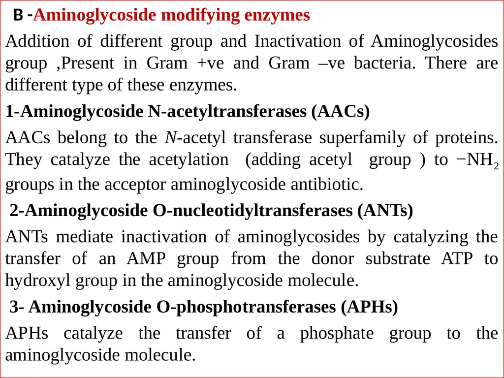
slide18. B -Aminoglycoside modifying enzymes
Addition of different group and Inactivation of Aminoglycosides group ,Present in Gram +ve and Gram –ve bacteria. There are different type of these enzymes.
1-Aminoglycoside N-acetyltransferases (AACs)
AACs belong to the N-acetyl transferase superfamily of proteins. They catalyze the acetylation (adding acetyl group ) to −NH2 groups in the acceptor aminoglycoside antibiotic.
2-Aminoglycoside O-nucleotidyltransferases (ANTs)
ANTs mediate inactivation of aminoglycosides by catalyzing the transfer of an AMP group from the donor substrate ATP to hydroxyl group in the aminoglycoside molecule.
3- Aminoglycoside O-phosphotransferases (APHs)
APHs catalyze the transfer of a phosphate group to the aminoglycoside molecule.<br>
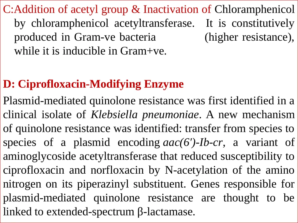
slide19. C:Addition of acetyl group & Inactivation of Chloramphenicol by chloramphenicol acetyltransferase. It is constitutively produced in Gram-ve bacteria (higher resistance), while it is inducible in Gram+ve.
D: Ciprofloxacin-Modifying Enzyme
Plasmid-mediated quinolone resistance was first identified in a clinical isolate of Klebsiella pneumoniae. A new mechanism of quinolone resistance was identified: transfer from species to species of a plasmid encoding aac(6′)-Ib-cr, a variant of aminoglycoside acetyltransferase that reduced susceptibility to ciprofloxacin and norfloxacin by N-acetylation of the amino nitrogen on its piperazinyl substituent. Genes responsible for plasmid-mediated quinolone resistance are thought to be linked to extended-spectrum β-lactamase.<br>
slide20. Step one : Antibiotic inactivation Interior of organism Cell wall Antibiotic Target site Binding Enzyme Enzyme Inactivating of antibiotic via enzymes<br>
slide21. Second step :Antibiotic inactivation Interior of organism Cell wall Antibiotic Target site Binding Enzyme Enzyme binding Enzymes bind to antibiotic molecules<br>
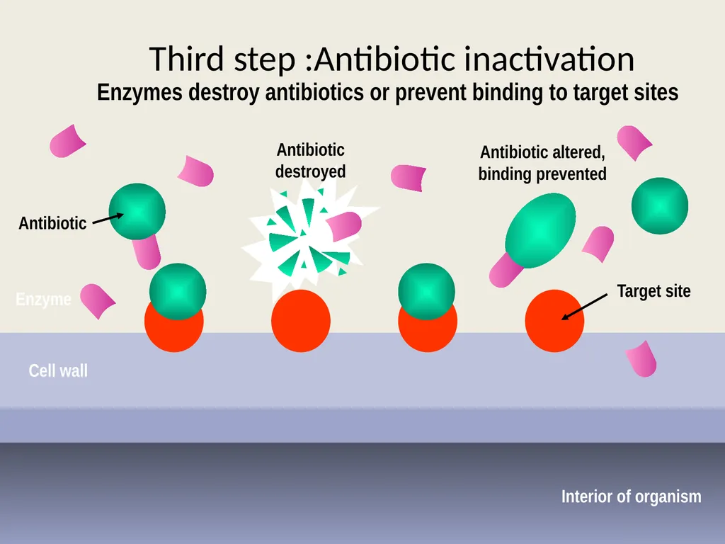
slide22. Third step :Antibiotic inactivation Interior of organism Cell wall Antibiotic Target site Enzyme Antibiotic destroyed Antibiotic altered,
binding prevented Enzymes destroy antibiotics or prevent binding to target sites<br>
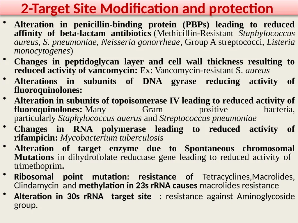
slide23. 2-Target Site Modification and protection Alteration in penicillin-binding protein (PBPs) leading to reduced affinity of beta-lactam antibiotics (Methicillin-Resistant Staphylococcus aureus, S. pneumoniae, Neisseria gonorrheae, Group A streptococci, Listeria monocytogenes)
Changes in peptidoglycan layer and cell wall thickness resulting to reduced activity of vancomycin: Ex: Vancomycin-resistant S. aureus
Alterations in subunits of DNA gyrase reducing activity of fluoroquinolones:
Alteration in subunits of topoisomerase IV leading to reduced activity of fluoroquinolones: Many Gram positive bacteria, particularly Staphylococcus auerus and Streptococcus pneumoniae
Changes in RNA polymerase leading to reduced activity of rifampicin: Mycobacterium tuberculosis
Alteration of target enzyme due to Spontaneous chromosomal Mutations in dihydrofolate reductase gene leading to reduced activity of trimethoprim.
Ribosomal point mutation: resistance of Tetracyclines,Macrolides, Clindamycin and methylation in 23s rRNA causes macrolides resistance
Alteration in 30s rRNA target site : resistance against Aminoglycoside group.<br>
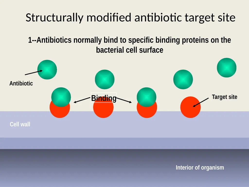
slide24. Structurally modified antibiotic target site Interior of organism Cell wall Target site Binding Antibiotic 1--Antibiotics normally bind to specific binding proteins on the bacterial cell surface<br>
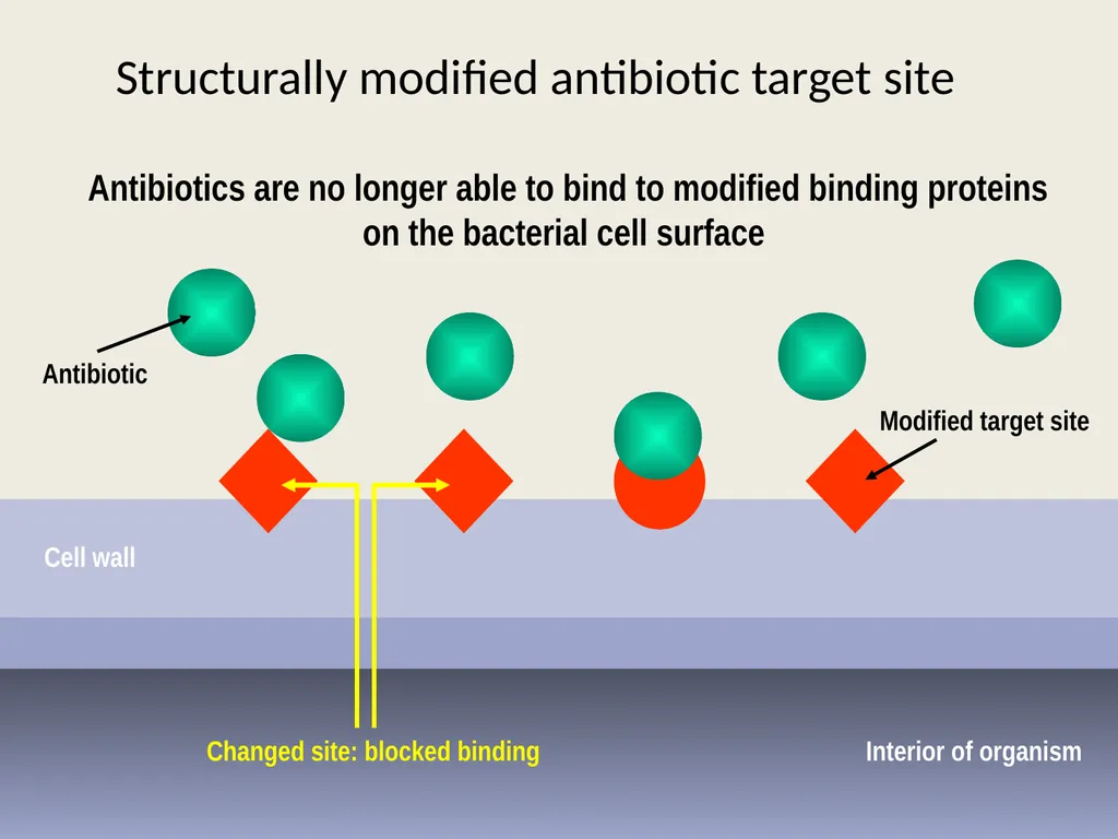
slide25. Structurally modified antibiotic target site Interior of organism Cell wall Modified target site Antibiotic Changed site: blocked binding Antibiotics are no longer able to bind to modified binding proteins on the bacterial cell surface<br>
slide26. 3-Prevention of drug accumulation in the bacterium(via Efflux pump or permeability barrier ) A- Efflux pumps are Cytoplasmic membrane transport proteins (Proteinaceous transporters) localized in the cytoplasmic membrane of all kinds of cells. They are active transporters, meaning that they require a source of chemical energy to perform their function. The genetic elements encoding efflux pumps may be encoded on chromosomes and/or plasmids( both intrinsic (natural) and acquired resistance respectively). In many cases, efflux pump genes are part of an operon, with a regulatory gene controlling expression. Expression of several efflux pumps in a given bacterial species may lead to a broad spectrum of resistance. Efflux systems that contribute to antibiotic resistance have been described from a number of clinically important bacteria, including Campylobacter jejuni, E. coli, Pseudomonas aeruginosa , and Acinitobacter spp.
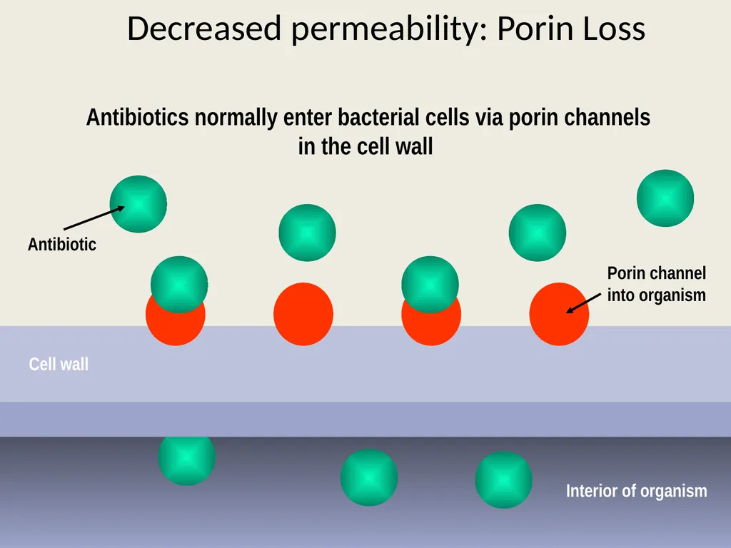
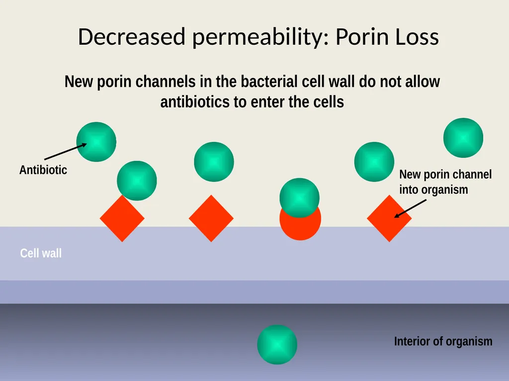
B- For the reducing permeability barrier : in this case the porin channels will change either by reducing number or change the shape or affinity of binding<br>
slide28. Decreased permeability: Porin Loss Interior of organism Cell wall Porin channel
into organism Antibiotic Antibiotics normally enter bacterial cells via porin channels in the cell wall<br>
slide29. Decreased permeability: Porin Loss Interior of organism Cell wall New porin channel
into organism Antibiotic New porin channels in the bacterial cell wall do not allow antibiotics to enter the cells<br>
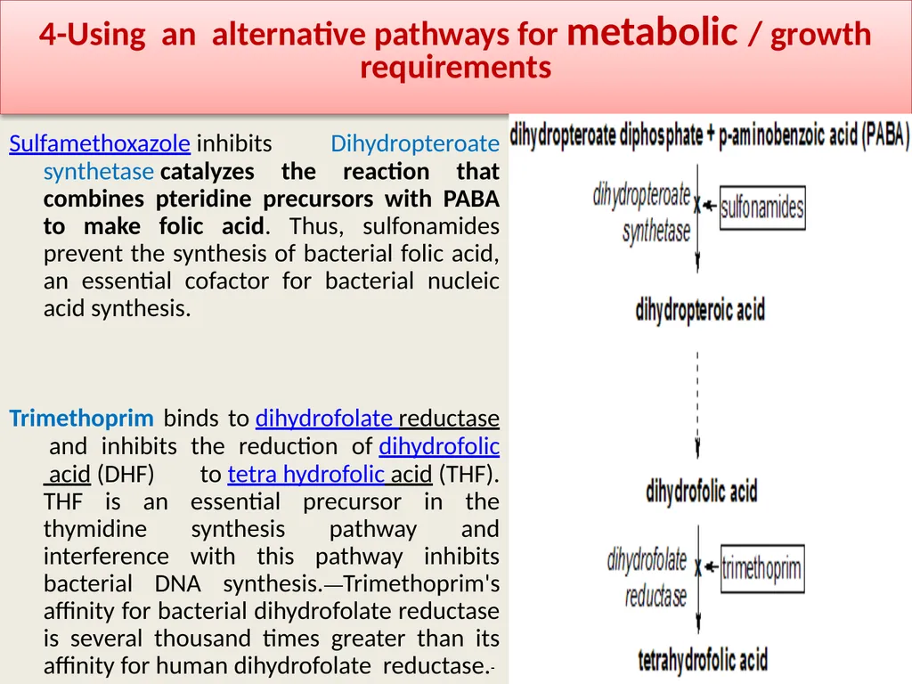
slide30. 4-Using an alternative pathways for metabolic / growth requirements Sulfamethoxazole inhibits Dihydropteroate synthetase catalyzes the reaction that combines pteridine precursors with PABA to make folic acid. Thus, sulfonamides prevent the synthesis of bacterial folic acid, an essential cofactor for bacterial nucleic acid synthesis.
Trimethoprim binds to dihydrofolate reductase and inhibits the reduction of dihydrofolic acid (DHF) to tetra hydrofolic acid (THF). THF is an essential precursor in the thymidine synthesis pathway and interference with this pathway inhibits bacterial DNA synthesis. Trimethoprim's affinity for bacterial dihydrofolate reductase is several thousand times greater than its affinity for human dihydrofolate reductase.<br>
slide31. sulfa drugs inhibit a step in the pathway to make folic acid, an essential vitamin that bacteria need for their everyday functions. But some resistant bacteria have developed different metabolic pathways that allow them to make folic acid even in the presence of these drugs . Or some microorganism produce large quantity of PABA (Para Amino Benzoic Acid ) thus overcome the inhibition of sulfa drug to folic acid cycle(Competition to enter the synthesis pathway)
Note :folic acid doesn’t synthesis in
human thus
we take as a tablet and
specially given to pregnant
women<br>
slide32. Quorum sensing is a process of cell–cell communication that allows bacteria to share information about cell density and adjust gene expression accordingly.
In this process the microbes communicate with each other and exchange signaling chemicals or so-called Autoinducers. when its colony reaches a critical density, the threshold of autoinduction is reached and gene expression starts.
These autoinducers allow bacterial population to coordinate gene expression for virulence factors, conjugation, apoptosis, mobility and Antimicrobial resistance.
QS signal molecules AHL, AIP, AI-2 & AI-3 have been identified in Gram-negative bacteria.
AI-2 QS –system is shared by Gram-positive bacteria.
Several QS inhibitors molecules has been synthesized which are analogues to AHL, AIP, and AI-2
QS inhibitors have been synthesized and have been isolated from several natural extracts such as garlic extract. 5- Quorum sensing<br>
slide2. In the previous lecture, we took about the mechanism by which the antimicrobial agents can act against the different pathogens. in this lecture, we will demonstrate the mechanisms of microbial resistance.
So we have:
Antibiotic action ----- against microorganism reaction
Antibiotic activity ------against microbial resistance
Antimicrobial resistance is the ability of microbes to grow and multiply in the presence of antimicrobial agents that would normally kill them or limit their growth. The concentration of drug at the site of infection must inhibit the organism and also remain below the level that is toxic to human cells.<br>
slide4. Sir Alexander Fleming introduction to resistance In his 1945 Nobel Prize lecture, Fleming himself warned of the danger of resistance –
“It is not difficult to make microbes resistant to penicillin in the laboratory by exposing them to concentrations not sufficient to kill them, and the same thing has occasionally happened in the body… …and by exposing his microbes to non-lethal quantities of the drug make them resistant.”<br>
slide5. Timeline of Antibiotic Resistance<br>
slide6. Resistant organisms lead to treatment failure
Increased mortality
Resistant bacteria may spread in Community
Added burden on healthcare costs
Threatens to return to the pre-antibiotic era
Selection pressure
Resistance might be directed by bacteria, viruses, fungi and even cancer cell can develop resistance Why resistance is a concern<br>
slide7. Mechanism Antibiotic Resistance Intrinsic (Natural) Acquired Genetic Methods Chromosomal Methods
Mutations Extra chromosomal Methods
Plasmids<br>
slide8. Intrinsic resistance might include Intrinsic and acquired Resistance Lack target: lacking cell wall ex: as in Mycoplasma; innately resistant to penicillin
Innate efflux pumps: Drug blocked from entering cell or ↑ export of drug (does not achieve an adequate internal concentration). Ex: as it happen in E. coli, P. aeruginosa
enzymatic antibiotic inactivation and modification .: ex: Cephalosporinase in Klebsiella spp
Extraordinary permeability barrier represented by the cell envelope of the Gram –ve bacteria ex: the envelope of Pseudomonas aeruginosa enables this bacteria to stand against many chemicals, dyes, disinfectants, and antibiotics.<br>
slide9. Acquired resistance Either by Mutations within the chromosome
It refers to the change in the DNA structure of the gene.
Bacterial mutation rates typically range from 1 in 10 million to 1 in a billion base substitutions per nucleotide per generation
Eg: Mycobacterium tuberculosis, Mycobacterium lepra, Methicillin resistance Staphylococcus aureus(MRSA).
Often chromosomal mutants have reduced susceptibility to different antibiotics and in most cases, the mutation occurs due to alteration in the target sit of antibiotic action.<br>
slide10. Or by extrachromosomal genetic elements: Plasmids are double helix DNA that exist in the cytoplasm. They can replicate independently from the chromosome, otherwise, they integrate with it and replicate once the chromosome replicated. Usually, they carry antibiotics resistance genes ( r-genes) and called R- plasmids.
These r-genes can be readily transferred from one R-plasmid to another plasmid or to chromosome.
Much of the drug resistance encountered in clinical practice is plasmid-mediated. Acquired resistance<br>
slide11. A: Transfer of R-genes from one bacterium to another
Conjugation : The main mechanism for spread of resistance.
The conjugative plasmids make a connecting tube between the 2 bacteria through which plasmid itself can pass.
Transduction: Less common method. The plasmid DNA enclosed in a bacteriophage and transferred to another bacterium of the same species. Seen in Staphylococci and Streptococci
Transformation: also less common. Free DNA is picked up from the environment (i.e.. From a cell belonging to closely related or same strain).
B: Transfer of R-genes between plasmids within the bacterium
By transposons: are sequences of DNA that can move around different positions within the genome of a single cell and between plasmid and chromosome, a process called transposition.
By Integrons: Integron is a mobile DNA element that can capture and carry genes, particularly those responsible for antibiotic resistance. Each Integron is packed with multiple gene cassettes, each consisting of a resistance gene attached to a small recognition site. Mechanisms of Resistance Gene Transfer<br>
slide12. mechanisms of antibiotic resistance Many mechanisms of antibiotic resistance have been developed in the last decades. In general the mechanisms of antimicrobial resistance are: 1- producing modifying enzymes
2-Target Site Modification and protection
3-Prevention of drug accumulation in the bacterium(via Efflux pump or permeability barrier )
4-Using an alternative pathways for metabolic / growth requirements
5-Quorum sensing<br>
slide16. 1-producing modifying enzymes that inactivate antibiotic These include either hydrolysis the antibiotic molecules rendering them inactive, or transfer and addition of chemical group and reduce their actions.
Hydrolysis and inactivation of -lactam antibiotics is one of this mechanism, ex: S. aureus, N. gonorrohoea , H.influenza, and most Gram-ve bacteria produce -lactamase which cleaves four-atom -lactam ring. Examples of β-lactamases are penicillinase aganist penicillins, cephalosporinase against cephalosporines, and carbapenemase against carbapenems. Penicillinase was the first β-lactamase to be identified: It was first isolated by Abraham and Chain in 1940 from Gram-negative E. coli even before penicillin entered clinical use.
Extended spectrum beta-lactamases (ESBLs) are enzymes that mediate resistance to extended-spectrum cephalosporins (ex: ceftazidime, cefotaxime, and ceftriaxone) and monobactams (ex: aztreonam) but do not affect cephamycins (ex: cefoxitin and Cefotetan) or carbapenems (ex: meropenem or imipenem).
Plasmid-encoding ESBLs related to TEM, SHV, and CTX-M β-lactamases family commonly expressed in members of family Enterobacteriaceae and other Gram-ve bacteria. Other less familiar genes within the ESBLs include: blaOXA, blaPER, blaVEB, blaCME, blaTLA, blaSFO and blaGES.<br>
slide18. B -Aminoglycoside modifying enzymes
Addition of different group and Inactivation of Aminoglycosides group ,Present in Gram +ve and Gram –ve bacteria. There are different type of these enzymes.
1-Aminoglycoside N-acetyltransferases (AACs)
AACs belong to the N-acetyl transferase superfamily of proteins. They catalyze the acetylation (adding acetyl group ) to −NH2 groups in the acceptor aminoglycoside antibiotic.
2-Aminoglycoside O-nucleotidyltransferases (ANTs)
ANTs mediate inactivation of aminoglycosides by catalyzing the transfer of an AMP group from the donor substrate ATP to hydroxyl group in the aminoglycoside molecule.
3- Aminoglycoside O-phosphotransferases (APHs)
APHs catalyze the transfer of a phosphate group to the aminoglycoside molecule.<br>
slide19. C:Addition of acetyl group & Inactivation of Chloramphenicol by chloramphenicol acetyltransferase. It is constitutively produced in Gram-ve bacteria (higher resistance), while it is inducible in Gram+ve.
D: Ciprofloxacin-Modifying Enzyme
Plasmid-mediated quinolone resistance was first identified in a clinical isolate of Klebsiella pneumoniae. A new mechanism of quinolone resistance was identified: transfer from species to species of a plasmid encoding aac(6′)-Ib-cr, a variant of aminoglycoside acetyltransferase that reduced susceptibility to ciprofloxacin and norfloxacin by N-acetylation of the amino nitrogen on its piperazinyl substituent. Genes responsible for plasmid-mediated quinolone resistance are thought to be linked to extended-spectrum β-lactamase.<br>
slide20. Step one : Antibiotic inactivation Interior of organism Cell wall Antibiotic Target site Binding Enzyme Enzyme Inactivating of antibiotic via enzymes<br>
slide21. Second step :Antibiotic inactivation Interior of organism Cell wall Antibiotic Target site Binding Enzyme Enzyme binding Enzymes bind to antibiotic molecules<br>
slide22. Third step :Antibiotic inactivation Interior of organism Cell wall Antibiotic Target site Enzyme Antibiotic destroyed Antibiotic altered,
binding prevented Enzymes destroy antibiotics or prevent binding to target sites<br>
slide23. 2-Target Site Modification and protection Alteration in penicillin-binding protein (PBPs) leading to reduced affinity of beta-lactam antibiotics (Methicillin-Resistant Staphylococcus aureus, S. pneumoniae, Neisseria gonorrheae, Group A streptococci, Listeria monocytogenes)
Changes in peptidoglycan layer and cell wall thickness resulting to reduced activity of vancomycin: Ex: Vancomycin-resistant S. aureus
Alterations in subunits of DNA gyrase reducing activity of fluoroquinolones:
Alteration in subunits of topoisomerase IV leading to reduced activity of fluoroquinolones: Many Gram positive bacteria, particularly Staphylococcus auerus and Streptococcus pneumoniae
Changes in RNA polymerase leading to reduced activity of rifampicin: Mycobacterium tuberculosis
Alteration of target enzyme due to Spontaneous chromosomal Mutations in dihydrofolate reductase gene leading to reduced activity of trimethoprim.
Ribosomal point mutation: resistance of Tetracyclines,Macrolides, Clindamycin and methylation in 23s rRNA causes macrolides resistance
Alteration in 30s rRNA target site : resistance against Aminoglycoside group.<br>
slide24. Structurally modified antibiotic target site Interior of organism Cell wall Target site Binding Antibiotic 1--Antibiotics normally bind to specific binding proteins on the bacterial cell surface<br>
slide25. Structurally modified antibiotic target site Interior of organism Cell wall Modified target site Antibiotic Changed site: blocked binding Antibiotics are no longer able to bind to modified binding proteins on the bacterial cell surface<br>
slide26. 3-Prevention of drug accumulation in the bacterium(via Efflux pump or permeability barrier ) A- Efflux pumps are Cytoplasmic membrane transport proteins (Proteinaceous transporters) localized in the cytoplasmic membrane of all kinds of cells. They are active transporters, meaning that they require a source of chemical energy to perform their function. The genetic elements encoding efflux pumps may be encoded on chromosomes and/or plasmids( both intrinsic (natural) and acquired resistance respectively). In many cases, efflux pump genes are part of an operon, with a regulatory gene controlling expression. Expression of several efflux pumps in a given bacterial species may lead to a broad spectrum of resistance. Efflux systems that contribute to antibiotic resistance have been described from a number of clinically important bacteria, including Campylobacter jejuni, E. coli, Pseudomonas aeruginosa , and Acinitobacter spp.
B- For the reducing permeability barrier : in this case the porin channels will change either by reducing number or change the shape or affinity of binding<br>
slide28. Decreased permeability: Porin Loss Interior of organism Cell wall Porin channel
into organism Antibiotic Antibiotics normally enter bacterial cells via porin channels in the cell wall<br>
slide29. Decreased permeability: Porin Loss Interior of organism Cell wall New porin channel
into organism Antibiotic New porin channels in the bacterial cell wall do not allow antibiotics to enter the cells<br>
slide30. 4-Using an alternative pathways for metabolic / growth requirements Sulfamethoxazole inhibits Dihydropteroate synthetase catalyzes the reaction that combines pteridine precursors with PABA to make folic acid. Thus, sulfonamides prevent the synthesis of bacterial folic acid, an essential cofactor for bacterial nucleic acid synthesis.
Trimethoprim binds to dihydrofolate reductase and inhibits the reduction of dihydrofolic acid (DHF) to tetra hydrofolic acid (THF). THF is an essential precursor in the thymidine synthesis pathway and interference with this pathway inhibits bacterial DNA synthesis. Trimethoprim's affinity for bacterial dihydrofolate reductase is several thousand times greater than its affinity for human dihydrofolate reductase.<br>
slide31. sulfa drugs inhibit a step in the pathway to make folic acid, an essential vitamin that bacteria need for their everyday functions. But some resistant bacteria have developed different metabolic pathways that allow them to make folic acid even in the presence of these drugs . Or some microorganism produce large quantity of PABA (Para Amino Benzoic Acid ) thus overcome the inhibition of sulfa drug to folic acid cycle(Competition to enter the synthesis pathway)
Note :folic acid doesn’t synthesis in
human thus
we take as a tablet and
specially given to pregnant
women<br>
slide32. Quorum sensing is a process of cell–cell communication that allows bacteria to share information about cell density and adjust gene expression accordingly.
In this process the microbes communicate with each other and exchange signaling chemicals or so-called Autoinducers. when its colony reaches a critical density, the threshold of autoinduction is reached and gene expression starts.
These autoinducers allow bacterial population to coordinate gene expression for virulence factors, conjugation, apoptosis, mobility and Antimicrobial resistance.
QS signal molecules AHL, AIP, AI-2 & AI-3 have been identified in Gram-negative bacteria.
AI-2 QS –system is shared by Gram-positive bacteria.
Several QS inhibitors molecules has been synthesized which are analogues to AHL, AIP, and AI-2
QS inhibitors have been synthesized and have been isolated from several natural extracts such as garlic extract. 5- Quorum sensing<br>