
Actinomycetes classification Domain:Bacteria

Description: Actinomycetes classification Domain:Bacteria Phylum:Actinobacteria Class:Actinobacteria Order:Actinomycetales Family:Actinomycetaceae Introduction The actinomycetes are very diverse and contain a variety of subdivisions, as well as
Related Topics
Download Presentation
"Actinomycetes classification Domain:Bacteria" is the property of its rightful owner. Permission is granted to download and print the materials on this website for personal, non-commercial use only, and to display it on your personal computer provided you do not modify the materials and that you retain all copyright notices contained in the materials. By downloading content from our website, you accept the terms of this agreement.
Presentation Transcript
slide1. Actinomycetes<br>
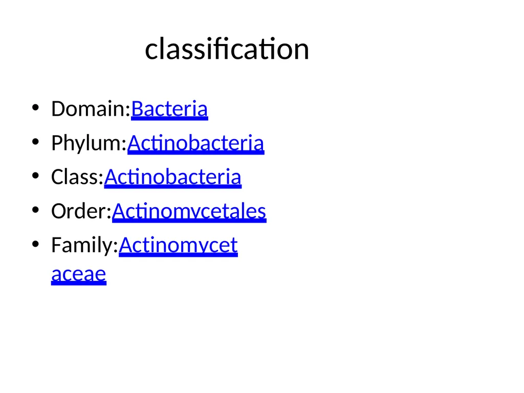
slide2. classification Domain:Bacteria
Phylum:Actinobacteria
Class:Actinobacteria
Order:Actinomycetales
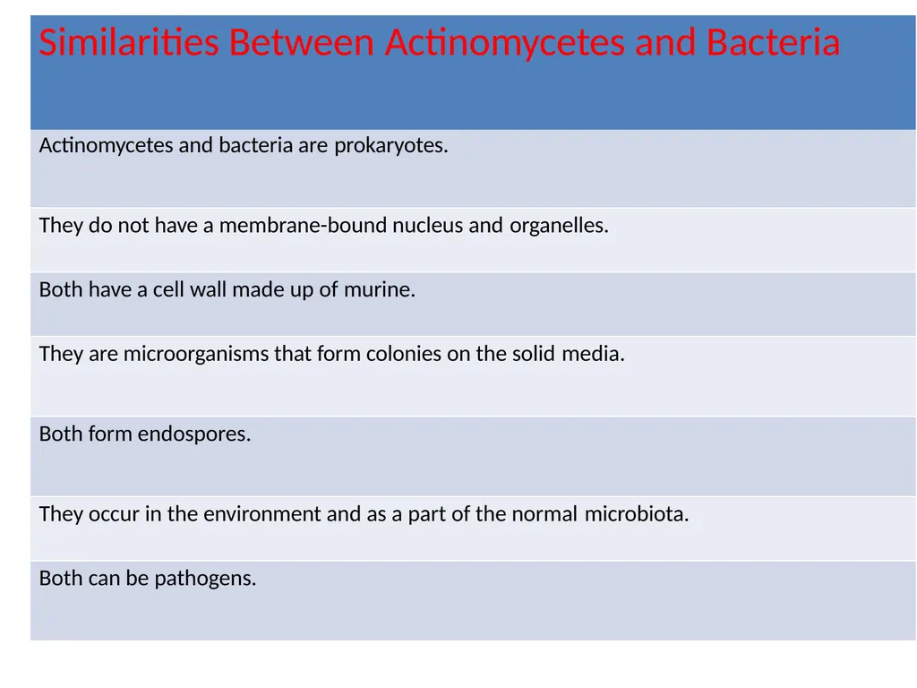
Family:Actinomycetaceae<br>
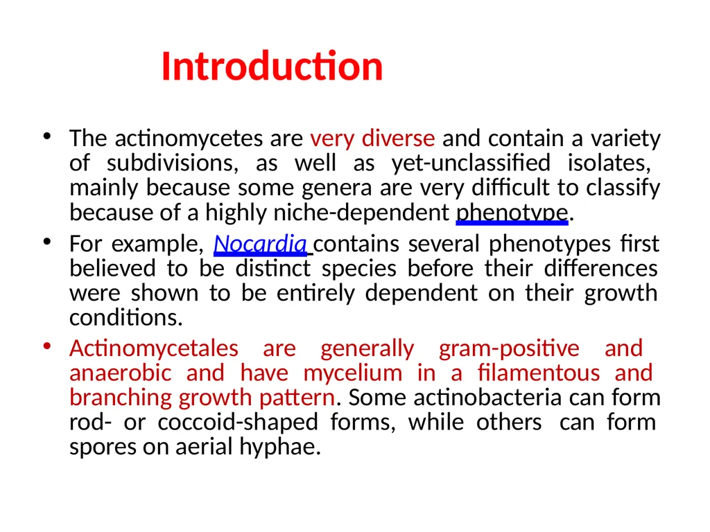
slide3. Introduction The actinomycetes are very diverse and contain a variety of subdivisions, as well as yet-unclassified isolates, mainly because some genera are very difficult to classify because of a highly niche-dependent phenotype.
For example, Nocardia contains several phenotypes first believed to be distinct species before their differences were shown to be entirely dependent on their growth conditions.
Actinomycetales are generally gram-positive and anaerobic and have mycelium in a filamentous and branching growth pattern. Some actinobacteria can form rod- or coccoid-shaped forms, while others can form spores on aerial hyphae.<br>
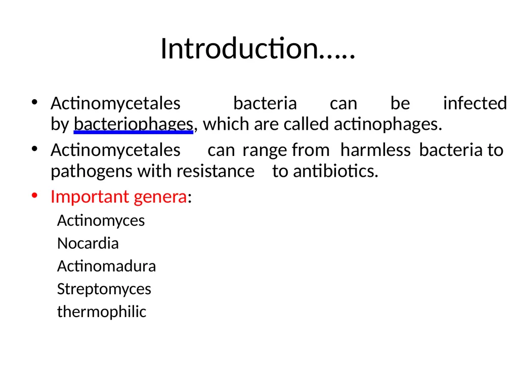
slide4. Introduction….. Actinomycetales bacteria can be infected by bacteriophages, which are called actinophages.
Actinomycetales can range from harmless bacteria to pathogens with resistance to antibiotics.
Important genera:
Actinomyces Nocardia Actinomadura Streptomyces thermophilic<br>
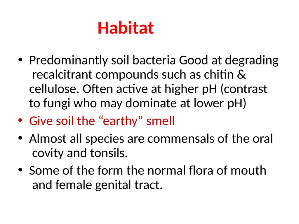
slide5. Habitat Predominantly soil bacteria Good at degrading recalcitrant compounds such as chitin & cellulose. Often active at higher pH (contrast to fungi who may dominate at lower pH)
Give soil the “earthy” smell
Almost all species are commensals of the oral covity and tonsils.
Some of the form the normal flora of mouth and female genital tract.<br>
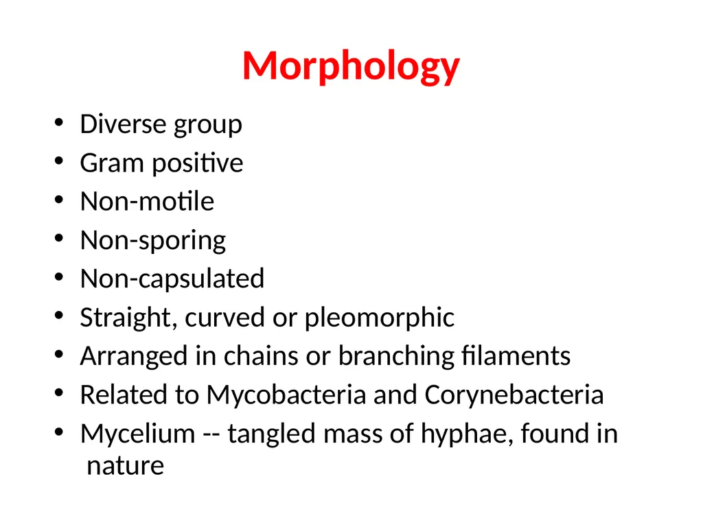
slide6. Morphology Diverse group
Gram positive
Non-motile
Non-sporing
Non-capsulated
Straight, curved or pleomorphic
Arranged in chains or branching filaments
Related to Mycobacteria and Corynebacteria
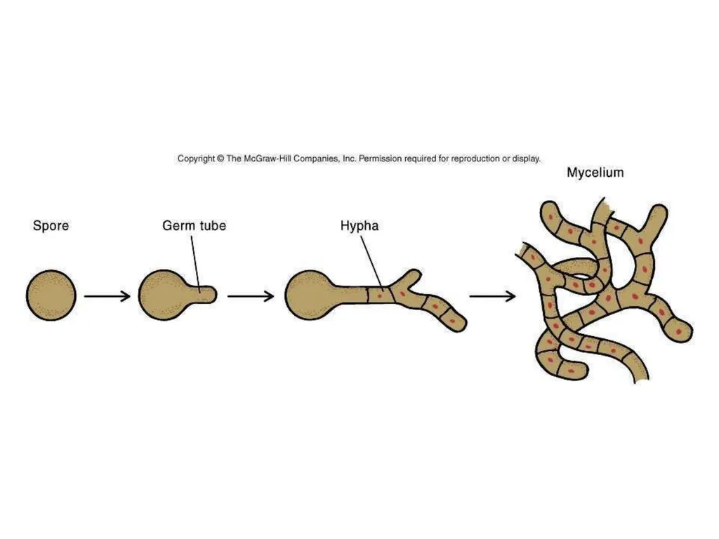
Mycelium -- tangled mass of hyphae, found in nature<br>
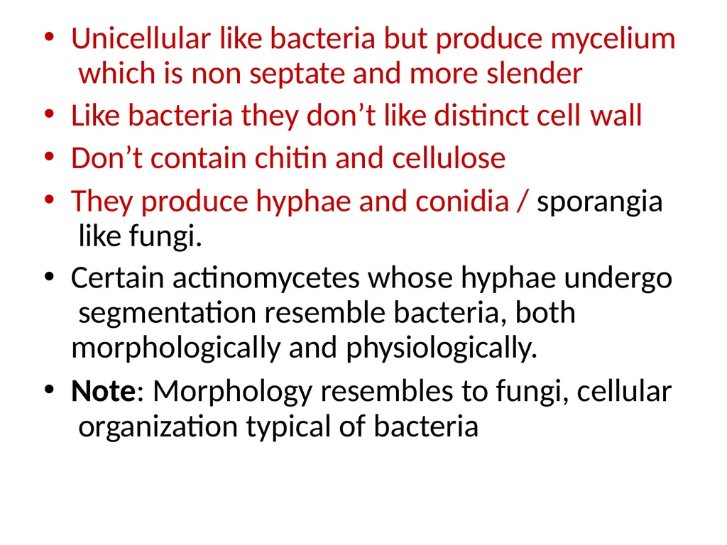
slide7. Unicellular like bacteria but produce mycelium which is non septate and more slender
Like bacteria they don’t like distinct cell wall
Don’t contain chitin and cellulose
They produce hyphae and conidia / sporangia like fungi.
Certain actinomycetes whose hyphae undergo segmentation resemble bacteria, both morphologically and physiologically.
Note: Morphology resembles to fungi, cellular organization typical of bacteria<br>
slide8. Morphology of Actinomycetes<br>
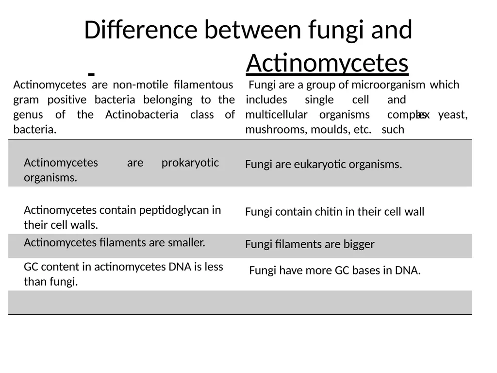
slide10. Difference between fungi and
Actinomycetes gram positive bacteria belonging to genus of the Actinobacteria class bacteria. Actinomycetes are non-motile filamentous Fungi are a group of microorganism which the includes
of single cell multicellular organisms and complex
such as yeast, mushrooms, moulds, etc. are prokaryotic Actinomycetes
organisms. Fungi are eukaryotic organisms. Actinomycetes contain peptidoglycan in their cell walls. Fungi contain chitin in their cell wall Actinomycetes filaments are smaller. Fungi filaments are bigger GC content in actinomycetes DNA is less than fungi. Fungi have more GC bases in DNA.<br>
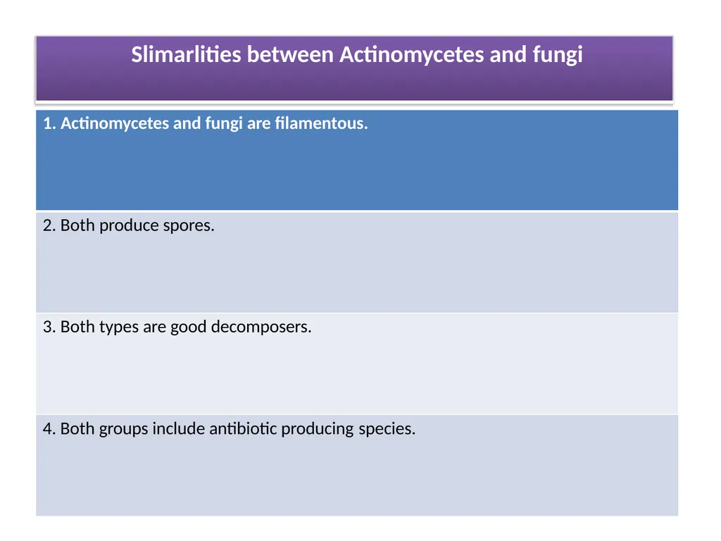
slide11. Slimarlities between Actinomycetes and fungi<br>
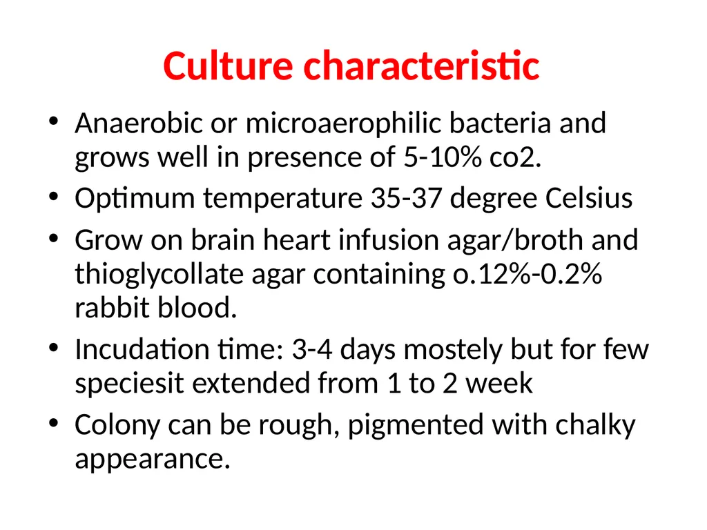
slide14. Culture characteristic Anaerobic or microaerophilic bacteria and grows well in presence of 5-10% co2.
Optimum temperature 35-37 degree Celsius
Grow on brain heart infusion agar/broth and thioglycollate agar containing o.12%-0.2% rabbit blood.
Incudation time: 3-4 days mostely but for few speciesit extended from 1 to 2 week
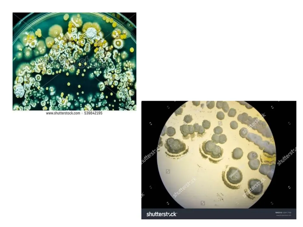
Colony can be rough, pigmented with chalky appearance.<br>
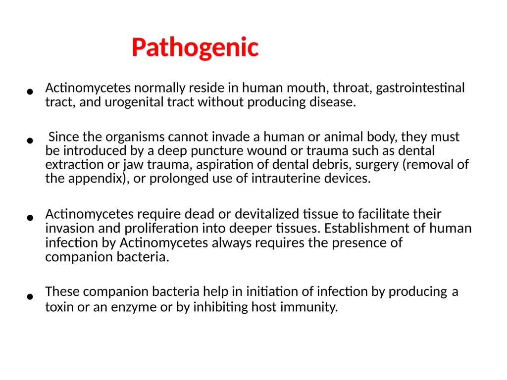
slide16. Pathogenic • Actinomycetes normally reside in human mouth, throat, gastrointestinal tract, and urogenital tract without producing disease. • Since the organisms cannot invade a human or animal body, they must be introduced by a deep puncture wound or trauma such as dental extraction or jaw trauma, aspiration of dental debris, surgery (removal of the appendix), or prolonged use of intrauterine devices. • Actinomycetes require dead or devitalized tissue to facilitate their invasion and proliferation into deeper tissues. Establishment of human infection by Actinomycetes always requires the presence of companion bacteria. • These companion bacteria help in initiation of infection by producing a toxin or an enzyme or by inhibiting host immunity.<br>
slide17. • Once the infection by Actinomycetes is established, the immune system of the infected human host stimulates an intense inflammation. Bacteria from the infected site may disseminate to distant organs of the body. • The Actinomycetes are particularly common type of bacteria found on moldy hay. Farmers may be routinely exposed to very high concentrations of Actinomyces and may inhale as many as 750,000
spores per minute. Frequent exposure to Actinomyces is the cause of Farmers Lung respiratory problems.<br>
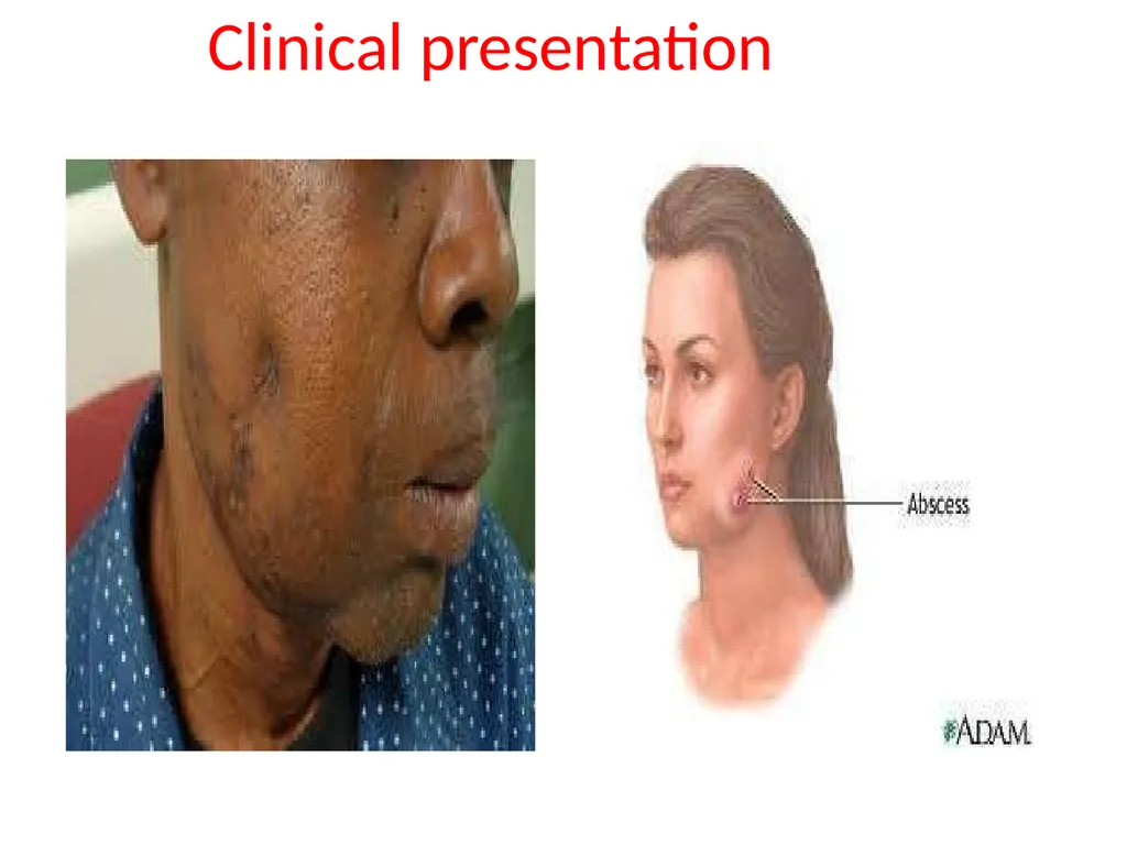
slide18. Clinical presentation<br>
slide19. Cervicofacial infection, which accounts for more than half of reported cases; the jaw is often involved. The disease is endogenous in origin; dental caries is a predisposing factor, and infection may follow tooth extractions or other dental procedures.<br>
slide20. Who Get Infected Men are affected more frequently than women, and in some regions the disease is more common in rural agricultural workers than in town dwellers, probably owing to lower standards of dental care in the former.<br>
slide21. Thoracic actinomycosis Thoracic actinomycosis commences in the lung, probably as a result of aspiration of actinomyces from the mouth.
Sinuses often appear on the chest wall, and the ribs and spine may be eroded.
Primary endobronchial actinomycosis is an uncommon complication of an inhaled foreign body.<br>
slide22. Abdominal Actinomyctes Abdominal cases commence in the appendix or, less frequently, in colonic diverticulae.
Pelvic actinomycosis occurs occasionally in women fitted with plastic intra-uterine contraceptive devices.<br>
slide23. Clinical significance Are part of the NF found in the cavities of humans and other animals.
All may cause actinomycosis or “lumpy jaw” which is a cervicofacial infection that used to occur following tooth extractions or dental surgery which provided traumatized tissue for growth of the microorganism which may also invade the bone. This is rare today because of prophylactic antibiotic therapy.
May cause thoracic or abdominal infections
May cause meningitis, endocarditis, or genital infections<br>
slide24. Every kind of infection is characterized by draining sinuses, usually containing characteristic granules which are colonies of bacteria that look like dense rosettes of club- shaped filaments in radial arrangement<br>
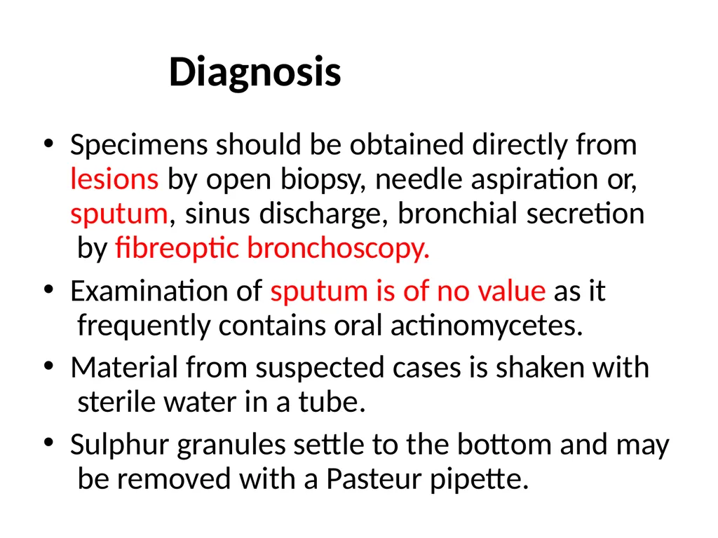
slide25. Diagnosis Specimens should be obtained directly from lesions by open biopsy, needle aspiration or, sputum, sinus discharge, bronchial secretion by fibreoptic bronchoscopy.
Examination of sputum is of no value as it frequently contains oral actinomycetes.
Material from suspected cases is shaken with sterile water in a tube.
Sulphur granules settle to the bottom and may be removed with a Pasteur pipette.<br>
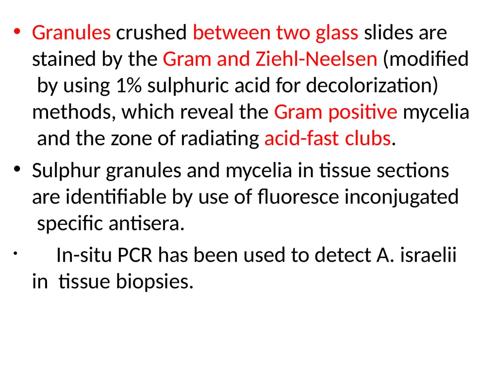
slide26. Granules crushed between two glass slides are stained by the Gram and Ziehl-Neelsen (modified by using 1% sulphuric acid for decolorization) methods, which reveal the Gram positive mycelia and the zone of radiating acid-fast clubs.
Sulphur granules and mycelia in tissue sections are identifiable by use of fluoresce inconjugated specific antisera.
In-situ PCR has been used to detect A. israelii in tissue biopsies.<br>
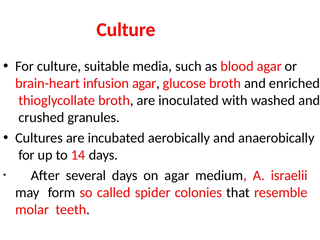
slide27. Culture For culture, suitable media, such as blood agar or brain-heart infusion agar, glucose broth and enriched thioglycollate broth, are inoculated with washed and crushed granules.
Cultures are incubated aerobically and anaerobically for up to 14 days.
After several days on agar medium, A. israelii may form so called spider colonies that resemble molar teeth.<br>
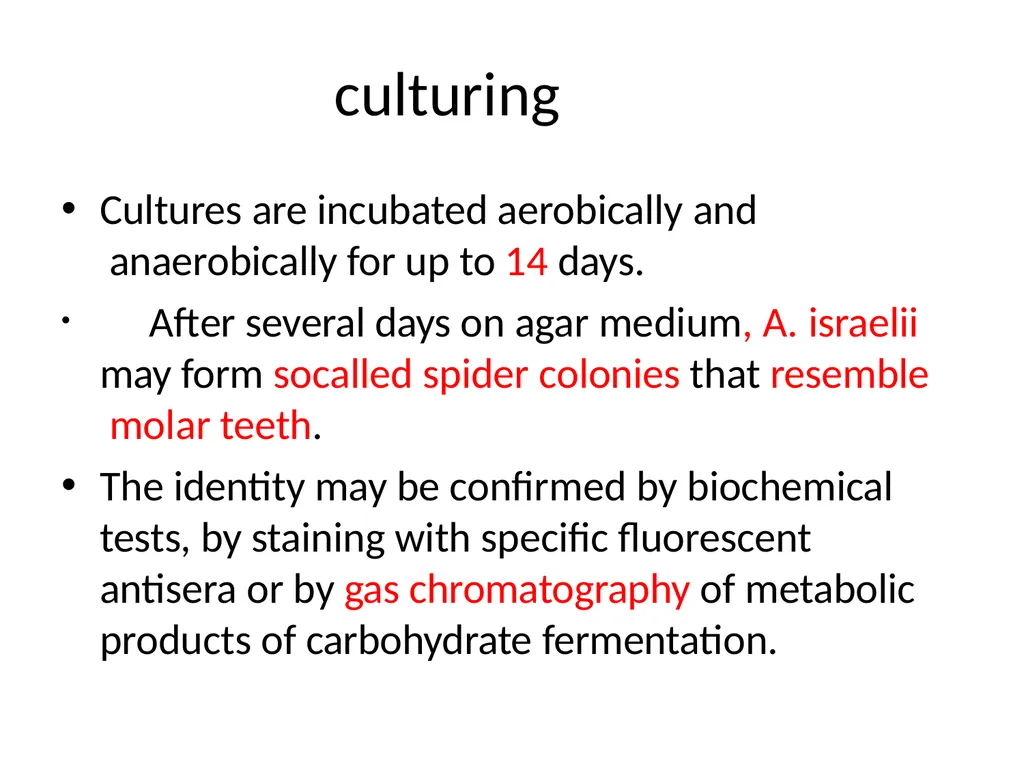
slide28. culturing Cultures are incubated aerobically and anaerobically for up to 14 days.
After several days on agar medium, A. israelii may form socalled spider colonies that resemble molar teeth.
The identity may be confirmed by biochemical tests, by staining with specific fluorescent antisera or by gas chromatography of metabolic products of carbohydrate fermentation.<br>
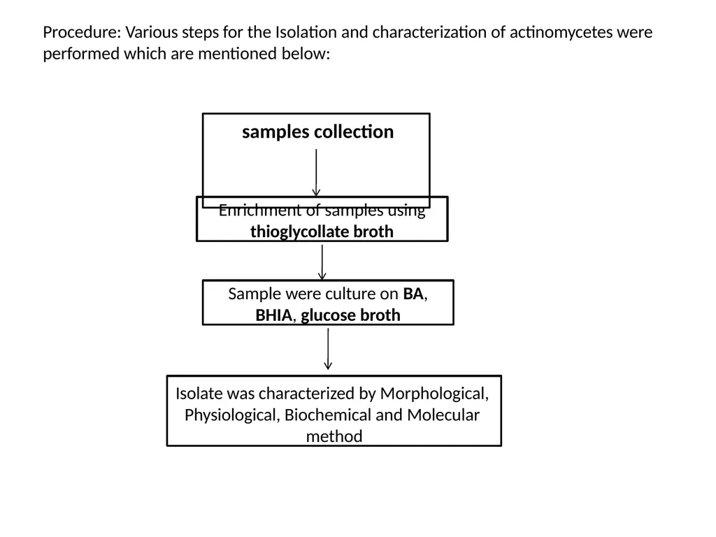
slide29. Procedure: Various steps for the Isolation and characterization of actinomycetes were performed which are mentioned below: samples collection Enrichment of samples using
thioglycollate broth Sample were culture on BA,
BHIA, glucose broth Isolate was characterized by Morphological, Physiological, Biochemical and Molecular method<br>
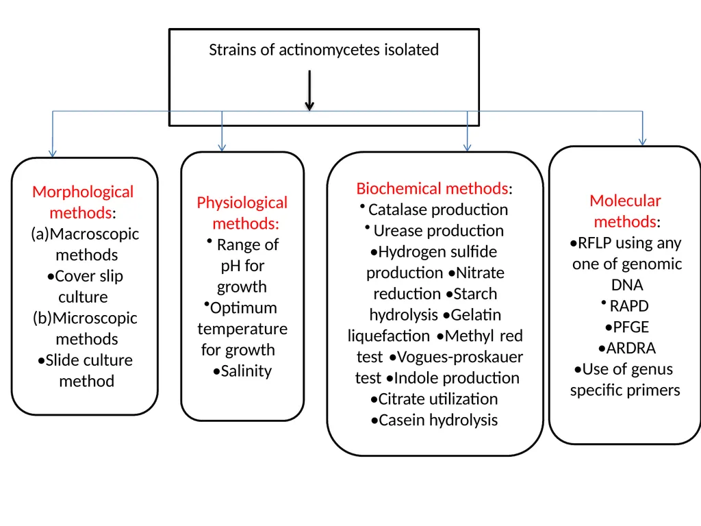
slide30. Strains of actinomycetes isolated Morphological methods: (a)Macroscopic methods
•Cover slip culture (b)Microscopic methods
•Slide culture method Physiological methods:
Range of pH for growth
Optimum temperature for growth
•Salinity Biochemical methods:
Catalase production
Urease production
•Hydrogen sulfide production •Nitrate reduction •Starch hydrolysis •Gelatin
liquefaction •Methyl red test •Vogues-proskauer test •Indole production
•Citrate utilization
•Casein hydrolysis Molecular methods:
•RFLP using any one of genomic DNA
RAPD
•PFGE
•ARDRA
•Use of genus specific primers<br>
slide2. classification Domain:Bacteria
Phylum:Actinobacteria
Class:Actinobacteria
Order:Actinomycetales
Family:Actinomycetaceae<br>
slide3. Introduction The actinomycetes are very diverse and contain a variety of subdivisions, as well as yet-unclassified isolates, mainly because some genera are very difficult to classify because of a highly niche-dependent phenotype.
For example, Nocardia contains several phenotypes first believed to be distinct species before their differences were shown to be entirely dependent on their growth conditions.
Actinomycetales are generally gram-positive and anaerobic and have mycelium in a filamentous and branching growth pattern. Some actinobacteria can form rod- or coccoid-shaped forms, while others can form spores on aerial hyphae.<br>
slide4. Introduction….. Actinomycetales bacteria can be infected by bacteriophages, which are called actinophages.
Actinomycetales can range from harmless bacteria to pathogens with resistance to antibiotics.
Important genera:
Actinomyces Nocardia Actinomadura Streptomyces thermophilic<br>
slide5. Habitat Predominantly soil bacteria Good at degrading recalcitrant compounds such as chitin & cellulose. Often active at higher pH (contrast to fungi who may dominate at lower pH)
Give soil the “earthy” smell
Almost all species are commensals of the oral covity and tonsils.
Some of the form the normal flora of mouth and female genital tract.<br>
slide6. Morphology Diverse group
Gram positive
Non-motile
Non-sporing
Non-capsulated
Straight, curved or pleomorphic
Arranged in chains or branching filaments
Related to Mycobacteria and Corynebacteria
Mycelium -- tangled mass of hyphae, found in nature<br>
slide7. Unicellular like bacteria but produce mycelium which is non septate and more slender
Like bacteria they don’t like distinct cell wall
Don’t contain chitin and cellulose
They produce hyphae and conidia / sporangia like fungi.
Certain actinomycetes whose hyphae undergo segmentation resemble bacteria, both morphologically and physiologically.
Note: Morphology resembles to fungi, cellular organization typical of bacteria<br>
slide8. Morphology of Actinomycetes<br>
slide10. Difference between fungi and
Actinomycetes gram positive bacteria belonging to genus of the Actinobacteria class bacteria. Actinomycetes are non-motile filamentous Fungi are a group of microorganism which the includes
of single cell multicellular organisms and complex
such as yeast, mushrooms, moulds, etc. are prokaryotic Actinomycetes
organisms. Fungi are eukaryotic organisms. Actinomycetes contain peptidoglycan in their cell walls. Fungi contain chitin in their cell wall Actinomycetes filaments are smaller. Fungi filaments are bigger GC content in actinomycetes DNA is less than fungi. Fungi have more GC bases in DNA.<br>
slide11. Slimarlities between Actinomycetes and fungi<br>
slide14. Culture characteristic Anaerobic or microaerophilic bacteria and grows well in presence of 5-10% co2.
Optimum temperature 35-37 degree Celsius
Grow on brain heart infusion agar/broth and thioglycollate agar containing o.12%-0.2% rabbit blood.
Incudation time: 3-4 days mostely but for few speciesit extended from 1 to 2 week
Colony can be rough, pigmented with chalky appearance.<br>
slide16. Pathogenic • Actinomycetes normally reside in human mouth, throat, gastrointestinal tract, and urogenital tract without producing disease. • Since the organisms cannot invade a human or animal body, they must be introduced by a deep puncture wound or trauma such as dental extraction or jaw trauma, aspiration of dental debris, surgery (removal of the appendix), or prolonged use of intrauterine devices. • Actinomycetes require dead or devitalized tissue to facilitate their invasion and proliferation into deeper tissues. Establishment of human infection by Actinomycetes always requires the presence of companion bacteria. • These companion bacteria help in initiation of infection by producing a toxin or an enzyme or by inhibiting host immunity.<br>
slide17. • Once the infection by Actinomycetes is established, the immune system of the infected human host stimulates an intense inflammation. Bacteria from the infected site may disseminate to distant organs of the body. • The Actinomycetes are particularly common type of bacteria found on moldy hay. Farmers may be routinely exposed to very high concentrations of Actinomyces and may inhale as many as 750,000
spores per minute. Frequent exposure to Actinomyces is the cause of Farmers Lung respiratory problems.<br>
slide18. Clinical presentation<br>
slide19. Cervicofacial infection, which accounts for more than half of reported cases; the jaw is often involved. The disease is endogenous in origin; dental caries is a predisposing factor, and infection may follow tooth extractions or other dental procedures.<br>
slide20. Who Get Infected Men are affected more frequently than women, and in some regions the disease is more common in rural agricultural workers than in town dwellers, probably owing to lower standards of dental care in the former.<br>
slide21. Thoracic actinomycosis Thoracic actinomycosis commences in the lung, probably as a result of aspiration of actinomyces from the mouth.
Sinuses often appear on the chest wall, and the ribs and spine may be eroded.
Primary endobronchial actinomycosis is an uncommon complication of an inhaled foreign body.<br>
slide22. Abdominal Actinomyctes Abdominal cases commence in the appendix or, less frequently, in colonic diverticulae.
Pelvic actinomycosis occurs occasionally in women fitted with plastic intra-uterine contraceptive devices.<br>
slide23. Clinical significance Are part of the NF found in the cavities of humans and other animals.
All may cause actinomycosis or “lumpy jaw” which is a cervicofacial infection that used to occur following tooth extractions or dental surgery which provided traumatized tissue for growth of the microorganism which may also invade the bone. This is rare today because of prophylactic antibiotic therapy.
May cause thoracic or abdominal infections
May cause meningitis, endocarditis, or genital infections<br>
slide24. Every kind of infection is characterized by draining sinuses, usually containing characteristic granules which are colonies of bacteria that look like dense rosettes of club- shaped filaments in radial arrangement<br>
slide25. Diagnosis Specimens should be obtained directly from lesions by open biopsy, needle aspiration or, sputum, sinus discharge, bronchial secretion by fibreoptic bronchoscopy.
Examination of sputum is of no value as it frequently contains oral actinomycetes.
Material from suspected cases is shaken with sterile water in a tube.
Sulphur granules settle to the bottom and may be removed with a Pasteur pipette.<br>
slide26. Granules crushed between two glass slides are stained by the Gram and Ziehl-Neelsen (modified by using 1% sulphuric acid for decolorization) methods, which reveal the Gram positive mycelia and the zone of radiating acid-fast clubs.
Sulphur granules and mycelia in tissue sections are identifiable by use of fluoresce inconjugated specific antisera.
In-situ PCR has been used to detect A. israelii in tissue biopsies.<br>
slide27. Culture For culture, suitable media, such as blood agar or brain-heart infusion agar, glucose broth and enriched thioglycollate broth, are inoculated with washed and crushed granules.
Cultures are incubated aerobically and anaerobically for up to 14 days.
After several days on agar medium, A. israelii may form so called spider colonies that resemble molar teeth.<br>
slide28. culturing Cultures are incubated aerobically and anaerobically for up to 14 days.
After several days on agar medium, A. israelii may form socalled spider colonies that resemble molar teeth.
The identity may be confirmed by biochemical tests, by staining with specific fluorescent antisera or by gas chromatography of metabolic products of carbohydrate fermentation.<br>
slide29. Procedure: Various steps for the Isolation and characterization of actinomycetes were performed which are mentioned below: samples collection Enrichment of samples using
thioglycollate broth Sample were culture on BA,
BHIA, glucose broth Isolate was characterized by Morphological, Physiological, Biochemical and Molecular method<br>
slide30. Strains of actinomycetes isolated Morphological methods: (a)Macroscopic methods
•Cover slip culture (b)Microscopic methods
•Slide culture method Physiological methods:
Range of pH for growth
Optimum temperature for growth
•Salinity Biochemical methods:
Catalase production
Urease production
•Hydrogen sulfide production •Nitrate reduction •Starch hydrolysis •Gelatin
liquefaction •Methyl red test •Vogues-proskauer test •Indole production
•Citrate utilization
•Casein hydrolysis Molecular methods:
•RFLP using any one of genomic DNA
RAPD
•PFGE
•ARDRA
•Use of genus specific primers<br>