02
The Case 22 year old otherwise healthy female with 2 days of abdominal pain
Surgical history of tonsillectomy at age 9
No other PMHx, no medications, NKDA, unremarkable FHx
No tobacco use, occasional alcohol, no recreational drug use<br>
04
Subjective Abdominal pain persisted throughout the night, gradually began to radiate to RLQ
Loss of appetite and nausea persisted, patient did not attempt to take in PO
Still no subjective fever but some chills<br>
05
Objective Eventually patient’s mother insists on taking patient to ED
Patient presents to ED and is found to be mildly hypertensive otherwise vitally stable but clearly uncomfortable
Physical exam by ED team notable for diffuse TTP<br>
06
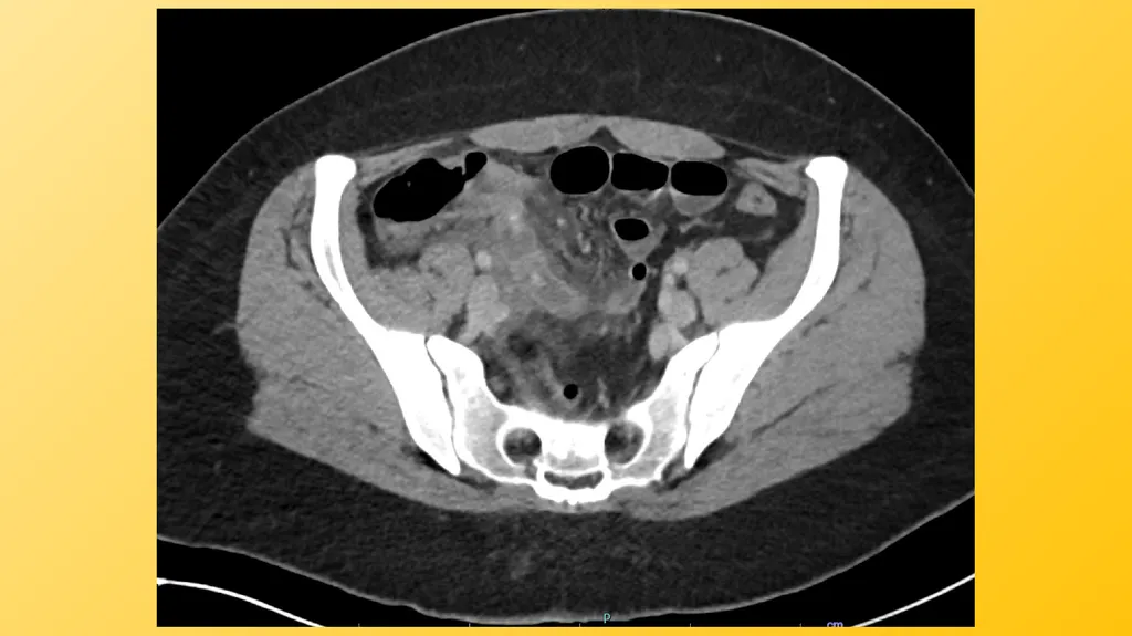
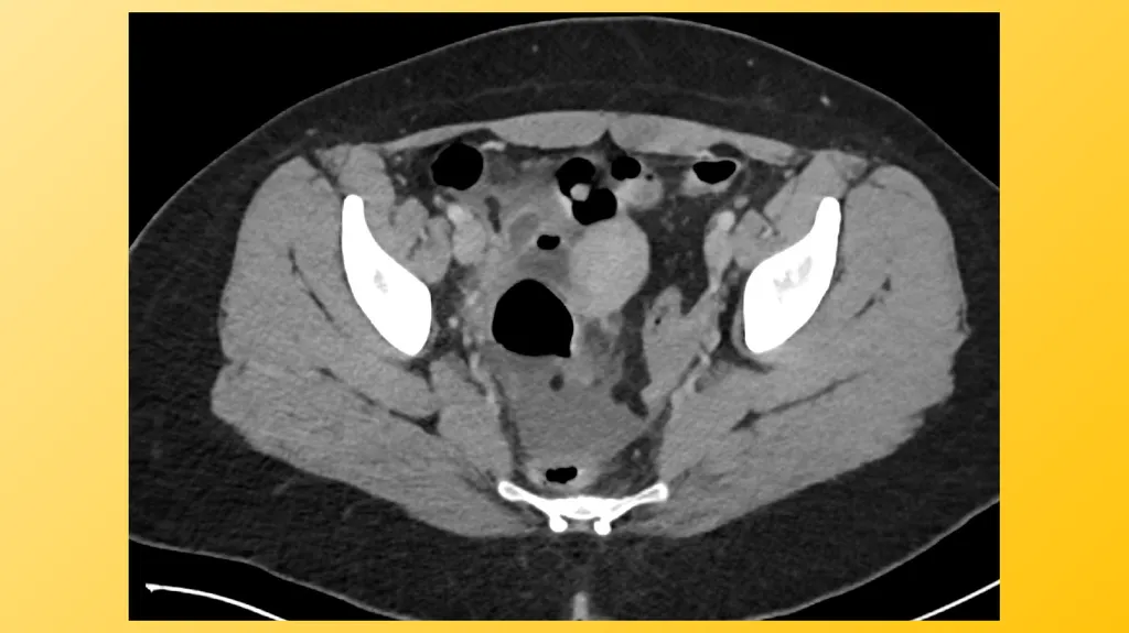
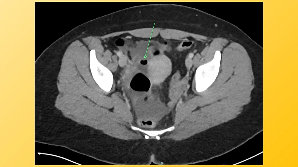
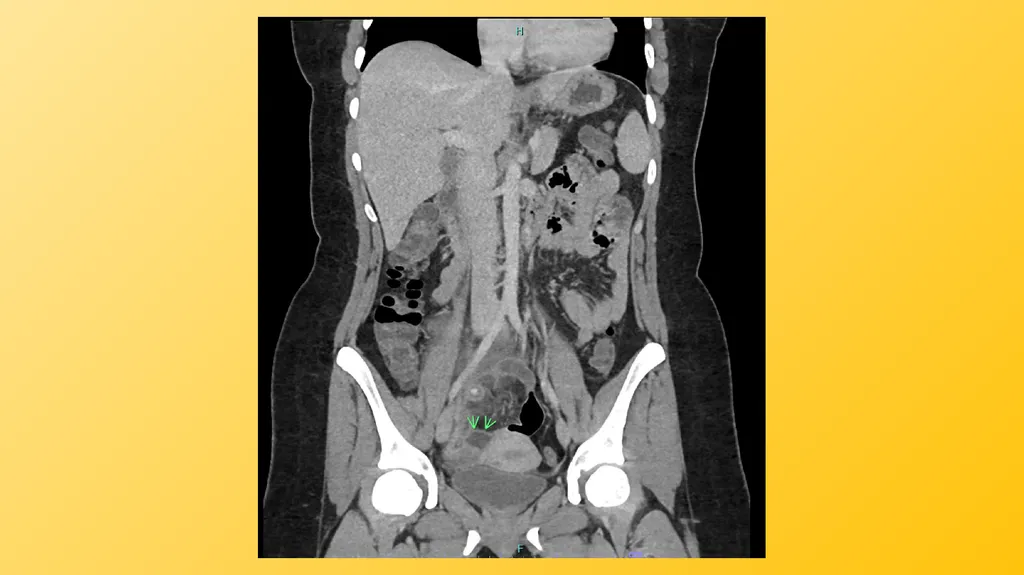
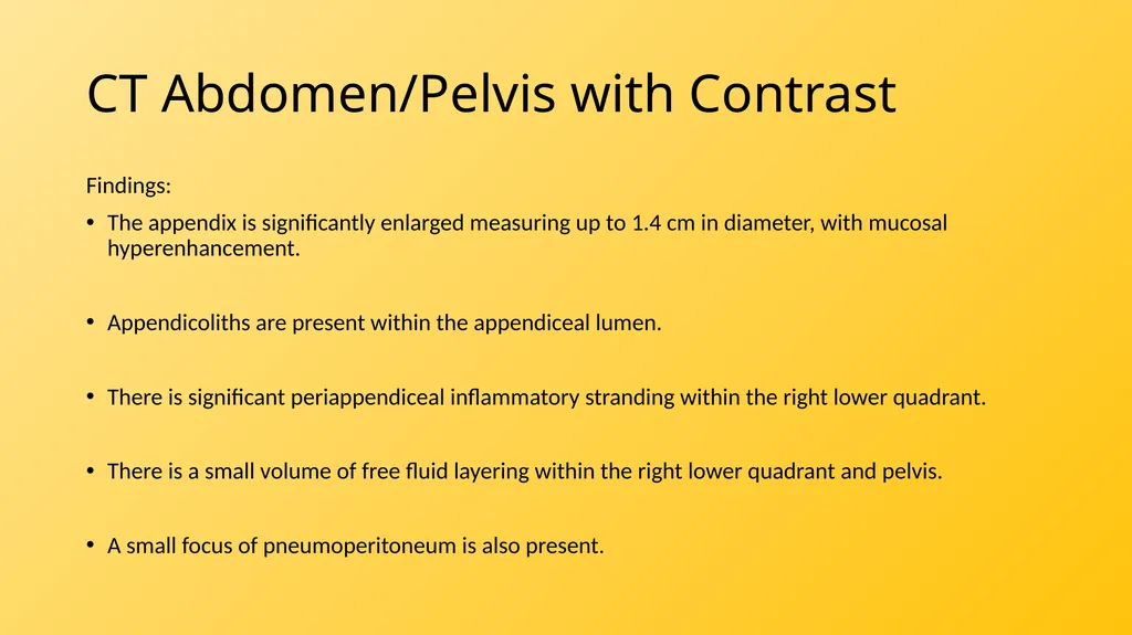
CT Abdomen/Pelvis with Contrast Findings:
The appendix is significantly enlarged measuring up to 1.4 cm in diameter, with mucosal hyperenhancement.
Appendicoliths are present within the appendiceal lumen.
There is significant periappendiceal inflammatory stranding within the right lower quadrant.
There is a small volume of free fluid layering within the right lower quadrant and pelvis.
A small focus of pneumoperitoneum is also present.<br>
07
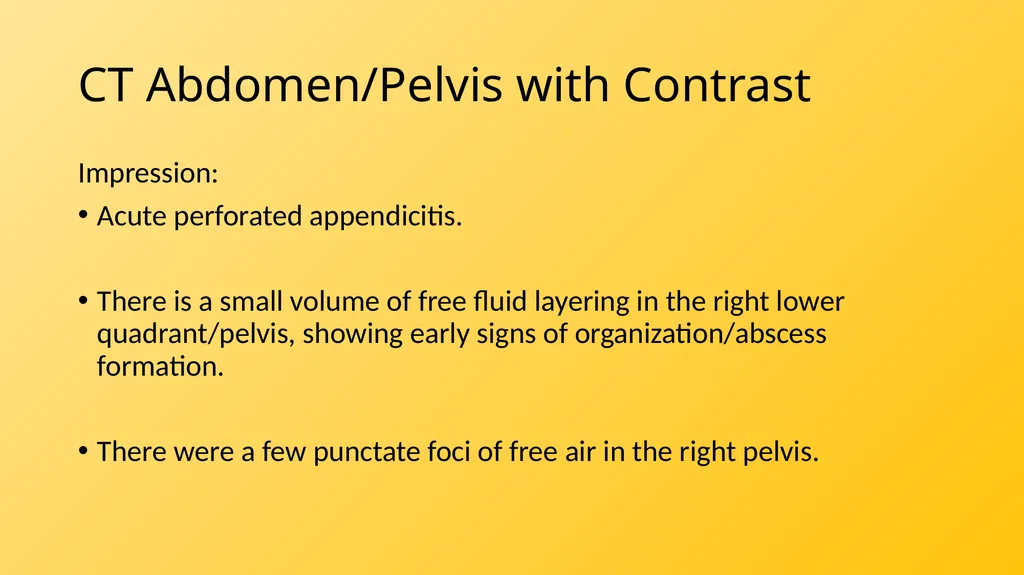
CT Abdomen/Pelvis with Contrast Impression:
Acute perforated appendicitis.
There is a small volume of free fluid layering in the right lower quadrant/pelvis, showing early signs of organization/abscess formation.
There were a few punctate foci of free air in the right pelvis.<br>
11
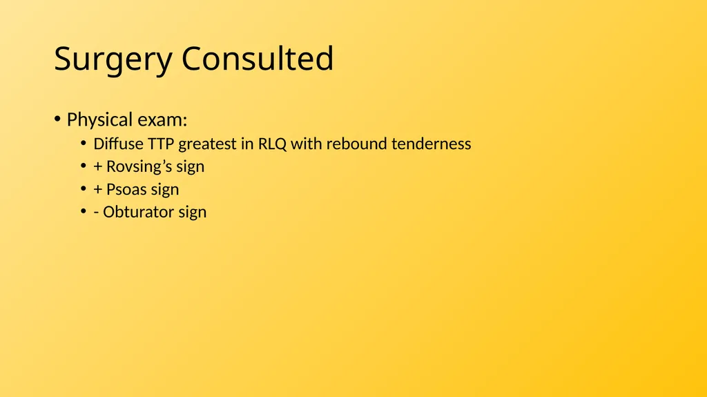
Surgery Consulted Physical exam:
Diffuse TTP greatest in RLQ with rebound tenderness
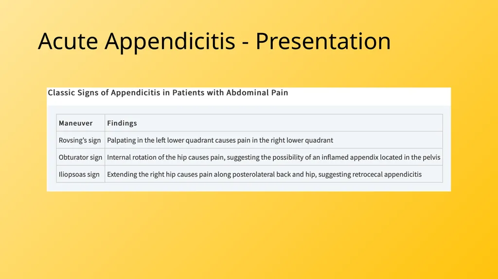
+ Rovsing’s sign
+ Psoas sign
- Obturator sign<br>
12
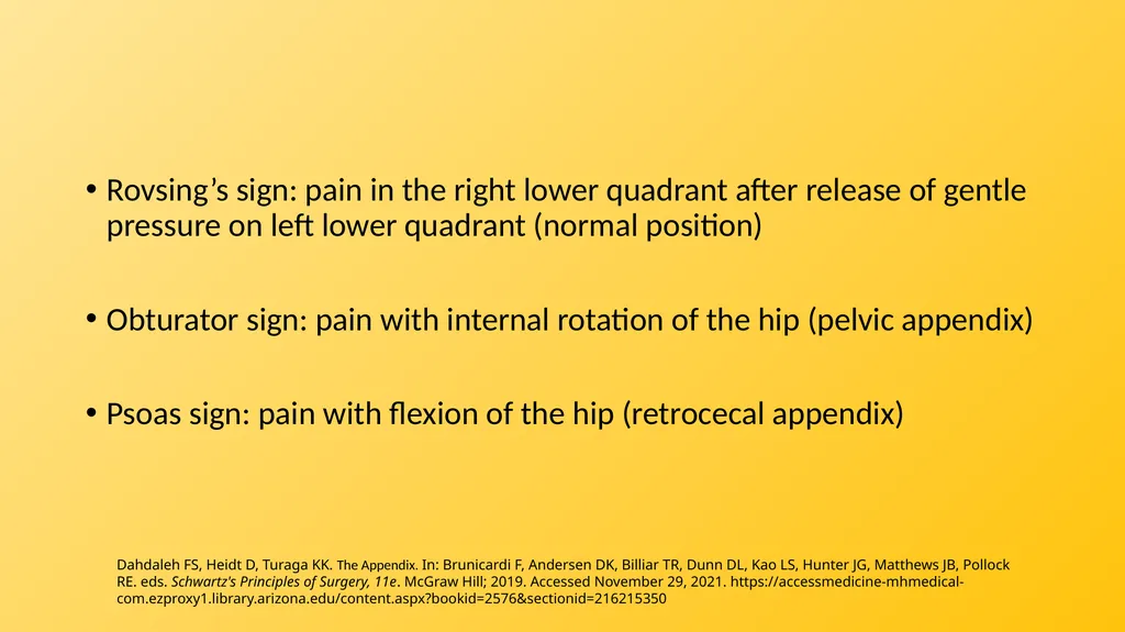
Rovsing’s sign: pain in the right lower quadrant after release of gentle pressure on left lower quadrant (normal position)
Obturator sign: pain with internal rotation of the hip (pelvic appendix)
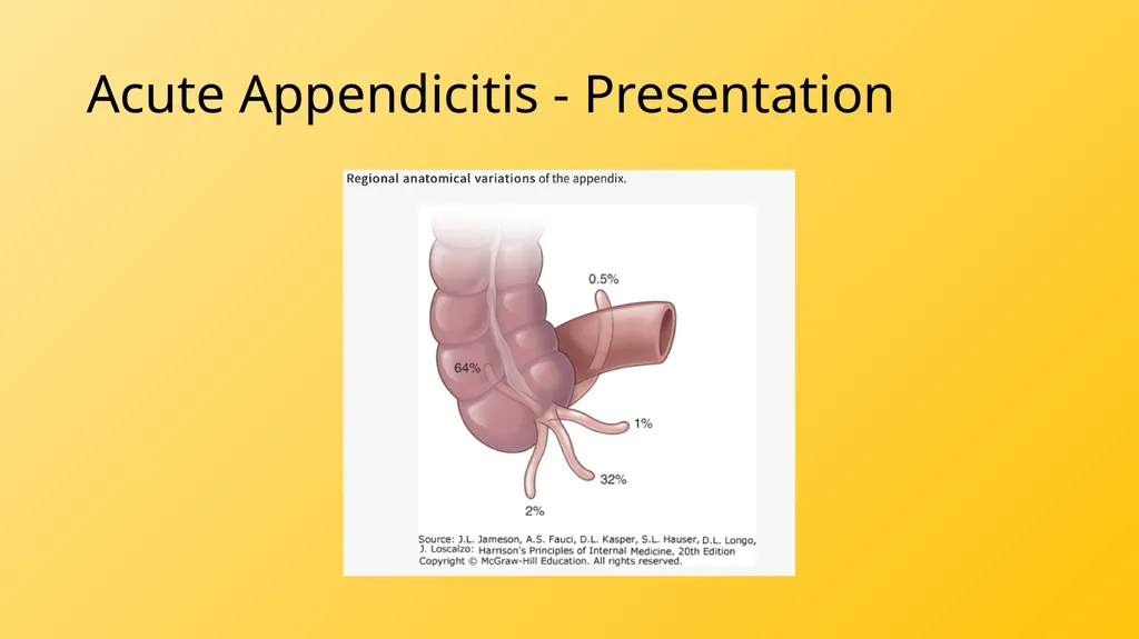
Psoas sign: pain with flexion of the hip (retrocecal appendix) Dahdaleh FS, Heidt D, Turaga KK. The Appendix. In: Brunicardi F, Andersen DK, Billiar TR, Dunn DL, Kao LS, Hunter JG, Matthews JB, Pollock RE. eds. Schwartz's Principles of Surgery, 11e. McGraw Hill; 2019. Accessed November 29, 2021. https://accessmedicine-mhmedical-com.ezproxy1.library.arizona.edu/content.aspx?bookid=2576§ionid=216215350<br>
13
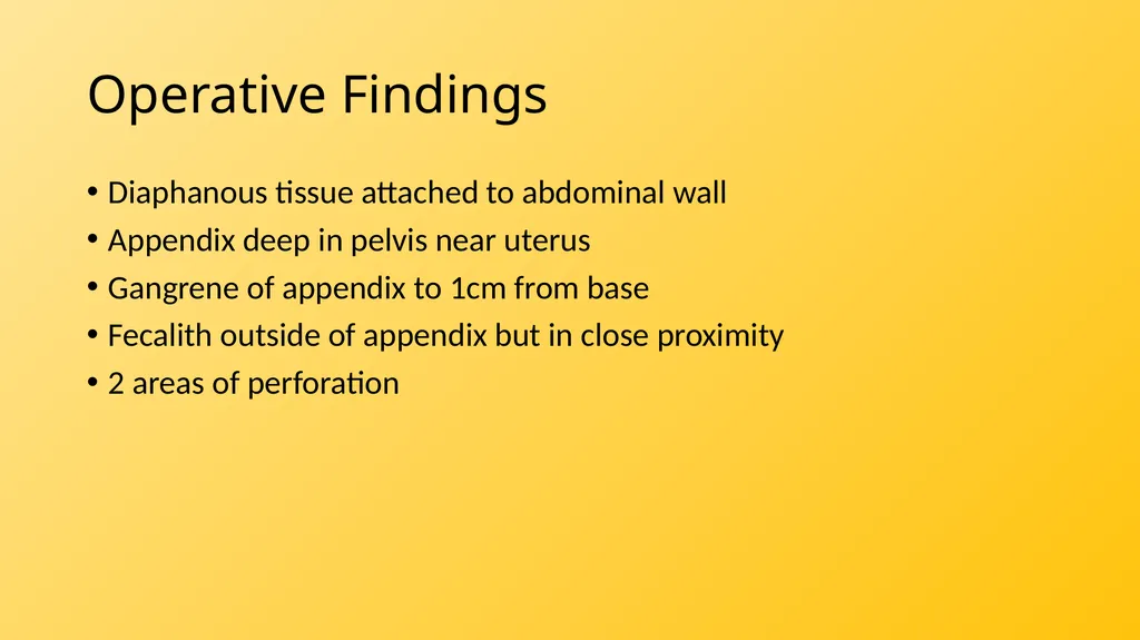
Operative Findings Diaphanous tissue attached to abdominal wall
Appendix deep in pelvis near uterus
Gangrene of appendix to 1cm from base
Fecalith outside of appendix but in close proximity
2 areas of perforation<br>
14
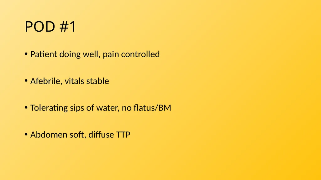
POD #1 Patient doing well, pain controlled
Afebrile, vitals stable
Tolerating sips of water, no flatus/BM
Abdomen soft, diffuse TTP<br>
15
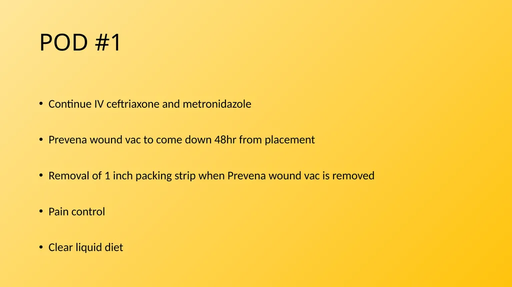
POD #1 Continue IV ceftriaxone and metronidazole
Prevena wound vac to come down 48hr from placement
Removal of 1 inch packing strip when Prevena wound vac is removed
Pain control
Clear liquid diet<br>
16
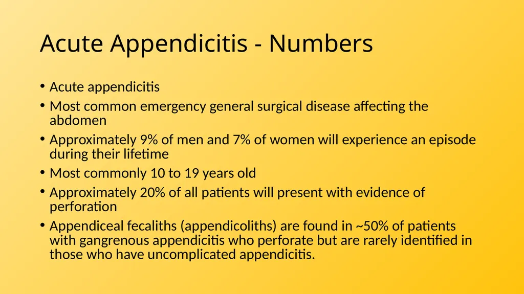
Acute Appendicitis - Numbers Acute appendicitis
Most common emergency general surgical disease affecting the abdomen
Approximately 9% of men and 7% of women will experience an episode during their lifetime
Most commonly 10 to 19 years old
Approximately 20% of all patients will present with evidence of perforation
Appendiceal fecaliths (appendicoliths) are found in ~50% of patients with gangrenous appendicitis who perforate but are rarely identified in those who have uncomplicated appendicitis.<br>
17
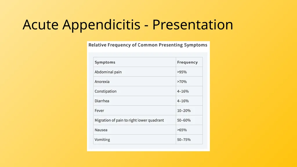
Acute Appendicitis - Presentation<br>
18
Acute Appendicitis - Presentation<br>
19
Acute Appendicitis - Presentation<br>
20
Acute Appendicitis - Differential<br>
21
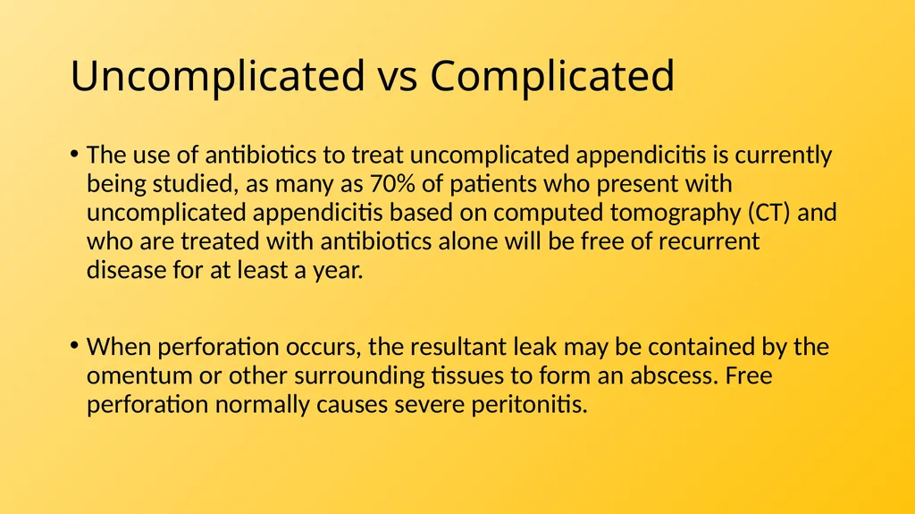
Uncomplicated vs Complicated The use of antibiotics to treat uncomplicated appendicitis is currently being studied, as many as 70% of patients who present with uncomplicated appendicitis based on computed tomography (CT) and who are treated with antibiotics alone will be free of recurrent disease for at least a year.
When perforation occurs, the resultant leak may be contained by the omentum or other surrounding tissues to form an abscess. Free perforation normally causes severe peritonitis.<br>
22
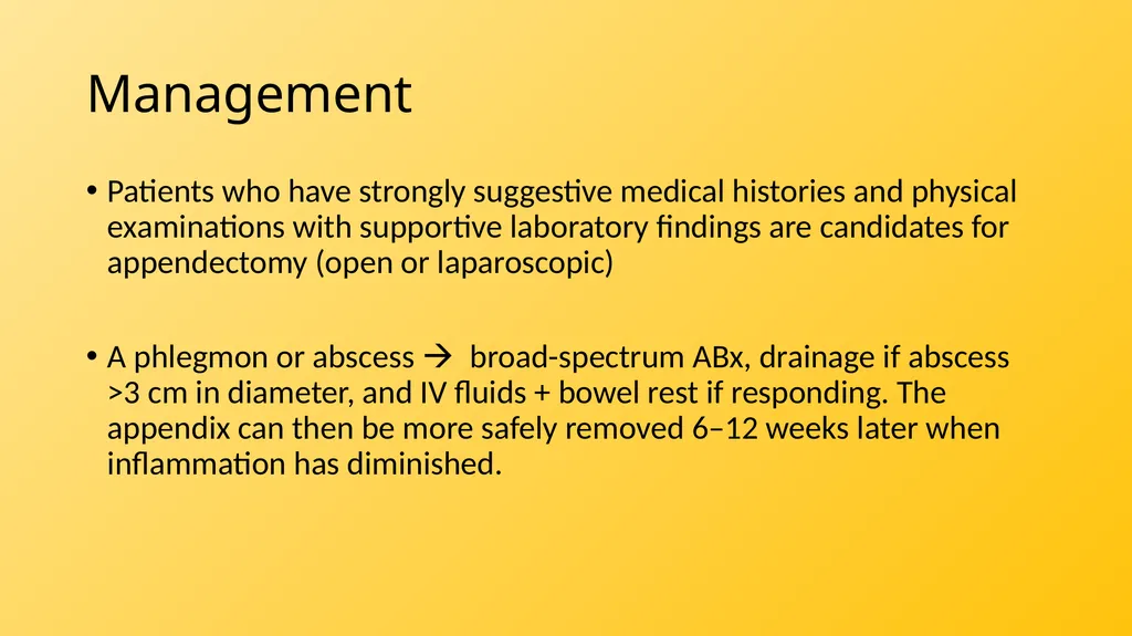
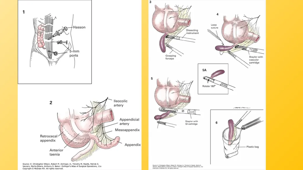
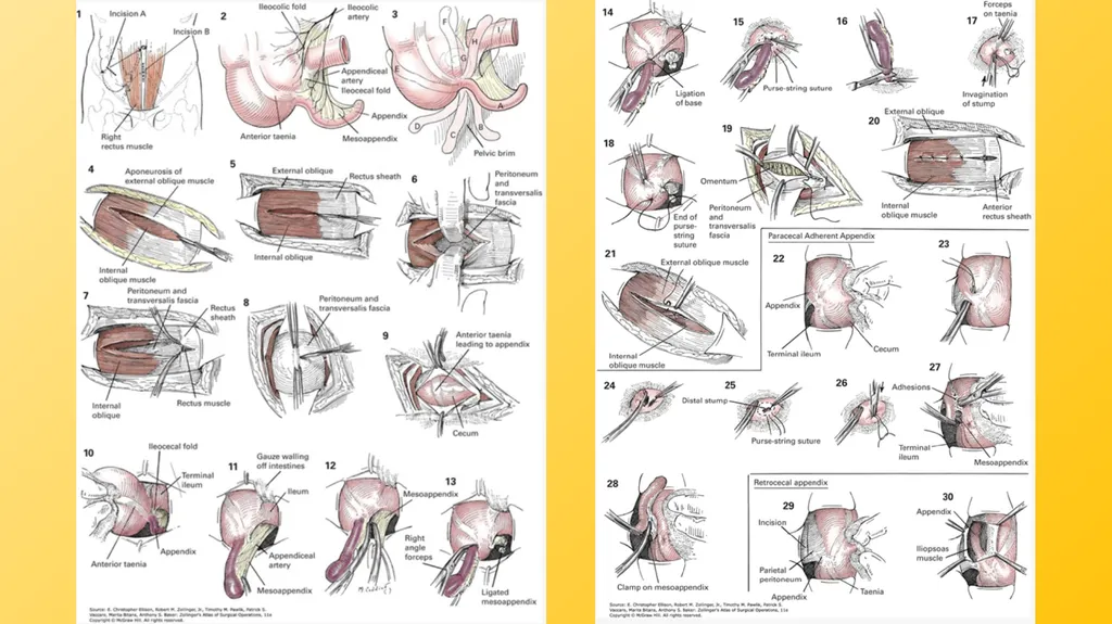
Management Patients who have strongly suggestive medical histories and physical examinations with supportive laboratory findings are candidates for appendectomy (open or laparoscopic)
A phlegmon or abscess broad-spectrum ABx, drainage if abscess >3 cm in diameter, and IV fluids + bowel rest if responding. The appendix can then be more safely removed 6–12 weeks later when inflammation has diminished.<br>
23
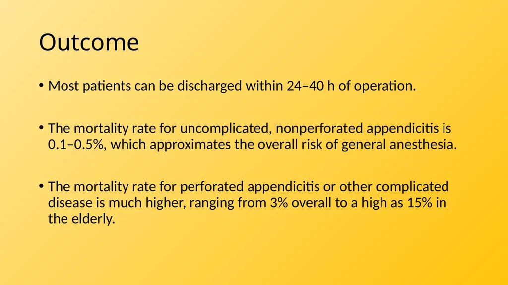
Outcome Most patients can be discharged within 24–40 h of operation.
The mortality rate for uncomplicated, nonperforated appendicitis is 0.1–0.5%, which approximates the overall risk of general anesthesia.
The mortality rate for perforated appendicitis or other complicated disease is much higher, ranging from 3% overall to a high as 15% in the elderly.<br>
24
References Dahdaleh FS, Heidt D, Turaga KK. The Appendix. In: Brunicardi F, Andersen DK, Billiar TR, Dunn DL, Kao LS, Hunter JG, Matthews JB, Pollock RE. eds. Schwartz's Principles of Surgery, 11e. McGraw Hill; 2019. Accessed November 29, 2021. https://accessmedicine-mhmedical-com.ezproxy1.library.arizona.edu/content.aspx?bookid=2576§ionid=216215350
Jacobs DO. Acute Appendicitis and Peritonitis. In: Jameson J, Fauci AS, Kasper DL, Hauser SL, Longo DL, Loscalzo J. eds. Harrison's Principles of Internal Medicine, 20e. McGraw Hill; 2018. Accessed November 30, 2021. https://accessmedicine-mhmedical-com.ezproxy1.library.arizona.edu/content.aspx?bookid=2129§ionid=192282881
Appendectomy. In: Ellison E, Zollinger, Jr. RM, Pawlik TM, Vaccaro PS, Bitans M, Baker AS. eds. Zollinger’s Atlas of Surgical Operations, 11e. McGraw Hill; 2022. Accessed November 30, 2021. https://accesssurgery-mhmedical-com.ezproxy3.library.arizona.edu/content.aspx?bookid=3101§ionid=260093695<br>