
Anatomy and Physiology CHAPTER 8 Case Study Part 1




Description: Anatomy and Physiology CHAPTER 8 Case Study Part 1 (1 of 3) It is 0300 and you are called to the scene of a 25-year-old male driver involved in a motor vehicle crash (MVC). You arrive on scene to find a compact sedan impacted into a
Related Topics
Download Presentation
"Anatomy and Physiology CHAPTER 8 Case Study Part 1" is the property of its rightful owner. Permission is granted to download and print the materials on this website for personal, non-commercial use only, and to display it on your personal computer provided you do not modify the materials and that you retain all copyright notices contained in the materials. By downloading content from our website, you accept the terms of this agreement.
Presentation Transcript
slide1. Anatomy and Physiology CHAPTER 8 Case Study<br>
slide2. Part 1 (1 of 3) It is 0300 and you are called to the scene of a 25-year-old male driver involved in a motor vehicle crash (MVC). You arrive on scene to find a compact sedan impacted into a telephone pole with significant front-end damage, a shattered windshield, and front airbags deployed. Bystanders moved the driver out of the vehicle thinking they were helping the patient. You find the driver on the pavement with rapid shallow respirations, facial cyanosis, accessory muscle use, absent radial pulses, rapid and weak carotid pulse, and pale skin.<br>
slide3. Part 1 (2 of 3) Although the patient can only speak in 2- to 3-word sentences, he states that his left chest hurts, and says, “I can’t breathe!” Bystanders state he was not wearing his seat belt. Your crew initiates spinal protection and places the patient on 15 L/min of oxygen via nonrebreathing mask. You assess his airway to be clear and note his increased work of breathing and absent radial pulses.<br>
slide4. Part 1 (3 of 3) What body structures are in the thoracic cavity and what is your current differential diagnosis related to the chest findings?
How would you proceed with your primary assessment considering your differential diagnosis?<br>
slide5. Part 2 (1 of 3) You continue your primary assessment by completing a rapid trauma assessment. Remarkable exam findings include jugular venous distention, absent lung sounds in the left chest, contusion to the left chest, and tachycardia.<br>
slide6. Part 2 (2 of 3)<br>
slide7. Part 2 (3 of 3) How has your differential diagnosis evolved?
What interventions should you consider?
Is the patient experiencing a shock state? If so, what type?<br>
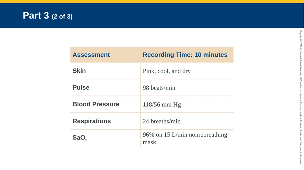
slide8. Part 3 (1 of 3) You perform a needle decompression of the left chest and hear a rush of air. You reassess your patient and find that his lung sounds are now present, clear and equal bilaterally; work of breathing has decreased; skin color is returning; radial pulses are present bilaterally; and the patient claims he can breathe much better now. You proceed with the rest of your rapid trauma assessment. Additional assessment findings include tender left upper quadrant, left shoulder pain, and left ankle deformity.<br>
slide9. Part 3 (2 of 3)<br>
slide10. Part 3 (3 of 3) What anatomic space was penetrated by your chest decompression needle? What two anatomic structures created this space?
Using your understanding of anatomy and physiology, explain how the pneumothorax occurred.
What might the left shoulder pain indicate?<br>
slide11. Part 4 (1 of 3) You perform spinal immobilization and rapid transport to a Level I trauma center. En route, your secondary assessment is unremarkable, and no other trauma injuries are observed. You initiate at least one large-bore IV with crystalloid solution hanging. He still complains of mild left chest pain, and states it is a 3 on a 0–10 visual analog scale. He complains that his left upper quadrant pain is an 8 and his left shoulder pain is a 5. His left shoulder remains unremarkable. Vital signs are reassessed. Lung sounds remain clear and equal bilaterally.<br>
slide12. Part 4 (2 of 3)<br>
slide13. Part 4 (3 of 3) What other concerns beyond the pneumothorax are in your differential diagnosis?
What body structures are in the left upper quadrant?
Describe your treatment for your patient.<br>
slide14. Part 5 (1 of 2) You keep the patient warm and continually reassess his chest and abdomen en route. His vital signs remain stable, and you consider administration of analgesics and antiemetics as needed per medical direction.<br>
slide15. Part 5 (2 of 2) Upon your arrival at the emergency department, the physician completes his exam. X-rays reveals a small left pneumothorax and left ankle fracture. A chest tube is placed in the left chest. CT scans of the abdomen and pelvis reveal a low-grade splenic rupture. The patient’s pain is managed, and he is transferred to the surgical ICU for continued care.<br>
slide2. Part 1 (1 of 3) It is 0300 and you are called to the scene of a 25-year-old male driver involved in a motor vehicle crash (MVC). You arrive on scene to find a compact sedan impacted into a telephone pole with significant front-end damage, a shattered windshield, and front airbags deployed. Bystanders moved the driver out of the vehicle thinking they were helping the patient. You find the driver on the pavement with rapid shallow respirations, facial cyanosis, accessory muscle use, absent radial pulses, rapid and weak carotid pulse, and pale skin.<br>
slide3. Part 1 (2 of 3) Although the patient can only speak in 2- to 3-word sentences, he states that his left chest hurts, and says, “I can’t breathe!” Bystanders state he was not wearing his seat belt. Your crew initiates spinal protection and places the patient on 15 L/min of oxygen via nonrebreathing mask. You assess his airway to be clear and note his increased work of breathing and absent radial pulses.<br>
slide4. Part 1 (3 of 3) What body structures are in the thoracic cavity and what is your current differential diagnosis related to the chest findings?
How would you proceed with your primary assessment considering your differential diagnosis?<br>
slide5. Part 2 (1 of 3) You continue your primary assessment by completing a rapid trauma assessment. Remarkable exam findings include jugular venous distention, absent lung sounds in the left chest, contusion to the left chest, and tachycardia.<br>
slide6. Part 2 (2 of 3)<br>
slide7. Part 2 (3 of 3) How has your differential diagnosis evolved?
What interventions should you consider?
Is the patient experiencing a shock state? If so, what type?<br>
slide8. Part 3 (1 of 3) You perform a needle decompression of the left chest and hear a rush of air. You reassess your patient and find that his lung sounds are now present, clear and equal bilaterally; work of breathing has decreased; skin color is returning; radial pulses are present bilaterally; and the patient claims he can breathe much better now. You proceed with the rest of your rapid trauma assessment. Additional assessment findings include tender left upper quadrant, left shoulder pain, and left ankle deformity.<br>
slide9. Part 3 (2 of 3)<br>
slide10. Part 3 (3 of 3) What anatomic space was penetrated by your chest decompression needle? What two anatomic structures created this space?
Using your understanding of anatomy and physiology, explain how the pneumothorax occurred.
What might the left shoulder pain indicate?<br>
slide11. Part 4 (1 of 3) You perform spinal immobilization and rapid transport to a Level I trauma center. En route, your secondary assessment is unremarkable, and no other trauma injuries are observed. You initiate at least one large-bore IV with crystalloid solution hanging. He still complains of mild left chest pain, and states it is a 3 on a 0–10 visual analog scale. He complains that his left upper quadrant pain is an 8 and his left shoulder pain is a 5. His left shoulder remains unremarkable. Vital signs are reassessed. Lung sounds remain clear and equal bilaterally.<br>
slide12. Part 4 (2 of 3)<br>
slide13. Part 4 (3 of 3) What other concerns beyond the pneumothorax are in your differential diagnosis?
What body structures are in the left upper quadrant?
Describe your treatment for your patient.<br>
slide14. Part 5 (1 of 2) You keep the patient warm and continually reassess his chest and abdomen en route. His vital signs remain stable, and you consider administration of analgesics and antiemetics as needed per medical direction.<br>
slide15. Part 5 (2 of 2) Upon your arrival at the emergency department, the physician completes his exam. X-rays reveals a small left pneumothorax and left ankle fracture. A chest tube is placed in the left chest. CT scans of the abdomen and pelvis reveal a low-grade splenic rupture. The patient’s pain is managed, and he is transferred to the surgical ICU for continued care.<br>