
Applied Anatomy and Physiology Learning Outcomes
AM
Published · 33 slides · 0 views
1 / 1


Description
Applied Anatomy and Physiology Learning Outcomes 1.1.a. The Structure and Function of the Skeletal System By the end of this topic you should Know the name and location of bones in the human body. Be able to describe the 6 functions of
Related Topics
Share
Embed code
Download this presentation From Below
"Applied Anatomy and Physiology Learning Outcomes" is the property of its rightful owner. Permission is granted to download and print the materials on this website for personal, non-commercial use only, and to display it on your personal computer provided you do not modify the materials and that you retain all copyright notices contained in the materials. By downloading content from our website, you accept the terms of this agreement.
Presentation Transcript
01
Applied Anatomy and Physiology<br>
02
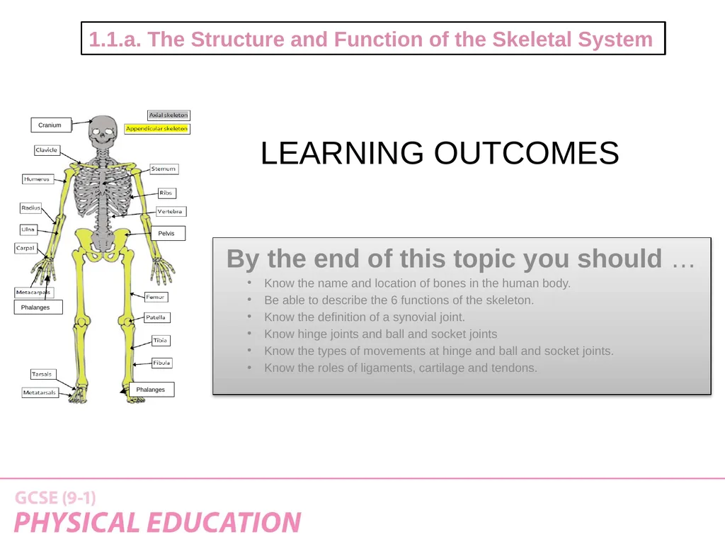
Learning Outcomes 1.1.a. The Structure and Function of the Skeletal System By the end of this topic you should …
Know the name and location of bones in the human body.
Be able to describe the 6 functions of the skeleton.
Know the definition of a synovial joint.
Know hinge joints and ball and socket joints
Know the types of movements at hinge and ball and socket joints.
Know the roles of ligaments, cartilage and tendons. Cranium Pelvis Phalanges Phalanges<br>
Know the name and location of bones in the human body.
Be able to describe the 6 functions of the skeleton.
Know the definition of a synovial joint.
Know hinge joints and ball and socket joints
Know the types of movements at hinge and ball and socket joints.
Know the roles of ligaments, cartilage and tendons. Cranium Pelvis Phalanges Phalanges<br>
03
The adult skeleton has 206 bones and provides the framework for all movement. PRACTICAL TASK:
Using the Labels and marker pen on your table, can you identify the location of the major bones on your ‘designated dummy!’ You need to know the location of the following bones:
Cranium
Vertebrae
Ribs
Sternum
Clavicle
Scapula
Humerus
Ulna
Radius
Carpals
Metacarpals Phalanges
Pelvis
Femur
Patella
Tibia
Fibula
Tarsals
Metatarsals Know the location of the major bones<br>
Using the Labels and marker pen on your table, can you identify the location of the major bones on your ‘designated dummy!’ You need to know the location of the following bones:
Cranium
Vertebrae
Ribs
Sternum
Clavicle
Scapula
Humerus
Ulna
Radius
Carpals
Metacarpals Phalanges
Pelvis
Femur
Patella
Tibia
Fibula
Tarsals
Metatarsals Know the location of the major bones<br>
04
So How did we do?! Who will be the winning table!
For each bone you correctly label you will earn yourself a point!
Most points WIN! Know the location of the major bones<br>
For each bone you correctly label you will earn yourself a point!
Most points WIN! Know the location of the major bones<br>
05
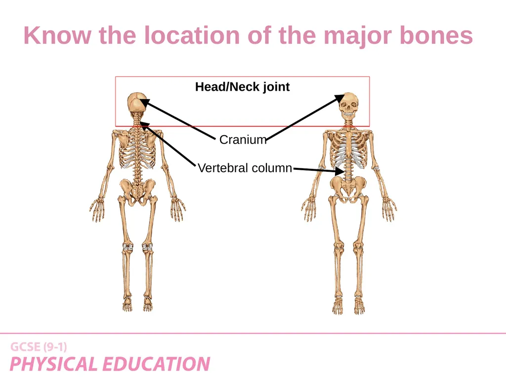
Cranium Vertebral column Head/Neck joint Know the location of the major bones<br>
06
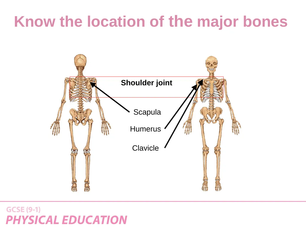
Scapula Humerus Shoulder joint Clavicle Know the location of the major bones<br>
07
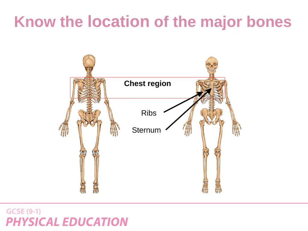
Ribs Sternum Chest region Know the location of the major bones<br>
08
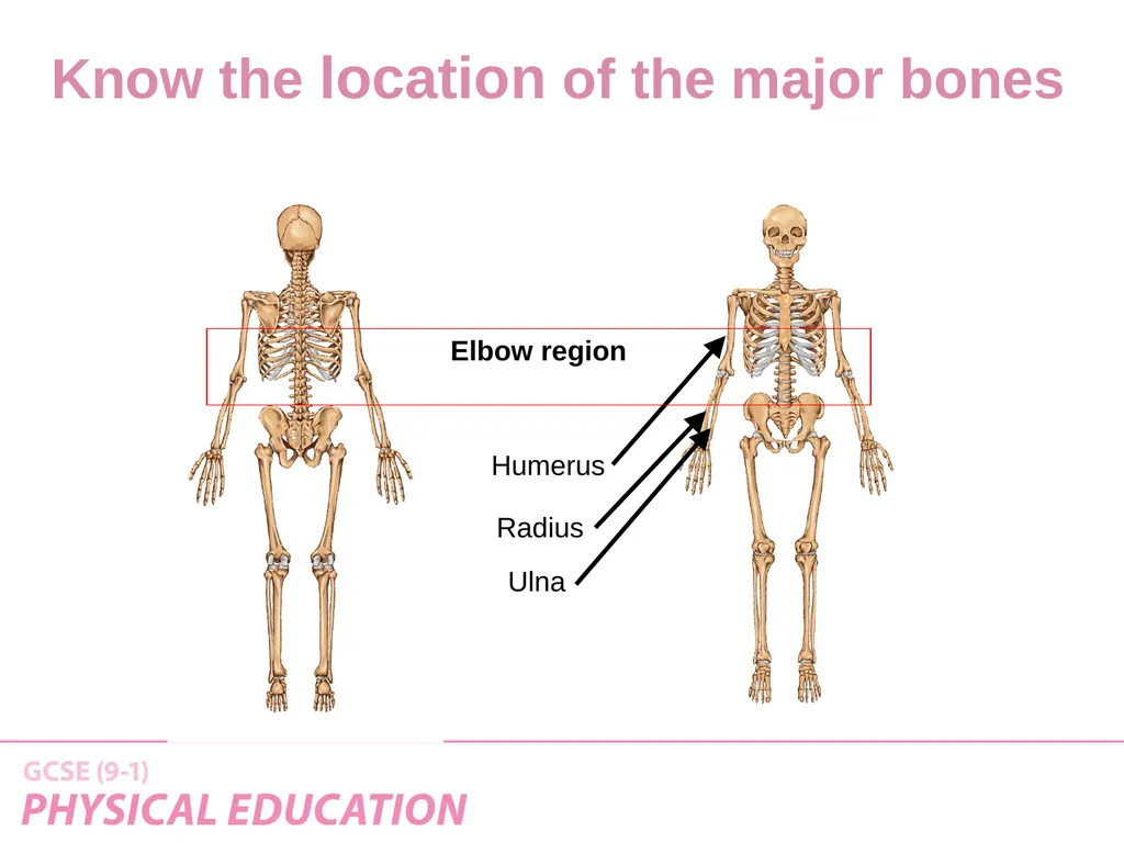
Humerus Radius Elbow region Ulna Know the location of the major bones<br>
09
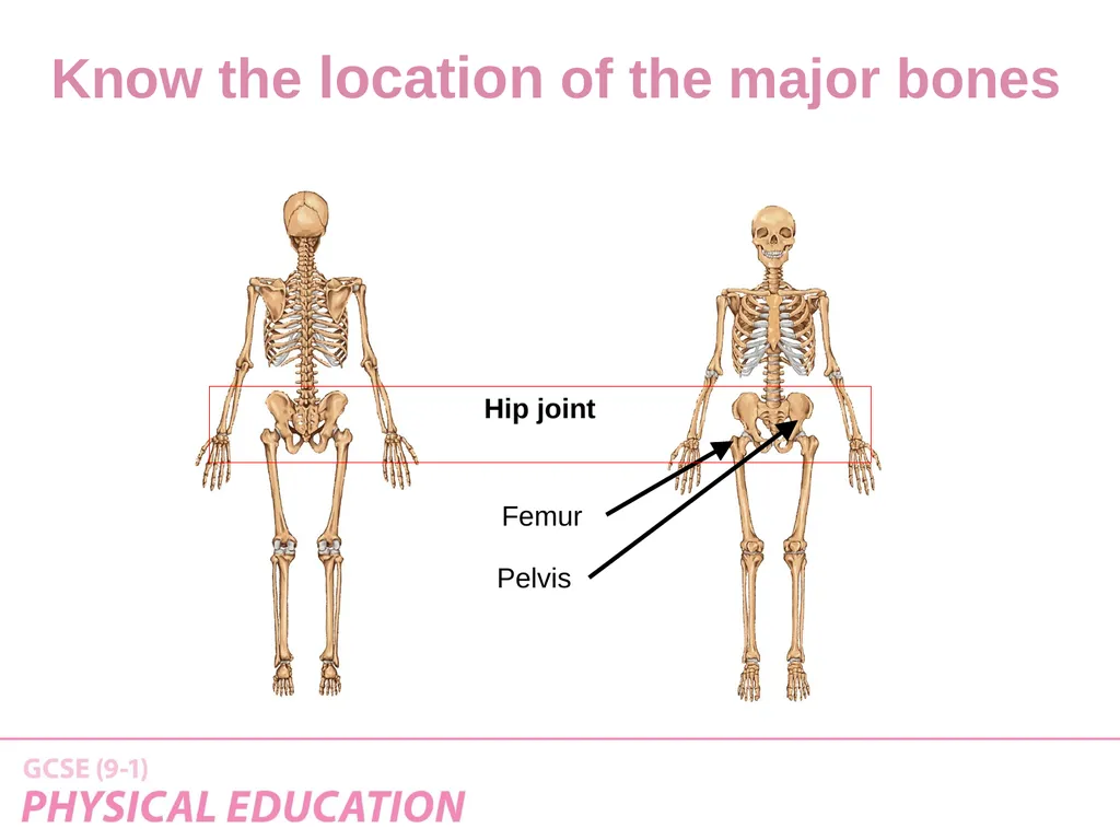
Femur Pelvis Hip joint Know the location of the major bones<br>
10
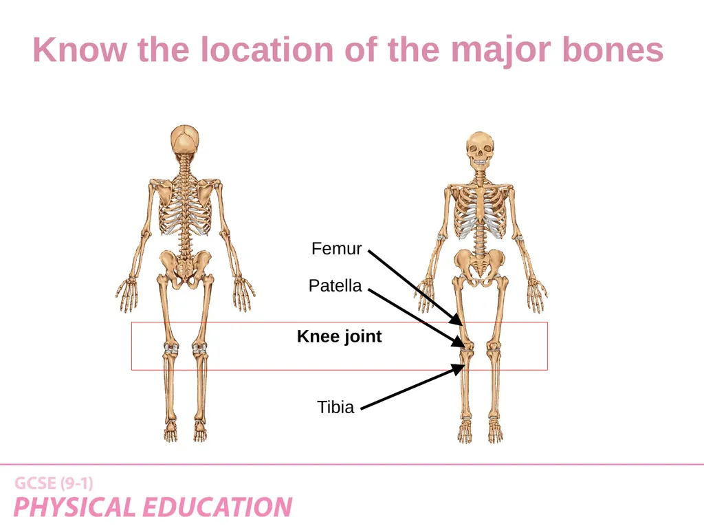
Patella Tibia Knee joint Femur Know the location of the major bones<br>
11
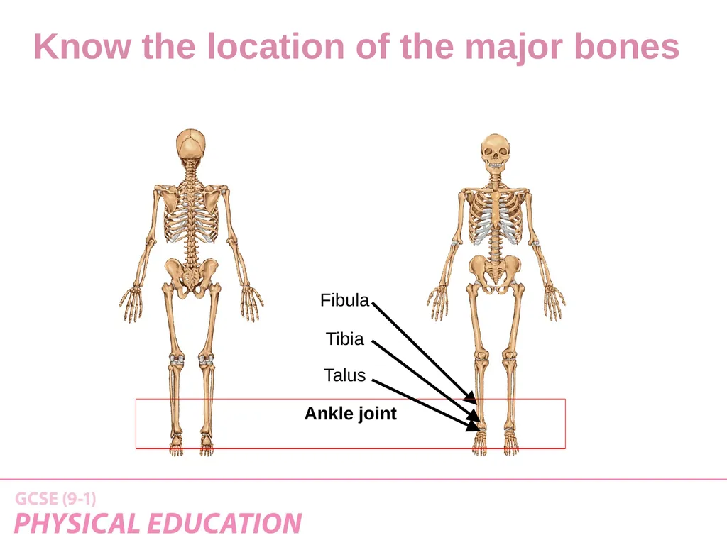
Fibula Tibia Ankle joint Talus Know the location of the major bones<br>
12
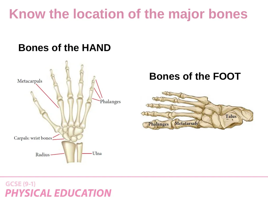
Bones of the HAND Bones of the FOOT Know the location of the major bones<br>
13
And Our Winners Are! Know the location of the major bones<br>
14
How might we remember these? https://www.youtube.com/watch?v=aUHh8uMdBso Did you get these?
1. To give Posture and Support to the body
2. To allow Movement of the body
3. To give Protection to the internal organs
4. To produce Blood (White and Red) Cells
5. To Store Minerals Main functions of the skeleton<br>
1. To give Posture and Support to the body
2. To allow Movement of the body
3. To give Protection to the internal organs
4. To produce Blood (White and Red) Cells
5. To Store Minerals Main functions of the skeleton<br>
15
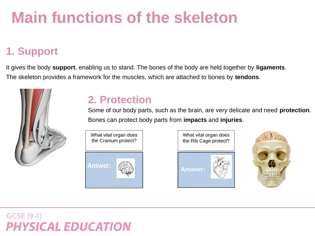
It gives the body support, enabling us to stand. The bones of the body are held together by ligaments.
The skeleton provides a framework for the muscles, which are attached to bones by tendons. 1. Support Some of our body parts, such as the brain, are very delicate and need protection.
Bones can protect body parts from impacts and injuries. 2. Protection What vital organ does the Cranium protect? What vital organ does the Rib Cage protect? Main functions of the skeleton<br>
The skeleton provides a framework for the muscles, which are attached to bones by tendons. 1. Support Some of our body parts, such as the brain, are very delicate and need protection.
Bones can protect body parts from impacts and injuries. 2. Protection What vital organ does the Cranium protect? What vital organ does the Rib Cage protect? Main functions of the skeleton<br>
16
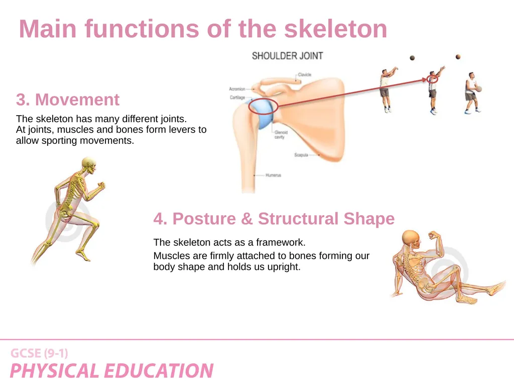
The skeleton has many different joints.
At joints, muscles and bones form levers to
allow sporting movements. 3. Movement The skeleton acts as a framework.
Muscles are firmly attached to bones forming our body shape and holds us upright. 4. Posture & Structural Shape Main functions of the skeleton<br>
At joints, muscles and bones form levers to
allow sporting movements. 3. Movement The skeleton acts as a framework.
Muscles are firmly attached to bones forming our body shape and holds us upright. 4. Posture & Structural Shape Main functions of the skeleton<br>
17
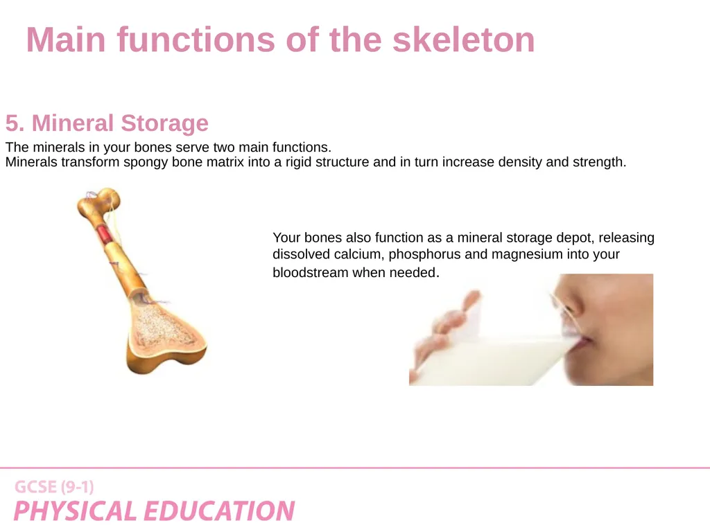
The minerals in your bones serve two main functions.
Minerals transform spongy bone matrix into a rigid structure and in turn increase density and strength. 5. Mineral Storage Your bones also function as a mineral storage depot, releasing dissolved calcium, phosphorus and magnesium into your bloodstream when needed. Main functions of the skeleton<br>
Minerals transform spongy bone matrix into a rigid structure and in turn increase density and strength. 5. Mineral Storage Your bones also function as a mineral storage depot, releasing dissolved calcium, phosphorus and magnesium into your bloodstream when needed. Main functions of the skeleton<br>
18
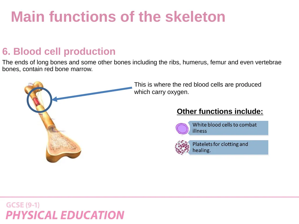
The ends of long bones and some other bones including the ribs, humerus, femur and even vertebrae bones, contain red bone marrow. 6. Blood cell production This is where the red blood cells are produced
which carry oxygen. Other functions include: Main functions of the skeleton<br>
19
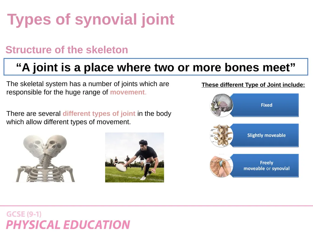
“A joint is a place where two or more bones meet” Structure of the skeleton The skeletal system has a number of joints which are responsible for the huge range of movement.
There are several different types of joint in the body which allow different types of movement. These different Type of Joint include: Types of synovial joint<br>
There are several different types of joint in the body which allow different types of movement. These different Type of Joint include: Types of synovial joint<br>
20
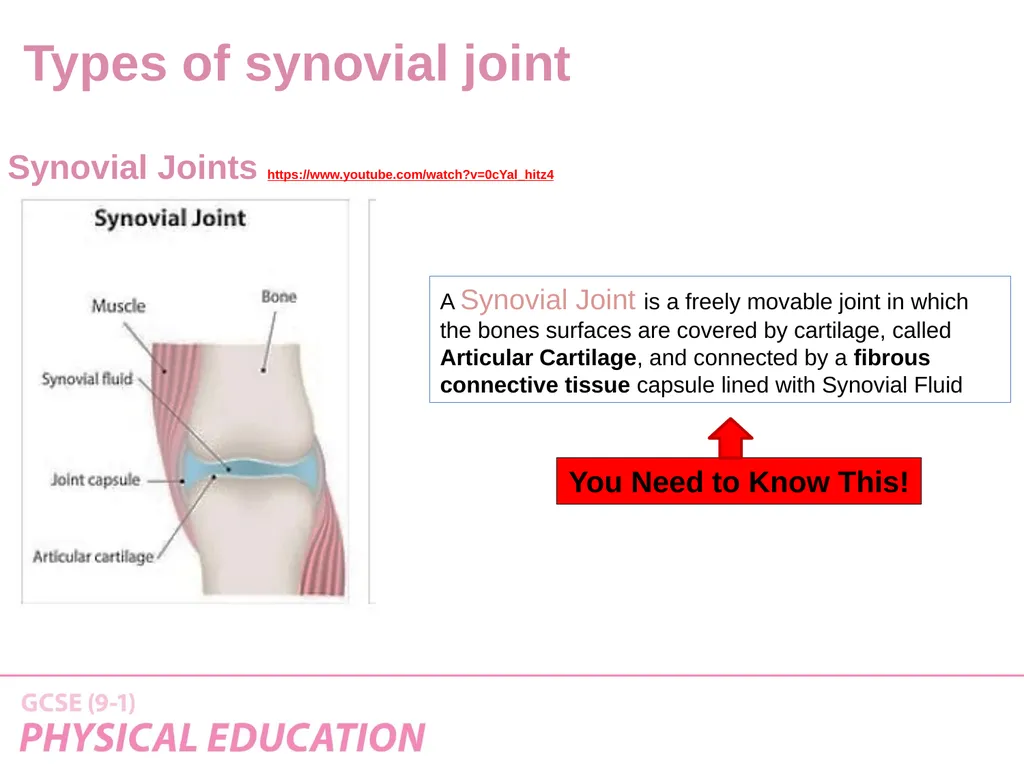
Synovial Joints https://www.youtube.com/watch?v=0cYal_hitz4 A Synovial Joint is a freely movable joint in which the bones surfaces are covered by cartilage, called Articular Cartilage, and connected by a fibrous connective tissue capsule lined with Synovial Fluid You Need to Know This! Types of synovial joint<br>
21
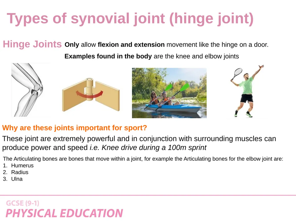
Only allow flexion and extension movement like the hinge on a door. Why are these joints important for sport? These joint are extremely powerful and in conjunction with surrounding muscles can produce power and speed i.e. Knee drive during a 100m sprint Examples found in the body are the knee and elbow joints Hinge Joints The Articulating bones are bones that move within a joint, for example the Articulating bones for the elbow joint are:
Humerus
Radius
Ulna Types of synovial joint (hinge joint)<br>
Humerus
Radius
Ulna Types of synovial joint (hinge joint)<br>
22
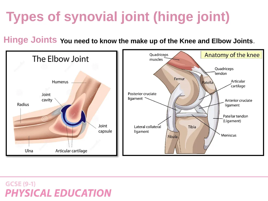
You need to know the make up of the Knee and Elbow Joints. Hinge Joints Types of synovial joint (hinge joint)<br>
23
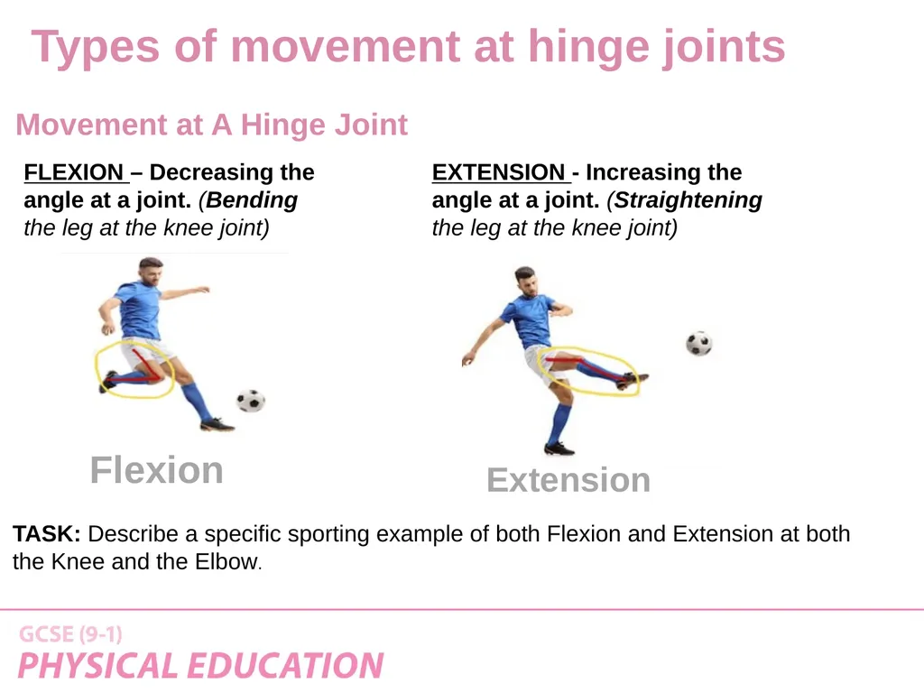
FLEXION – Decreasing the angle at a joint. (Bending the leg at the knee joint) EXTENSION - Increasing the angle at a joint. (Straightening the leg at the knee joint) Movement at A Hinge Joint Flexion Extension TASK: Describe a specific sporting example of both Flexion and Extension at both
the Knee and the Elbow. Types of movement at hinge joints<br>
the Knee and the Elbow. Types of movement at hinge joints<br>
24
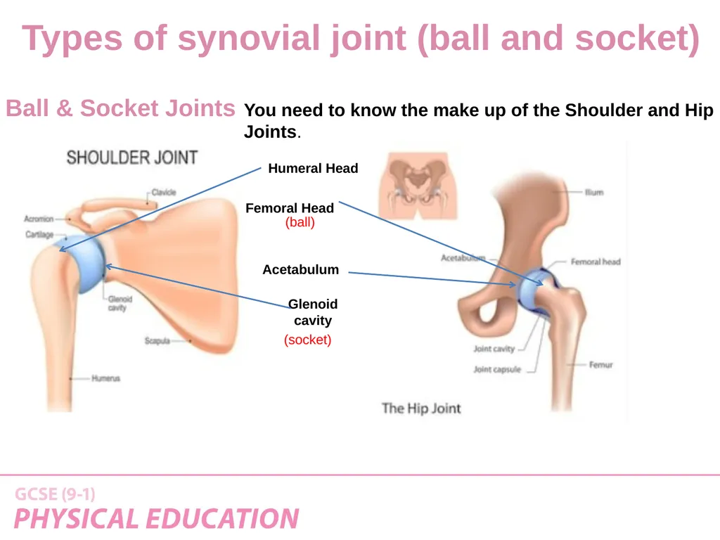
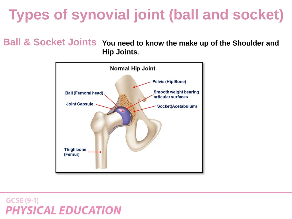
You need to know the make up of the Shoulder and Hip Joints. Ball & Socket Joints Humeral Head Glenoid cavity Acetabulum (socket) Femoral Head (ball) Types of synovial joint (ball and socket)<br>
25
You need to know the make up of the Shoulder and Hip Joints. Ball & Socket Joints Types of synovial joint (ball and socket)<br>
26
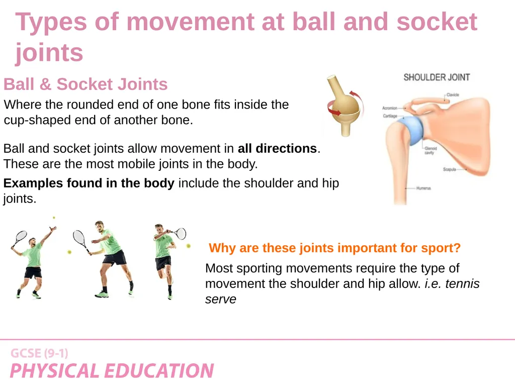
Where the rounded end of one bone fits inside the
cup-shaped end of another bone. Ball and socket joints allow movement in all directions.
These are the most mobile joints in the body.
Examples found in the body include the shoulder and hip joints. Why are these joints important for sport? Most sporting movements require the type of movement the shoulder and hip allow. i.e. tennis serve Ball & Socket Joints Types of movement at ball and socket joints<br>
cup-shaped end of another bone. Ball and socket joints allow movement in all directions.
These are the most mobile joints in the body.
Examples found in the body include the shoulder and hip joints. Why are these joints important for sport? Most sporting movements require the type of movement the shoulder and hip allow. i.e. tennis serve Ball & Socket Joints Types of movement at ball and socket joints<br>
27
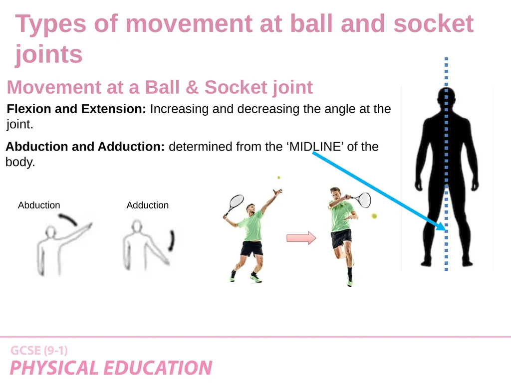
Flexion and Extension: Increasing and decreasing the angle at the joint. Movement at a Ball & Socket joint Abduction and Adduction: determined from the ‘MIDLINE’ of the body. Abduction Adduction Types of movement at ball and socket joints<br>
28
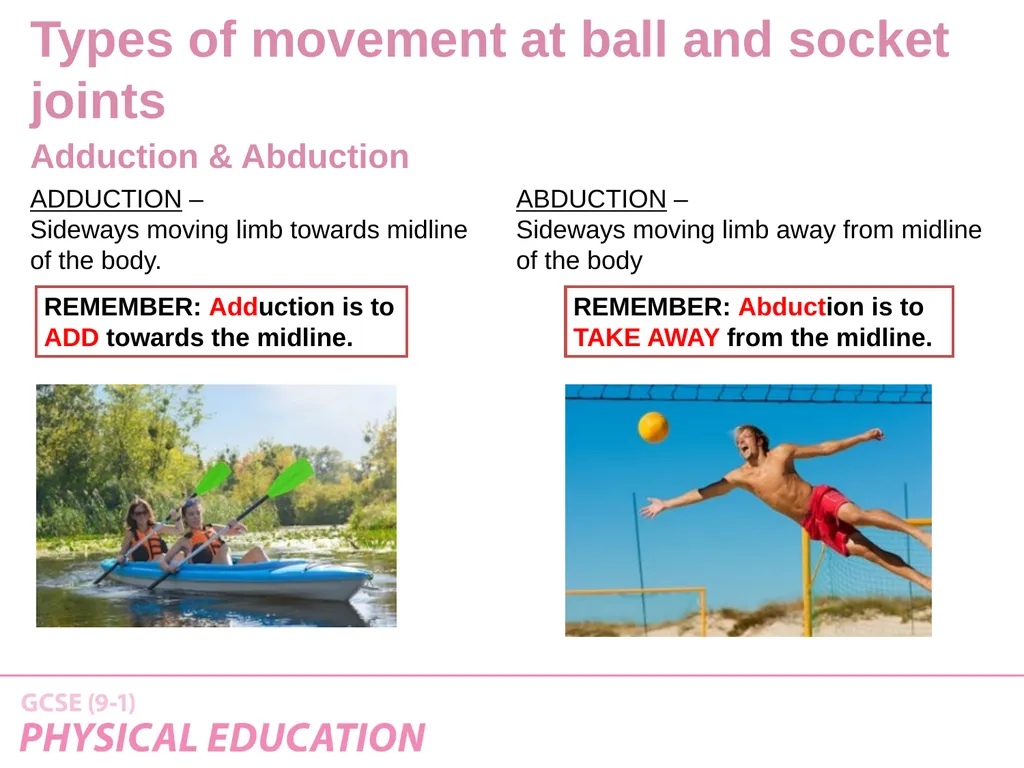
ABDUCTION –
Sideways moving limb away from midline of the body ADDUCTION –
Sideways moving limb towards midline of the body. REMEMBER: Adduction is to ADD towards the midline. REMEMBER: Abduction is to TAKE AWAY from the midline. Adduction & Abduction Types of movement at ball and socket joints<br>
Sideways moving limb away from midline of the body ADDUCTION –
Sideways moving limb towards midline of the body. REMEMBER: Adduction is to ADD towards the midline. REMEMBER: Abduction is to TAKE AWAY from the midline. Adduction & Abduction Types of movement at ball and socket joints<br>
29
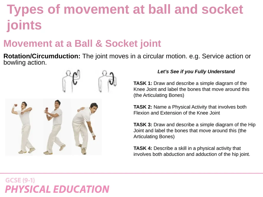
Rotation/Circumduction: The joint moves in a circular motion. e.g. Service action or bowling action. Movement at a Ball & Socket joint Let’s See if you Fully Understand
TASK 1: Draw and describe a simple diagram of the Knee Joint and label the bones that move around this (the Articulating Bones)
TASK 2: Name a Physical Activity that involves both Flexion and Extension of the Knee Joint
TASK 3: Draw and describe a simple diagram of the Hip Joint and label the bones that move around this (the Articulating Bones)
TASK 4: Describe a skill in a physical activity that involves both abduction and adduction of the hip joint. Types of movement at ball and socket joints<br>
TASK 1: Draw and describe a simple diagram of the Knee Joint and label the bones that move around this (the Articulating Bones)
TASK 2: Name a Physical Activity that involves both Flexion and Extension of the Knee Joint
TASK 3: Draw and describe a simple diagram of the Hip Joint and label the bones that move around this (the Articulating Bones)
TASK 4: Describe a skill in a physical activity that involves both abduction and adduction of the hip joint. Types of movement at ball and socket joints<br>
30
Structure of a synovial joint Other components of joints<br>
31
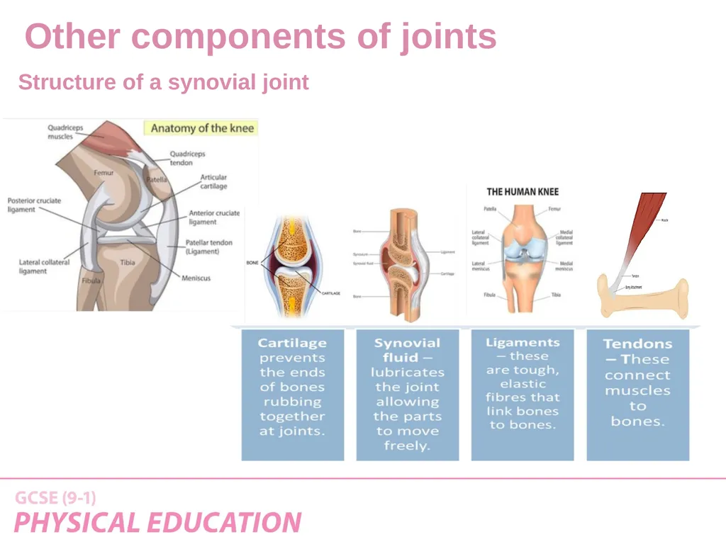
A Synovial Joint
is a freely movable joint in which the bones surfaces are covered by cartilage, called Articular Cartilage, and connected by a
fibrous connective tissue capsule lined with Synovial Fluid Structure of a synovial joint YOU NEED TO KNOW THESE!
Ligaments:
Bands of connective tissue that connect bone to bone & stabilise moment.
Cartilage:
Designed to reduce friction and act as a shock absorber for the joint
Synovial fluid:
Acts like an oil and lubricates the joint.
Tendons:
Attach Muscle to Bone (example - Achilles tendon) Other components of joints<br>
is a freely movable joint in which the bones surfaces are covered by cartilage, called Articular Cartilage, and connected by a
fibrous connective tissue capsule lined with Synovial Fluid Structure of a synovial joint YOU NEED TO KNOW THESE!
Ligaments:
Bands of connective tissue that connect bone to bone & stabilise moment.
Cartilage:
Designed to reduce friction and act as a shock absorber for the joint
Synovial fluid:
Acts like an oil and lubricates the joint.
Tendons:
Attach Muscle to Bone (example - Achilles tendon) Other components of joints<br>
32
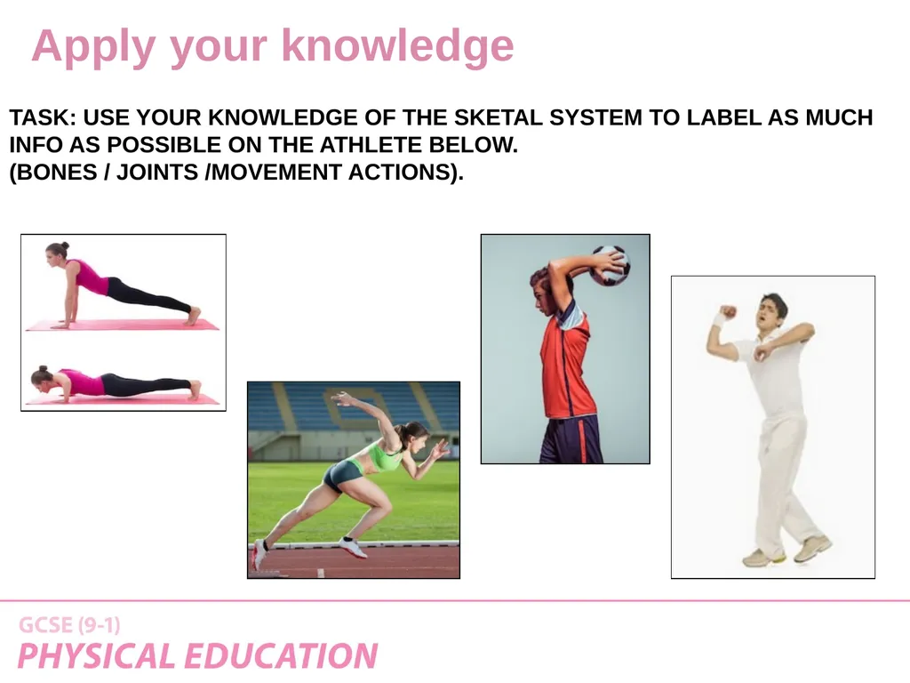
TASK: USE YOUR KNOWLEDGE OF THE SKETAL SYSTEM TO LABEL AS MUCH INFO AS POSSIBLE ON THE ATHLETE BELOW.
(BONES / JOINTS /MOVEMENT ACTIONS). Apply your knowledge<br>
(BONES / JOINTS /MOVEMENT ACTIONS). Apply your knowledge<br>
33
OCR Resources: the small print
OCR’s resources are provided to support the delivery of OCR qualifications, but in no way constitute an endorsed teaching method that is required by the Board, and the decision to use them lies with the individual teacher. Whilst every effort is made to ensure the accuracy of the content, OCR cannot be held responsible for any errors or omissions within these resources.
Our documents are updated over time. Whilst every effort is made to check all documents, there may be contradictions between published support and the specification, therefore please use the information on the latest specification at all times. Where changes are made to specifications these will be indicated within the document, there will be a new version number indicated, and a summary of the changes. If you do notice a discrepancy between the specification and a resource please contact us at:
resources.feedback@ocr.org.uk.
© OCR 2018 - This resource may be freely copied and distributed, as long as the OCR logo and this message remain intact and OCR is acknowledged as the originator of this work. OCR acknowledges the use of the following content:
Please get in touch if you want to discuss the accessibility of resources we offer to support delivery of our qualifications: resources.feedback@ocr.org.uk<br>
Our documents are updated over time. Whilst every effort is made to check all documents, there may be contradictions between published support and the specification, therefore please use the information on the latest specification at all times. Where changes are made to specifications these will be indicated within the document, there will be a new version number indicated, and a summary of the changes. If you do notice a discrepancy between the specification and a resource please contact us at:
resources.feedback@ocr.org.uk.
© OCR 2018 - This resource may be freely copied and distributed, as long as the OCR logo and this message remain intact and OCR is acknowledged as the originator of this work. OCR acknowledges the use of the following content:
Please get in touch if you want to discuss the accessibility of resources we offer to support delivery of our qualifications: resources.feedback@ocr.org.uk<br>