Bacillus anthracis Dr. Priyanka Chaturvedi Assistant Professor BACILLUS ANTHRACIS Causative agent of anthrax. Used as a BIOLOGICAL WEAPON. First bacterium to be isolated in pure culture by Robert Koch and Kochs postulate were made based on
"Bacillus anthracis Dr. Priyanka Chaturvedi" is the property of its rightful owner. Permission is granted to
download and print the materials on this website for personal, non-commercial use only, and to display it
on your personal computer provided you do not modify the materials and that you retain all copyright
notices contained in the materials. By downloading content from our website, you accept the terms of this
agreement.
Presentation Transcript
01
Bacillus anthracis Dr. Priyanka Chaturvedi
Assistant Professor<br>
02
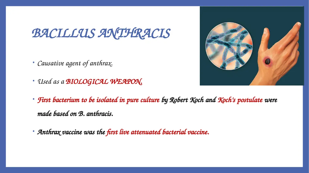
BACILLUS ANTHRACIS Causative agent of anthrax.
Used as a BIOLOGICAL WEAPON.
First bacterium to be isolated in pure culture by Robert Koch and Koch’s postulate were made based on B. anthracis.
Anthrax vaccine was the first live attenuated bacterial vaccine.<br>
03
It was the first pathogenic bacterium seen under microscope (by Pollender, 1849).
It was the first communicable disease shown to be transmitted by inoculation of infected blood. BACILLUS ANTHRACIS<br>
04
Introduction Bacillus anthracis causes ANTHRAX
It is a non-motile aerobic Gram positive bacteria.
Large rectangular capsulated rods arranged in chains with non- bulging spores.
Pathogenic species:-
B. anthracis
B. cereus
Bacillus species are generally motile (with peritrichous flagella) and non-capsulated except Bacillus anthracis.<br>
05
Virulence Factors and Pathogenesis Pathogenesis of anthrax is due to two important virulence factors-
Anthrax toxin and
Capsule.<br>
06
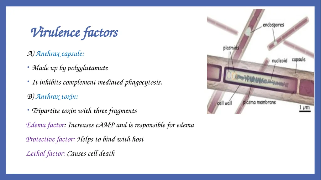
Virulence factors A) Anthrax capsule:
Made up by polyglutamate
It inhibits complement mediated phagocytosis.
B) Anthrax toxin:
Tripartite toxin with three fragments
Edema factor: Increases cAMP and is responsible for edema
Protective factor: Helps to bind with host
Lethal factor: Causes cell death<br>
07
Transmission Modes of transmission:
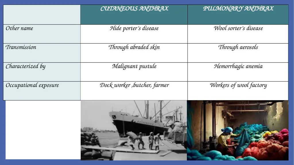
Inoculation of spores (most common mode) : Spores enter through the abraded skin. Seen in people with occupational exposure to animals
Inhalation of spores
Ingestion of carcasses of animals dying of anthrax containing spores (manifested as bloody diarrhea).<br>
08
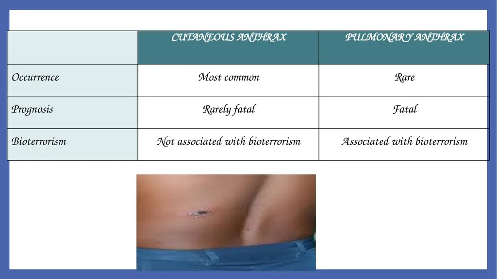
Clinical Types There are mainly three types of human anthrax:
Cutaneous anthrax
Pulmonary anthrax
Intestinal anthrax<br>
09
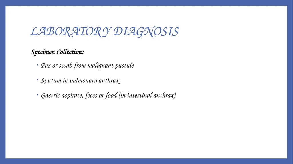
LABORATORY DIAGNOSIS Specimen Collection:
Pus or swab from malignant pustule
Sputum in pulmonary anthrax
Gastric aspirate, feces or food (in intestinal anthrax)<br>
10
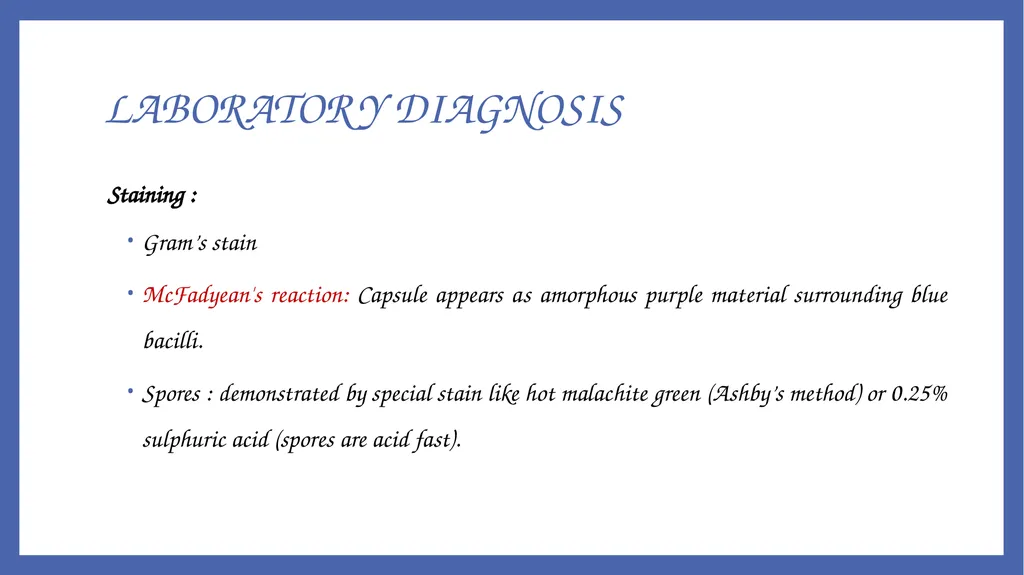
Staining :
Gram’s stain
McFadyean's reaction: Capsule appears as amorphous purple material surrounding blue bacilli.
Spores : demonstrated by special stain like hot malachite green (Ashby’s method) or 0.25% sulphuric acid (spores are acid fast). LABORATORY DIAGNOSIS<br>
11
Mcfadyean's reaction- amorphous purple capsule surrounding blue bacilli (Polychrome Methylene Blue Stain) LABORATORY DIAGNOSIS<br>
12
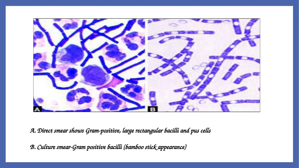
A. Direct smear shows Gram-positive, large rectangular bacilli and pus cells B. Culture smear-Gram positive bacilli (bamboo stick appearance)<br>
13
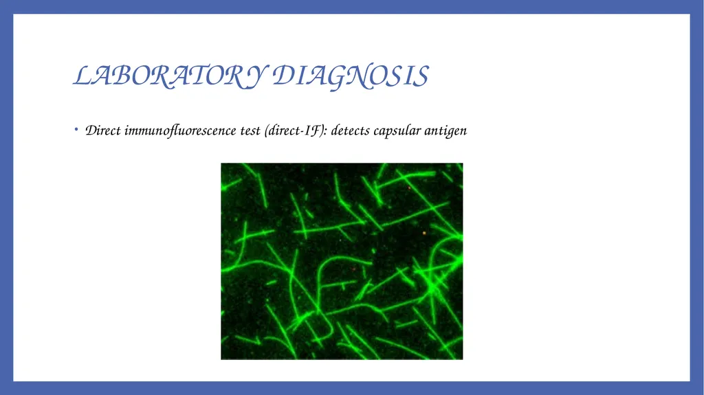
Direct immunofluorescence test (direct-IF): detects capsular antigen LABORATORY DIAGNOSIS<br>
14
CULTURE Bacillus anthracis is aerobic, non-fastidious, grows in ordinary media and has a wide temperature range (12- 450C) of growth.
Sporulation is promoted at 25-300C and in presence of unfavorable conditions.<br>
15
Nutrient agar: Colonies are 2-3 mm in size, irregular, round, opaque, greyish white with a frosted glass appearance.
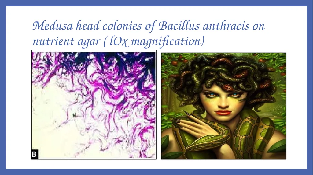
Medusa head appearance: When colonies are viewed under low power microscope, the edge of colony which is composed of long interlacing chains of bacilli, appears as locks of matted hair. CULTURE<br>
16
Medusa head colonies of Bacillus anthracis on nutrient agar ( lOx magnification)<br>
17
Blood agar: Dry wrinkled, non-hemolytic colonies
Gelatin stab agar: inverted fir tree appearance (due to liquefaction of gelatin which occurs maximum at surface, and then slows down towards the bottom CULTURE<br>
18
C. Non-hemolytic dry wrinkled colonies of Bacillus anthracis on blood agar;
D. Gelatin stab agar-inverted fir tree appearance CULTURE<br>
19
Selective media:
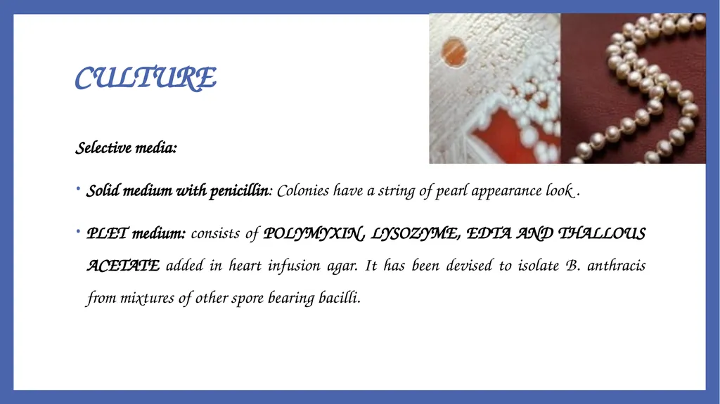
Solid medium with penicillin: Colonies have a string of pearl appearance look .
PLET medium: consists of POLYMYXIN, LYSOZYME, EDTA AND THALLOUS ACETATE added in heart infusion agar. It has been devised to isolate B. anthracis from mixtures of other spore bearing bacilli. CULTURE<br>
20
BIOCHEMICAL REACTIONS Catalase positive
Glucose, maltose, sucrose – fermented – with production of Acid, but no gas
Nitrates – coverts to nitrites<br>
21
Serology :
ELISA
Immunodiffusion in gel method
Molecular methods :-
PCR<br>
22
TREATMENT Anthrax can be successfully treated if the disease is promptly recognized and appropriate therapy is initiated early .
Antibiotic regimen – Ciprofloxacin or Doxycycline, plus Clindamycin for 60 days<br>
23
Antibiotics for postexposure prophylaxis:
Raxibacumab: It is a monoclonal antibody that neutralizes anthrax toxin (protective antigen). It is intended for the prophylaxis and treatment of inhalational anthrax. TREATMENT<br>
24
Prevention Disposal of animal carcasses by burning or by deep burial in lime pits.
Decontamination ( usually by autoclaving) of animal products.
Protective clothing and gloves for handling potentially infectious materials.
Live Attenuated, Non-capsulated Spore Vaccine (Stern Vaccine): It is extensively used in animals
Alum Precipitated Toxoid Vaccine.<br>