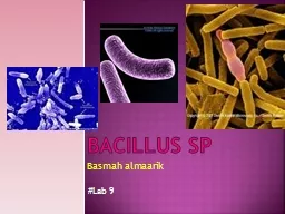
PPT-Figure Figure. Direct fluorescent-antibody (DFA) staining of Bacillus anthracis

Author : ruby | Published Date : 2023-07-08

De BK Bragg SL Sanden GN Wilson KE Diem LA Marston CK et al TwoComponent Direct FluorescentAntibody Assay for Rapid Identification of Bacillus anthracis Emerg Infect

Presentation Embed Code

Download Presentation

Download Presentation The PPT/PDF document "Figure Figure. Direct fluoresce..." is the property of its rightful owner. Permission is granted to download and print the materials on this website for personal, non-commercial use only, and to display it on your personal computer provided you do not modify the materials and that you retain all copyright notices contained in the materials. By downloading content from our website, you accept the terms of this agreement.

Figure Figure. Direct fluorescent-antibody (DFA) staining of Bacillus anthracis: Transcript


De BK Bragg SL Sanden GN Wilson KE Diem LA Marston CK et al TwoComponent Direct FluorescentAntibody Assay for Rapid Identification of Bacillus anthracis Emerg Infect Dis 200281010601065 httpsdoiorg103201eid0810020392. Pavan. Kumar . Akulakrishna. (. M.Tech. , SERC). SR No: 06-02-01-10-51-12-1-09469. Under Guidance of : Prof. Deepak . D’souza. .. Fundamental Question . Is 2 way DFA more powerful than a one way DFA?. . sp. Basmah. . almaarik. #Lab . 9. Bacillus culture. Spore forming facultative anaerobic bacteria.. Grow every where cause it produce spores.. Grow in wide rage 12-45 ºC. Spore formation is best seen in 25 – 30 ºC. Rajeev . Alur. (Penn), Loris D’Antoni (Penn), Sumit Gulwani (MSR), . Bjoern. Hartmann (Berkeley), . Dileep. . Kini. (UIUC), Mahesh . Viswanathan. (UIUC). Motivations. Grading is a tedious and time consuming task. Fluorescence Technology. Josh Leung. James Weis. February 18th, 2010. Bio 1220, Gary Wessel. Fluorescence: How does it work?. Fluorescence vs Phosphorescence. -Time delay – microsecond vs min. Photon absorbed → photon released. Lab #4 . Medgar Evers College. Bio 261 Microbiology. Prof. Santos. Aim #1 exercise 14. Capsular staining. Capsule- extracellular layer composed of polysaccharides found in some bacteria that plays a role in immunity and adhesion.. Staining. Coloring organism with a dye. Microorganisms must be . FIXED. to microscopic slide first. Kills microbe. Attaches organism to the slide. Preserves various parts with minimal distortion. Steps to fix:. Gram-Positive Bacilli Bacteria Dr. Alan N. Talabani Lecturer at College of Medicine/University of Sulaimani Gram-Positive Bacteria I- Gram Positive bacteria A- Gram positive cocci B- Gram positive rods SDTICEIECTEApproved for public relecise tDistribution UnlimitedPlasmids Pasteur and Anthrax into healthy animals 18 and 15years later the anthrax bacillus wasobserved microscopically by Dela-fond 2 Du Author: Neal Chamberlain. Citation: Neal Chamberlain. 2015. Bacillus anthracis: string of pearls reaction.. Publication Date: August 2015. . RESEARCH ARTICLE Open Access Isolation and identification of avirulent strains of Bacillus anthracis from environmental samples in Central Java, Indonesia Ully Apriliana 1,2 , Hendra Wibawa 1 , Endan : It is important to record the fluorescent image of the gel prior to EZBlue staining. EZBlue quenches the fluorescent signal. GlycoProfile III can also be used on PVDF membranes. After gel electroph Jernigan JA, Stephens DS, Ashford DA, Omenaca C, Topiel MS, Galbraith M, et al. Bioterrorism-Related Inhalational Anthrax: The First 10 Cases Reported in the United States. Emerg Infect Dis. 2001;7(6):933-944. https://doi.org/10.3201/eid0706.010604. #Lab . 9. Bacillus culture. Spore forming facultative anaerobic bacteria.. Grow every where cause it produce spores.. Grow in wide rage 12-45 ºC. Spore formation is best seen in 25 – 30 ºC. Grow on NA and BAP. Fixatives used in histopathology. 2. Staining. Differential staining. Gram’s staining. Vital staining. Trypan. blue- dead cells take up the stain . i.e. stain positively and live cells . Exclude the stain or stain negatively.

Download Document

Here is the link to download the presentation.
"Figure Figure. Direct fluorescent-antibody (DFA) staining of Bacillus anthracis"The content belongs to its owner. You may download and print it for personal use, without modification, and keep all copyright notices. By downloading, you agree to these terms.

Related Documents