
BLS 2015 Cardiovascular Emergencies Cardiovascular
DP
Published · 58 slides · 0 views

1 / 1


Description
BLS 2015 Cardiovascular Emergencies Cardiovascular Emergencies Many different presentations! 45-year-old male with crushing substernal chest pain 50-year-old female with a sudden onset of sharp chest pain and shortness of breath 75-year-old
Related Topics
Share
Embed code
Download this presentation From Below
"BLS 2015 Cardiovascular Emergencies Cardiovascular" is the property of its rightful owner. Permission is granted to download and print the materials on this website for personal, non-commercial use only, and to display it on your personal computer provided you do not modify the materials and that you retain all copyright notices contained in the materials. By downloading content from our website, you accept the terms of this agreement.
Presentation Transcript
01
BLS 2015
Cardiovascular Emergencies<br>
02
Cardiovascular Emergencies Many different presentations!
45-year-old male with crushing substernal chest pain
50-year-old female with a sudden onset of sharp chest pain and shortness of breath
75-year-old male with hypertension, tachycardia, and shortness of breath
Each of these patients has a cardiovascular emergency – let’s learn more!<br>
45-year-old male with crushing substernal chest pain
50-year-old female with a sudden onset of sharp chest pain and shortness of breath
75-year-old male with hypertension, tachycardia, and shortness of breath
Each of these patients has a cardiovascular emergency – let’s learn more!<br>
03
Introduction Cardiovascular Emergencies
Leading cause of death in the US
Approximately 600,000 people per year
9 out of 10 cardiac arrest victims die before they get to the hospital
Many more people suffer disability
Cost runs into the billions<br>
Leading cause of death in the US
Approximately 600,000 people per year
9 out of 10 cardiac arrest victims die before they get to the hospital
Many more people suffer disability
Cost runs into the billions<br>
04
Practical Skills Perform focused history using SAMPLE/OPQRST
Assist patient in taking prescribed nitroglycerin
Perform proper technique for auscultating the chest using a stethoscope
Assist a patient with ventilations using a BVM
Demonstrate the ability to care for acute coronary syndrome, CHF, aortic dissection, cardiogenic shock
Demonstrate proper use of AED during CPR To receive CBT or OTEP credit, a trained skills evaluator must evaluate your ability to perform the following hands-on practical skills<br>
Assist patient in taking prescribed nitroglycerin
Perform proper technique for auscultating the chest using a stethoscope
Assist a patient with ventilations using a BVM
Demonstrate the ability to care for acute coronary syndrome, CHF, aortic dissection, cardiogenic shock
Demonstrate proper use of AED during CPR To receive CBT or OTEP credit, a trained skills evaluator must evaluate your ability to perform the following hands-on practical skills<br>
05
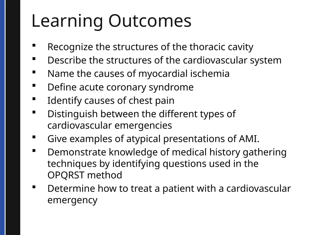
Learning Outcomes Recognize the structures of the thoracic cavity
Describe the structures of the cardiovascular system
Name the causes of myocardial ischemia
Define acute coronary syndrome
Identify causes of chest pain
Distinguish between the different types of cardiovascular emergencies
Give examples of atypical presentations of AMI.
Demonstrate knowledge of medical history gathering techniques by identifying questions used in the OPQRST method
Determine how to treat a patient with a cardiovascular emergency<br>
Describe the structures of the cardiovascular system
Name the causes of myocardial ischemia
Define acute coronary syndrome
Identify causes of chest pain
Distinguish between the different types of cardiovascular emergencies
Give examples of atypical presentations of AMI.
Demonstrate knowledge of medical history gathering techniques by identifying questions used in the OPQRST method
Determine how to treat a patient with a cardiovascular emergency<br>
06
TERMS AND DEFINITIONS<br>
07
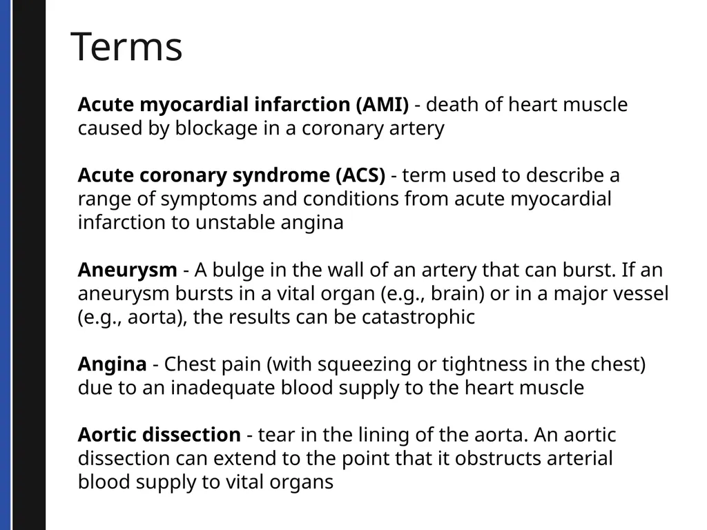
Terms Acute myocardial infarction (AMI) - death of heart muscle caused by blockage in a coronary artery
Acute coronary syndrome (ACS) - term used to describe a range of symptoms and conditions from acute myocardial infarction to unstable angina
Aneurysm - A bulge in the wall of an artery that can burst. If an aneurysm bursts in a vital organ (e.g., brain) or in a major vessel (e.g., aorta), the results can be catastrophic
Angina - Chest pain (with squeezing or tightness in the chest) due to an inadequate blood supply to the heart muscle
Aortic dissection - tear in the lining of the aorta. An aortic dissection can extend to the point that it obstructs arterial blood supply to vital organs<br>
Acute coronary syndrome (ACS) - term used to describe a range of symptoms and conditions from acute myocardial infarction to unstable angina
Aneurysm - A bulge in the wall of an artery that can burst. If an aneurysm bursts in a vital organ (e.g., brain) or in a major vessel (e.g., aorta), the results can be catastrophic
Angina - Chest pain (with squeezing or tightness in the chest) due to an inadequate blood supply to the heart muscle
Aortic dissection - tear in the lining of the aorta. An aortic dissection can extend to the point that it obstructs arterial blood supply to vital organs<br>
08
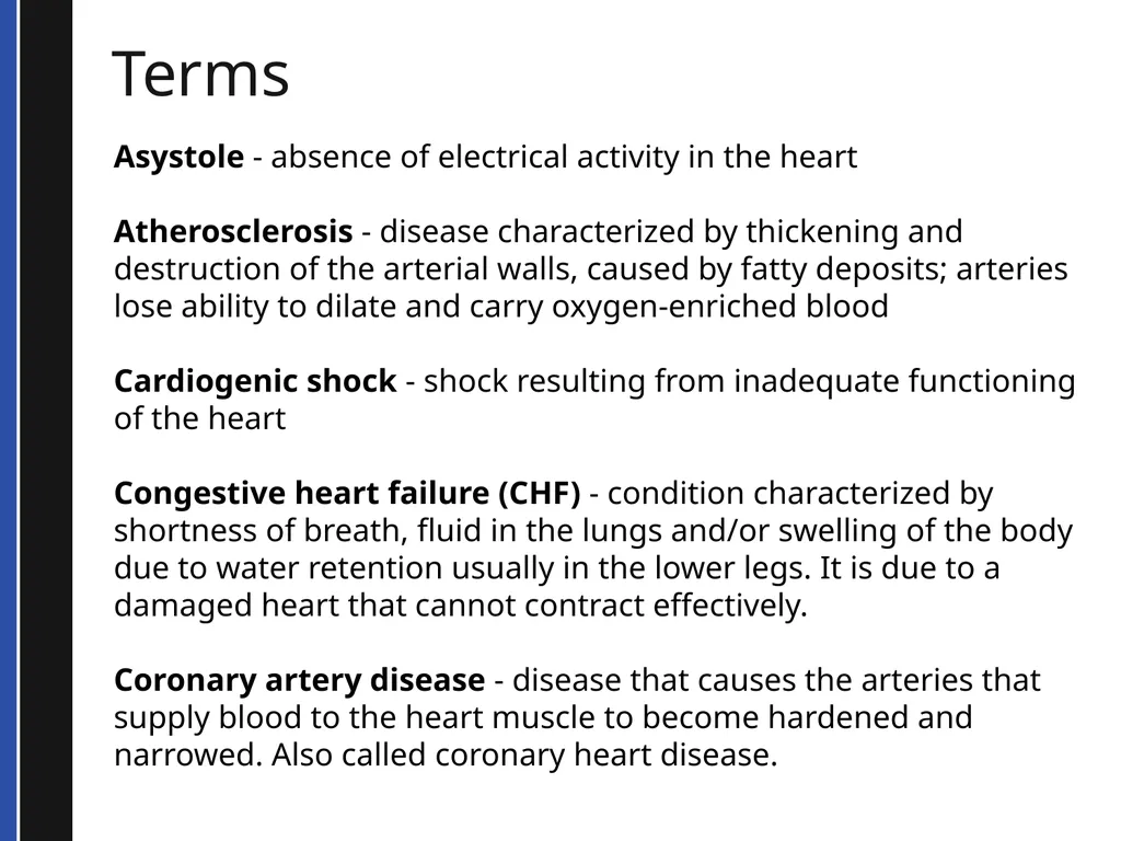
Terms Asystole - absence of electrical activity in the heart
Atherosclerosis - disease characterized by thickening and destruction of the arterial walls, caused by fatty deposits; arteries lose ability to dilate and carry oxygen-enriched blood
Cardiogenic shock - shock resulting from inadequate functioning of the heart
Congestive heart failure (CHF) - condition characterized by shortness of breath, fluid in the lungs and/or swelling of the body due to water retention usually in the lower legs. It is due to a damaged heart that cannot contract effectively.
Coronary artery disease - disease that causes the arteries that supply blood to the heart muscle to become hardened and narrowed. Also called coronary heart disease.<br>
Atherosclerosis - disease characterized by thickening and destruction of the arterial walls, caused by fatty deposits; arteries lose ability to dilate and carry oxygen-enriched blood
Cardiogenic shock - shock resulting from inadequate functioning of the heart
Congestive heart failure (CHF) - condition characterized by shortness of breath, fluid in the lungs and/or swelling of the body due to water retention usually in the lower legs. It is due to a damaged heart that cannot contract effectively.
Coronary artery disease - disease that causes the arteries that supply blood to the heart muscle to become hardened and narrowed. Also called coronary heart disease.<br>
09
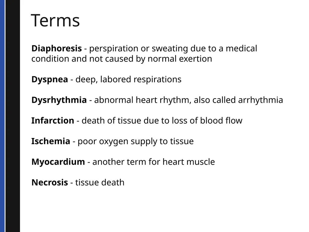
Terms Diaphoresis - perspiration or sweating due to a medical condition and not caused by normal exertion
Dyspnea - deep, labored respirations
Dysrhythmia - abnormal heart rhythm, also called arrhythmia
Infarction - death of tissue due to loss of blood flow
Ischemia - poor oxygen supply to tissue
Myocardium - another term for heart muscle
Necrosis - tissue death<br>
Dyspnea - deep, labored respirations
Dysrhythmia - abnormal heart rhythm, also called arrhythmia
Infarction - death of tissue due to loss of blood flow
Ischemia - poor oxygen supply to tissue
Myocardium - another term for heart muscle
Necrosis - tissue death<br>
10
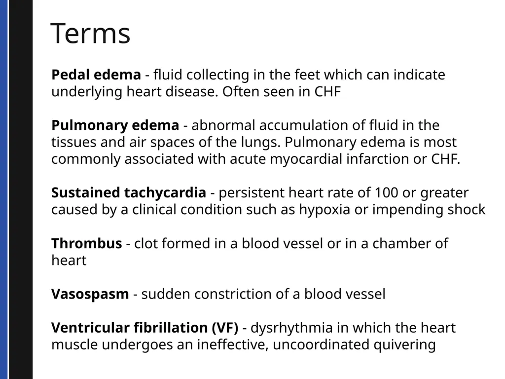
Terms Pedal edema - fluid collecting in the feet which can indicate underlying heart disease. Often seen in CHF
Pulmonary edema - abnormal accumulation of fluid in the tissues and air spaces of the lungs. Pulmonary edema is most commonly associated with acute myocardial infarction or CHF.
Sustained tachycardia - persistent heart rate of 100 or greater caused by a clinical condition such as hypoxia or impending shock
Thrombus - clot formed in a blood vessel or in a chamber of heart
Vasospasm - sudden constriction of a blood vessel
Ventricular fibrillation (VF) - dysrhythmia in which the heart muscle undergoes an ineffective, uncoordinated quivering<br>
Pulmonary edema - abnormal accumulation of fluid in the tissues and air spaces of the lungs. Pulmonary edema is most commonly associated with acute myocardial infarction or CHF.
Sustained tachycardia - persistent heart rate of 100 or greater caused by a clinical condition such as hypoxia or impending shock
Thrombus - clot formed in a blood vessel or in a chamber of heart
Vasospasm - sudden constriction of a blood vessel
Ventricular fibrillation (VF) - dysrhythmia in which the heart muscle undergoes an ineffective, uncoordinated quivering<br>
11
ANATOMY<br>
12
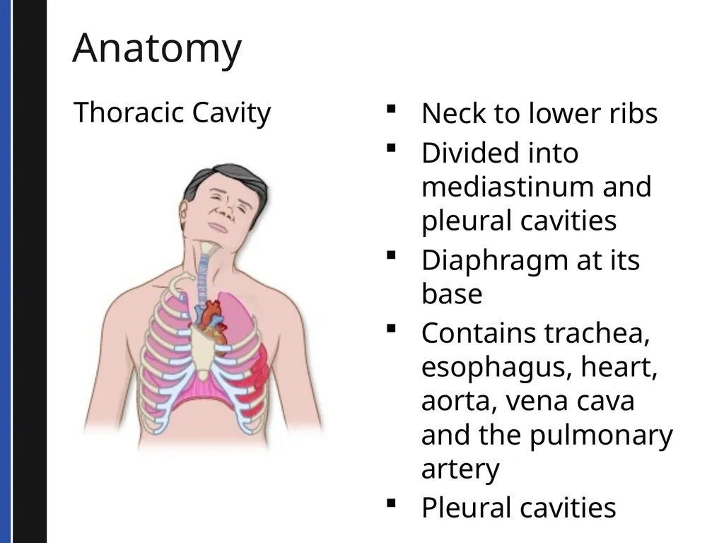
Anatomy Thoracic Cavity Neck to lower ribs
Divided into mediastinum and pleural cavities
Diaphragm at its base
Contains trachea, esophagus, heart, aorta, vena cava and the pulmonary artery
Pleural cavities<br>
Divided into mediastinum and pleural cavities
Diaphragm at its base
Contains trachea, esophagus, heart, aorta, vena cava and the pulmonary artery
Pleural cavities<br>
13
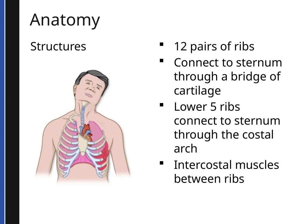
Anatomy Structures 12 pairs of ribs
Connect to sternum through a bridge of cartilage
Lower 5 ribs connect to sternum through the costal arch
Intercostal muscles between ribs<br>
Connect to sternum through a bridge of cartilage
Lower 5 ribs connect to sternum through the costal arch
Intercostal muscles between ribs<br>
14
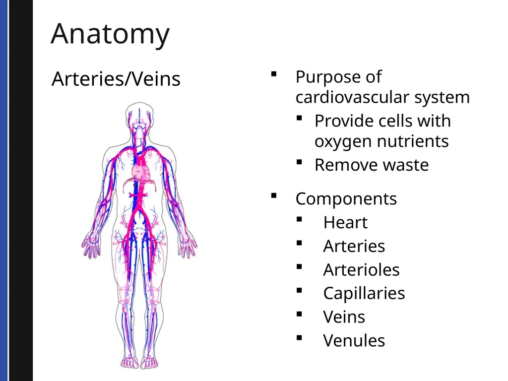
Anatomy Arteries/Veins Purpose of cardiovascular system
Provide cells with oxygen nutrients
Remove waste
Components
Heart
Arteries
Arterioles
Capillaries
Veins
Venules<br>
Provide cells with oxygen nutrients
Remove waste
Components
Heart
Arteries
Arterioles
Capillaries
Veins
Venules<br>
15
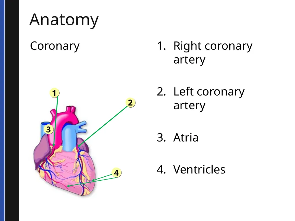
Anatomy Coronary Right coronary artery
Left coronary artery
Atria
Ventricles 2 1 3 4<br>
Left coronary artery
Atria
Ventricles 2 1 3 4<br>
16
PATHOPHYSIOLOGY<br>
17
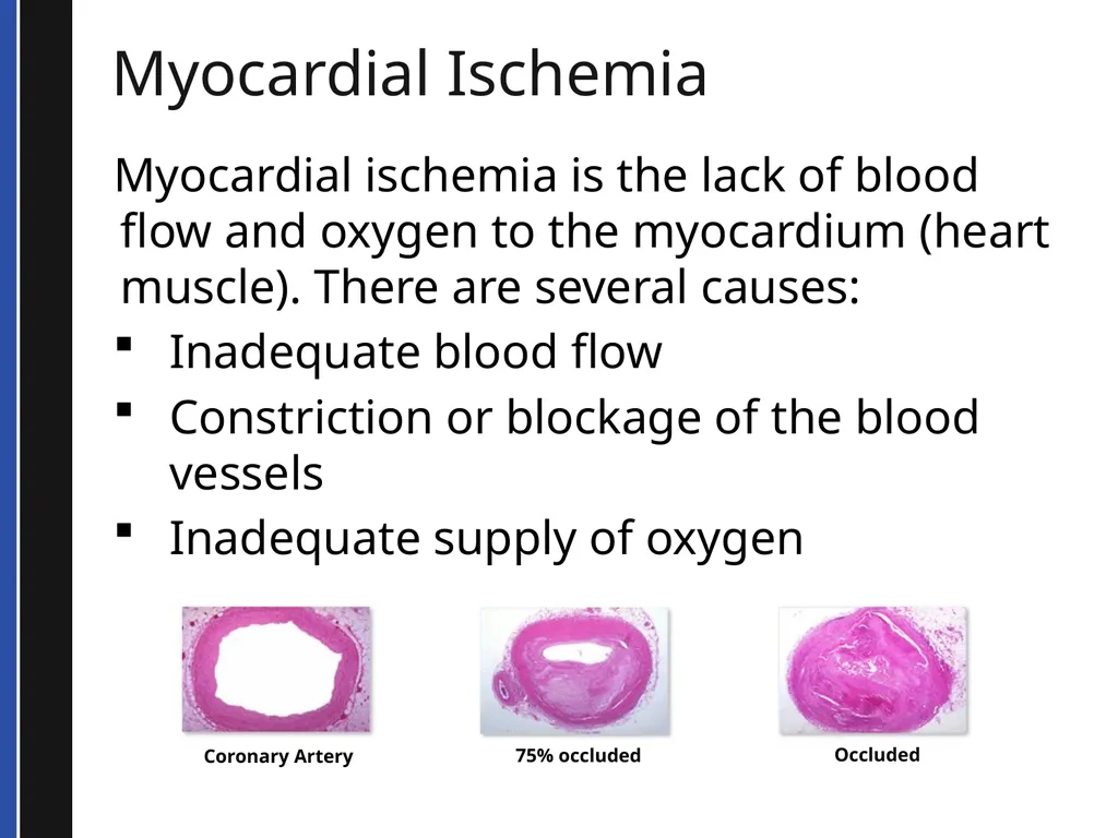
Myocardial Ischemia Myocardial ischemia is the lack of blood flow and oxygen to the myocardium (heart muscle). There are several causes:
Inadequate blood flow
Constriction or blockage of the blood vessels
Inadequate supply of oxygen Coronary Artery 75% occluded Occluded<br>
Inadequate blood flow
Constriction or blockage of the blood vessels
Inadequate supply of oxygen Coronary Artery 75% occluded Occluded<br>
18
Coronary Thrombosis One of the major causes of myocardial ischemia
Recent scientific research has expanded our understanding
This research has discovered the steps to thrombosis formation<br>
Recent scientific research has expanded our understanding
This research has discovered the steps to thrombosis formation<br>
19
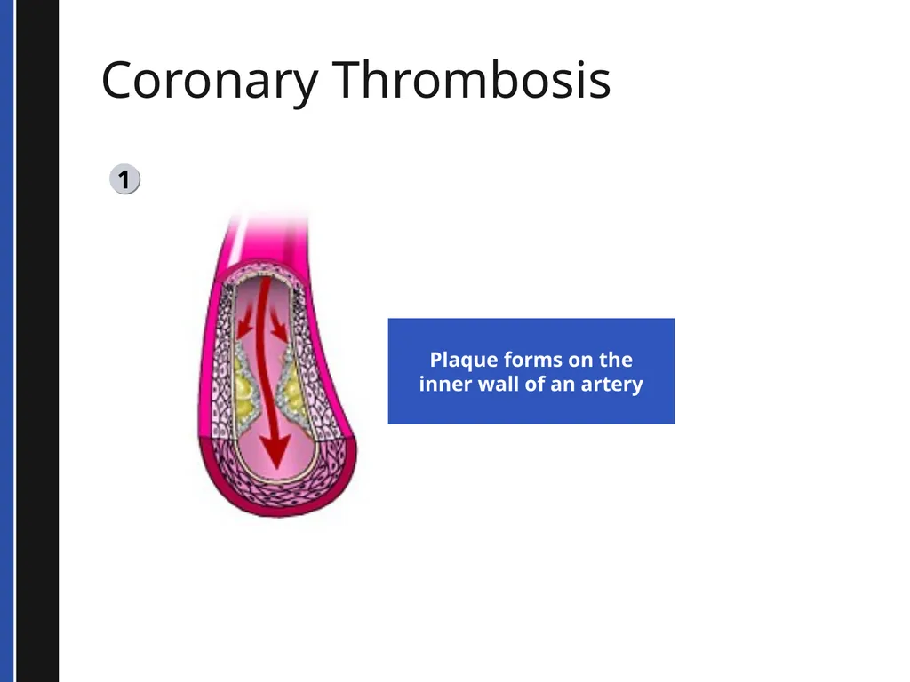
Coronary Thrombosis 1 Plaque forms on the
inner wall of an artery<br>
inner wall of an artery<br>
20
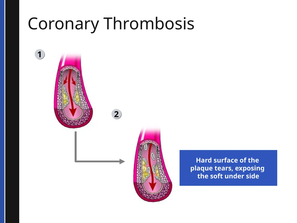
Coronary Thrombosis 1 2 Hard surface of the
plaque tears, exposing
the soft under side<br>
plaque tears, exposing
the soft under side<br>
21
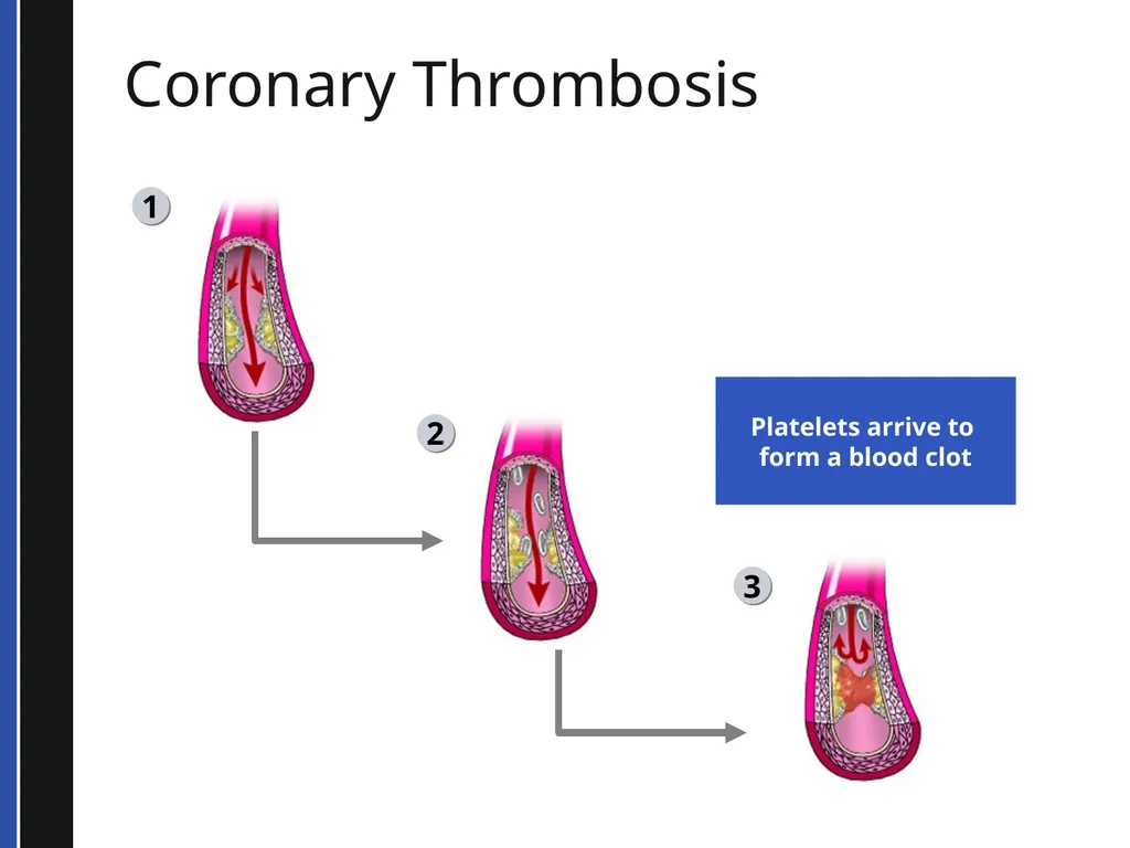
Coronary Thrombosis 1 2 3 Platelets arrive to
form a blood clot<br>
form a blood clot<br>
22
Atherosclerosis Can also cause myocardial ischemia and chest pain
No disruption of plaque
Stable plaque that increases in size over time
Decreases blood flow past the obstruction
Example: angina<br>
No disruption of plaque
Stable plaque that increases in size over time
Decreases blood flow past the obstruction
Example: angina<br>
23
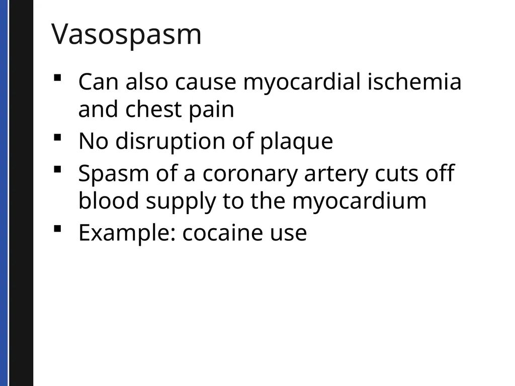
Vasospasm Can also cause myocardial ischemia and chest pain
No disruption of plaque
Spasm of a coronary artery cuts off blood supply to the myocardium
Example: cocaine use<br>
No disruption of plaque
Spasm of a coronary artery cuts off blood supply to the myocardium
Example: cocaine use<br>
24
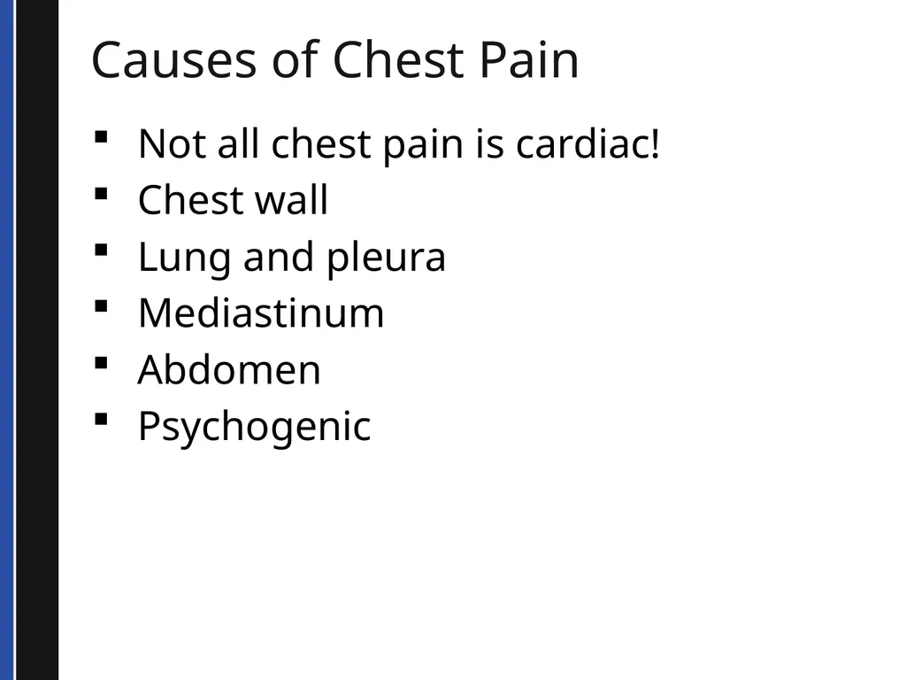
Causes of Chest Pain Not all chest pain is cardiac!
Chest wall
Lung and pleura
Mediastinum
Abdomen
Psychogenic<br>
Chest wall
Lung and pleura
Mediastinum
Abdomen
Psychogenic<br>
25
ACUTE CORONARY SYNDROMES<br>
26
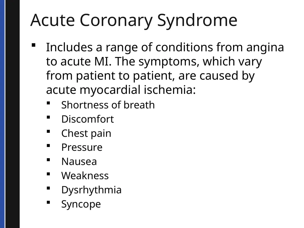
Acute Coronary Syndrome Includes a range of conditions from angina to acute MI. The symptoms, which vary from patient to patient, are caused by acute myocardial ischemia:
Shortness of breath
Discomfort
Chest pain
Pressure
Nausea
Weakness
Dysrhythmia
Syncope<br>
Shortness of breath
Discomfort
Chest pain
Pressure
Nausea
Weakness
Dysrhythmia
Syncope<br>
27
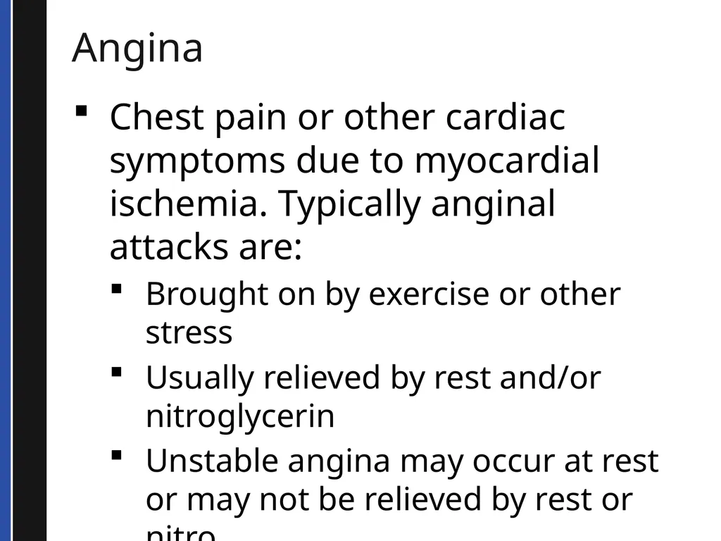
Angina Chest pain or other cardiac symptoms due to myocardial ischemia. Typically anginal attacks are:
Brought on by exercise or other stress
Usually relieved by rest and/or nitroglycerin
Unstable angina may occur at rest or may not be relieved by rest or nitro<br>
Brought on by exercise or other stress
Usually relieved by rest and/or nitroglycerin
Unstable angina may occur at rest or may not be relieved by rest or nitro<br>
28
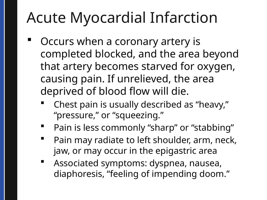
Acute Myocardial Infarction Occurs when a coronary artery is completed blocked, and the area beyond that artery becomes starved for oxygen, causing pain. If unrelieved, the area deprived of blood flow will die.
Chest pain is usually described as “heavy,” “pressure,” or “squeezing.”
Pain is less commonly “sharp” or “stabbing”
Pain may radiate to left shoulder, arm, neck, jaw, or may occur in the epigastric area
Associated symptoms: dyspnea, nausea, diaphoresis, “feeling of impending doom.”<br>
Chest pain is usually described as “heavy,” “pressure,” or “squeezing.”
Pain is less commonly “sharp” or “stabbing”
Pain may radiate to left shoulder, arm, neck, jaw, or may occur in the epigastric area
Associated symptoms: dyspnea, nausea, diaphoresis, “feeling of impending doom.”<br>
29
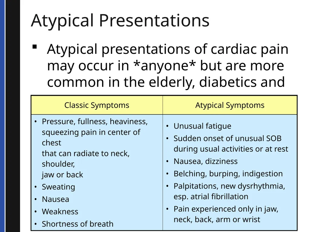
Atypical Presentations Atypical presentations of cardiac pain may occur in *anyone* but are more common in the elderly, diabetics and women<br>
30
OTHER CARDIOVASCULAR
EMEGENCIES<br>
31
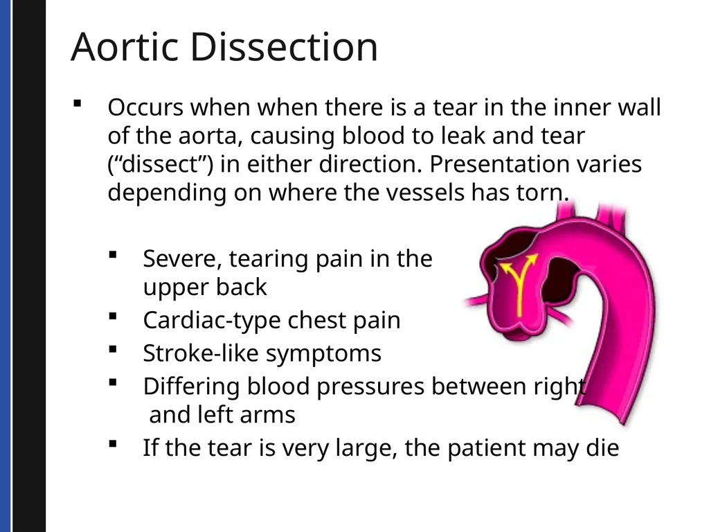
Aortic Dissection Occurs when when there is a tear in the inner wall of the aorta, causing blood to leak and tear (“dissect”) in either direction. Presentation varies depending on where the vessels has torn.
Severe, tearing pain in the upper back
Cardiac-type chest pain
Stroke-like symptoms
Differing blood pressures between right and left arms
If the tear is very large, the patient may die<br>
Severe, tearing pain in the upper back
Cardiac-type chest pain
Stroke-like symptoms
Differing blood pressures between right and left arms
If the tear is very large, the patient may die<br>
32
CHF Occurs when the heart is too weak to adequately circulate blood (often due to an MI).
Left-sided heart failure causes fluid to back up into the lungs.
Right-sided heart failure causes fluid to back up into the venous system.
Many patients have a combination of both types of failure. Presentation includes:
Dyspnea
Pulmonary edema (heard as rales)
Chest pain
Hypertension
Swollen ankles (if there is a component of right-sided heart failure)<br>
Left-sided heart failure causes fluid to back up into the lungs.
Right-sided heart failure causes fluid to back up into the venous system.
Many patients have a combination of both types of failure. Presentation includes:
Dyspnea
Pulmonary edema (heard as rales)
Chest pain
Hypertension
Swollen ankles (if there is a component of right-sided heart failure)<br>
33
Cardiogenic Shock Occurs when heart is seriously weakened and cannot pump enough blood to perfuse body. This may occur after an MI. Presentation is similar to other types of shock:
Hypotension
Chest pain
Bradycardic/tachycardic/other arrhythmias
Restless, anxious
Pale, cool skin
Possibly altered LOC<br>
Hypotension
Chest pain
Bradycardic/tachycardic/other arrhythmias
Restless, anxious
Pale, cool skin
Possibly altered LOC<br>
34
Pulmonary Embolus Pulmonary embolus occurs when a blood clot travels from somewhere in the body (usually the legs or pelvis) and lodges in the pulmonary vasculature. Presentation varies by the size of the clot. A very small clot may cause mild dyspnea, a very large clot may cause sudden death. Other presentations include:
Dyspnea
Chest pain, often sharp in nature
Usually, clear lung sounds (remember, this is a vascular problem, not a lung tissue problem!)
Tachycardia
Hypotension
Pale, cool skin
Low oxygen saturations (check before administering oxygen)<br>
Dyspnea
Chest pain, often sharp in nature
Usually, clear lung sounds (remember, this is a vascular problem, not a lung tissue problem!)
Tachycardia
Hypotension
Pale, cool skin
Low oxygen saturations (check before administering oxygen)<br>
35
Hypertensive Emergencies Very high blood pressure associated with neurologic findings.
Generally higher than 180/120
Unilateral weakness
Visual disturbances
Severe headache
Seizure<br>
Generally higher than 180/120
Unilateral weakness
Visual disturbances
Severe headache
Seizure<br>
36
PATIENT CARE<br>
37
Initial Assessment Guides initial treatment
Quickly assess a patient
Make a decision SICK
or
NOT SICK<br>
Quickly assess a patient
Make a decision SICK
or
NOT SICK<br>
38
SICK/NOT SICK SICK patient is one who can die quickly unless you initiate aggressive BLS and ALS treatment and rapid transport
NOT SICK patient is one who can be ill or injured, but not severely enough to need immediate ALS care<br>
NOT SICK patient is one who can be ill or injured, but not severely enough to need immediate ALS care<br>
39
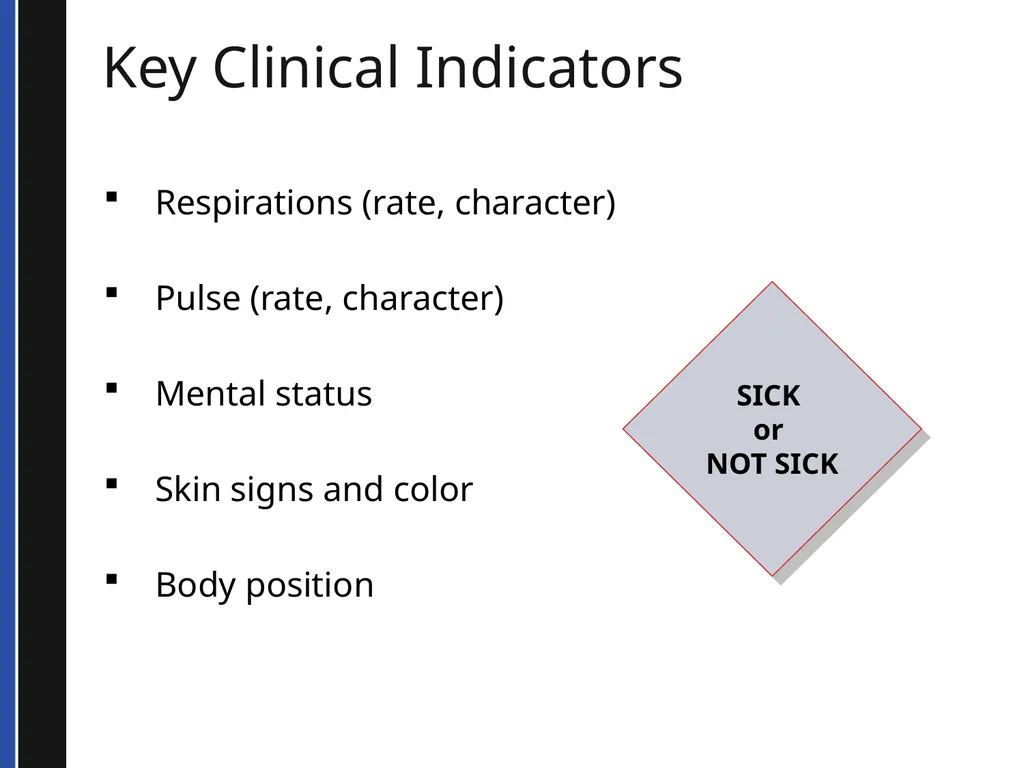
Key Clinical Indicators Respirations (rate, character)
Pulse (rate, character)
Mental status
Skin signs and color
Body position SICK
or
NOT SICK<br>
Pulse (rate, character)
Mental status
Skin signs and color
Body position SICK
or
NOT SICK<br>
40
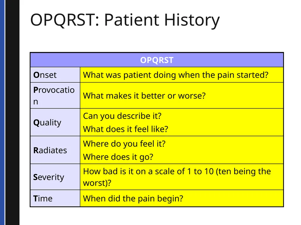
OPQRST: Patient History<br>
41
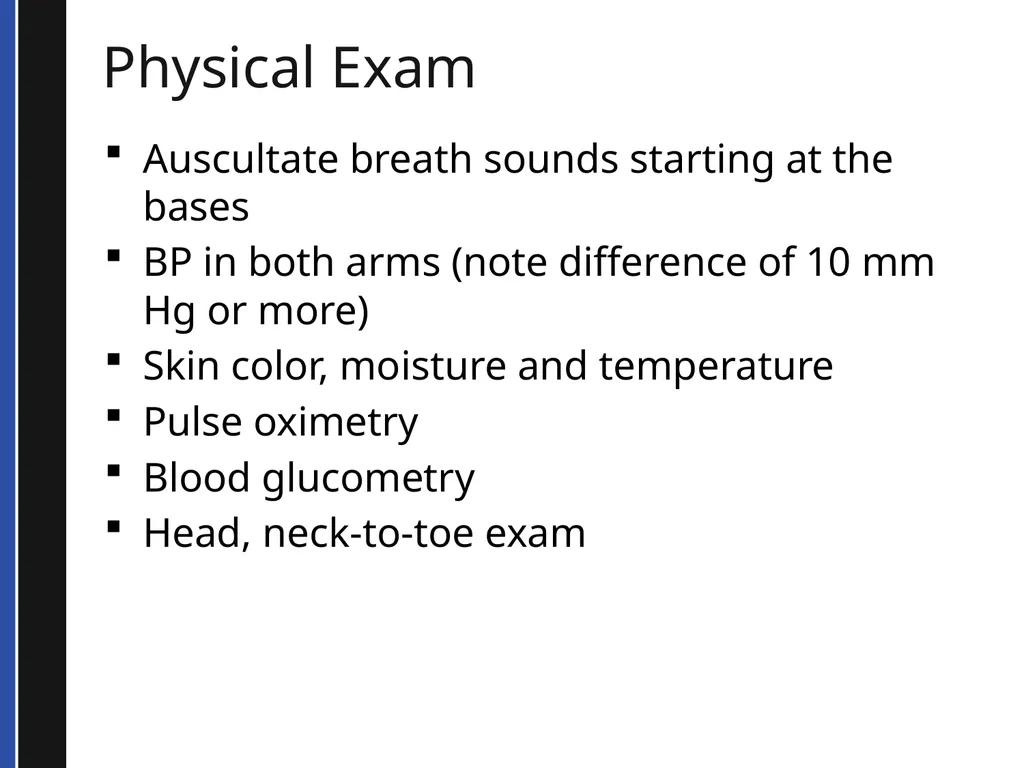
Physical Exam Auscultate breath sounds starting at the bases
BP in both arms (note difference of 10 mm Hg or more)
Skin color, moisture and temperature
Pulse oximetry
Blood glucometry
Head, neck-to-toe exam<br>
BP in both arms (note difference of 10 mm Hg or more)
Skin color, moisture and temperature
Pulse oximetry
Blood glucometry
Head, neck-to-toe exam<br>
42
LUNG SOUNDS<br>
43
Lung Sounds Listen at six locations on the back and four locations on the front
Start in the back at the base first in sitting patient
Instruct patient to take a deep breath through the mouth then exhale
Listen to one or two inspiration/expiration cycles per location
Move to the other side and compare
Try to avoid listening through clothing<br>
Start in the back at the base first in sitting patient
Instruct patient to take a deep breath through the mouth then exhale
Listen to one or two inspiration/expiration cycles per location
Move to the other side and compare
Try to avoid listening through clothing<br>
44
Lung Sounds - Example Normal lung sounds – recorded over the left anterior upper chest of a 15 year old male adolescent
(click the sound button to play)<br>
(click the sound button to play)<br>
45
Lung Sounds - Example Rales – These crackles and bronchial breathing were recorded posteriorly over the consolidated left lower lung of a 65-year-old male with CHF
(click the sound button to play)<br>
(click the sound button to play)<br>
46
Lung Sounds - Example Rales – These late inspiratory fine crackles were recorded over the right posterior lower lung of a 55 year old woman with CHF
(click the sound button to play)<br>
(click the sound button to play)<br>
47
Lung Sounds - Example Wheeze (a) – This expiratory wheezing was recorded over the right anterior upper chest of an 8 year old boy with asthma
(click the sound button to play)<br>
(click the sound button to play)<br>
48
Lung Sounds - Example Wheeze (b) – These wheezing & coarse crackles were recorded over right posterior lower lung of an 8 month old boy with viral bronchiolitis
(click the sound button to play)<br>
(click the sound button to play)<br>
49
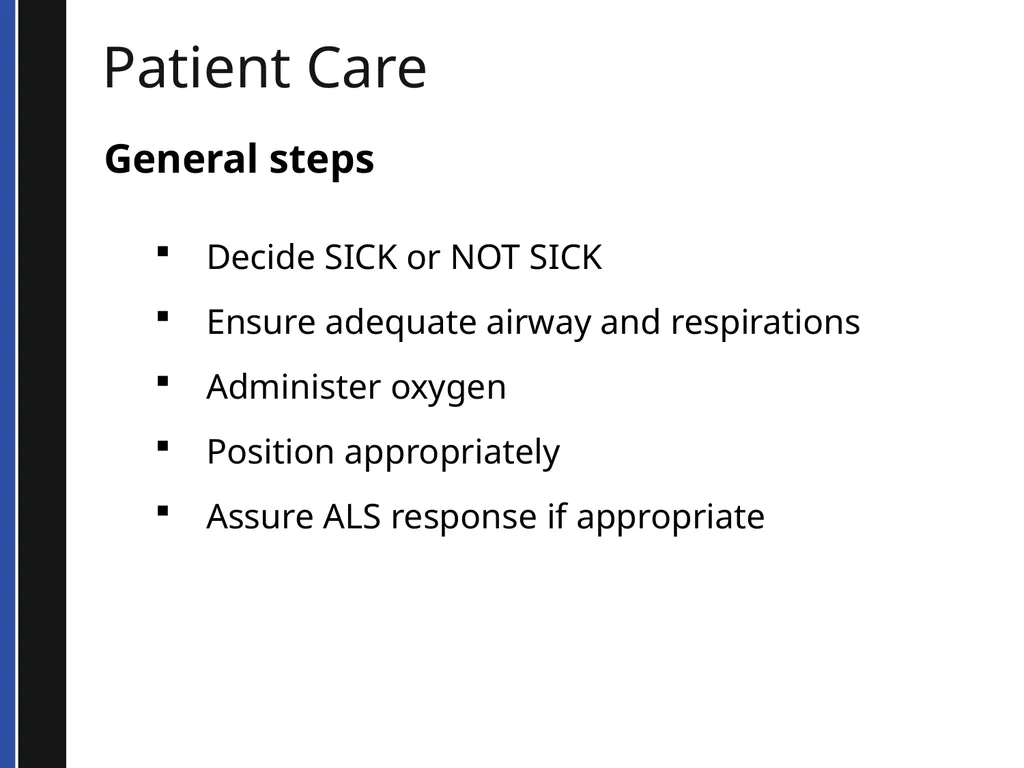
Patient Care General steps
Decide SICK or NOT SICK
Ensure adequate airway and respirations
Administer oxygen
Position appropriately
Assure ALS response if appropriate<br>
Decide SICK or NOT SICK
Ensure adequate airway and respirations
Administer oxygen
Position appropriately
Assure ALS response if appropriate<br>
50
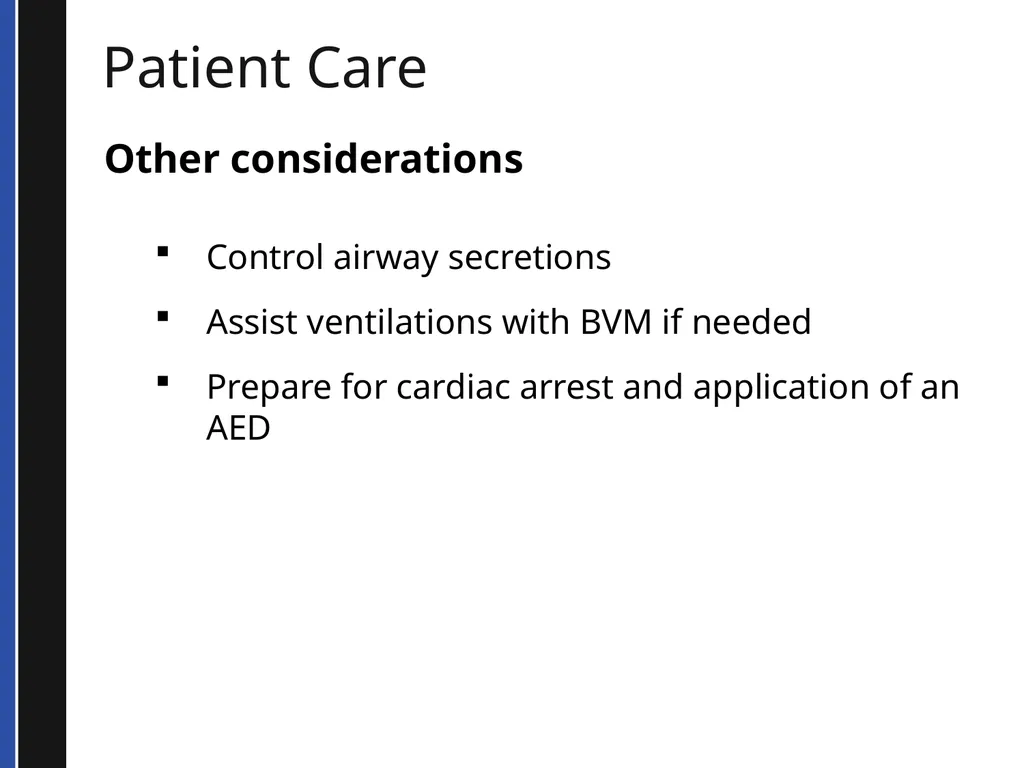
Patient Care Other considerations
Control airway secretions
Assist ventilations with BVM if needed
Prepare for cardiac arrest and application of an AED<br>
Control airway secretions
Assist ventilations with BVM if needed
Prepare for cardiac arrest and application of an AED<br>
51
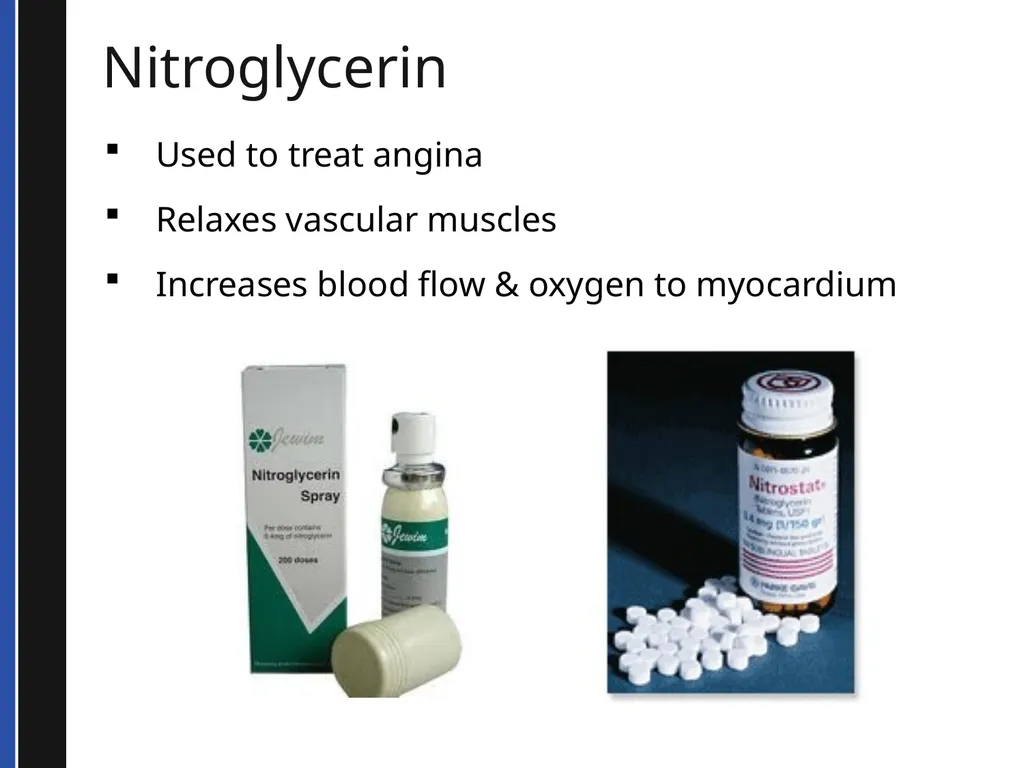
Nitroglycerin Used to treat angina
Relaxes vascular muscles
Increases blood flow & oxygen to myocardium<br>
Relaxes vascular muscles
Increases blood flow & oxygen to myocardium<br>
52
Nitroglycerin You may assist a patient in taking prescribed nitroglycerin if:
Pain is the same for which nitroglycerin is normally taken
Patient’s BP is greater than 100 mmHg systolic
*Follow your local protocol if different.<br>
Pain is the same for which nitroglycerin is normally taken
Patient’s BP is greater than 100 mmHg systolic
*Follow your local protocol if different.<br>
53
Nitroglycerin “Assisting” a patient with nitroglycerin means you can:
Locate the container
Open it
Offer a pill to the patient<br>
Locate the container
Open it
Offer a pill to the patient<br>
54
Aspirin in the Field Why give aspirin to someone with acute coronary syndrome (angina or MI)?
Anti-inflammatory properties
Helps protect inflamed heart muscle
Reduces body’s production of prostaglandins
May reduce risk of blood clots, heart attacks, strokes
Anti-platelet effect takes an hour to “kick in”<br>
Anti-inflammatory properties
Helps protect inflamed heart muscle
Reduces body’s production of prostaglandins
May reduce risk of blood clots, heart attacks, strokes
Anti-platelet effect takes an hour to “kick in”<br>
55
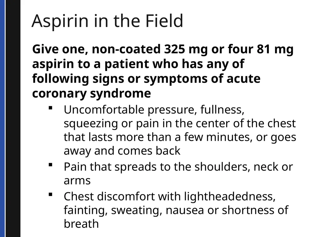
Aspirin in the Field Give one, non-coated 325 mg or four 81 mg aspirin to a patient who has any of following signs or symptoms of acute coronary syndrome
Uncomfortable pressure, fullness, squeezing or pain in the center of the chest that lasts more than a few minutes, or goes away and comes back
Pain that spreads to the shoulders, neck or arms
Chest discomfort with lightheadedness, fainting, sweating, nausea or shortness of breath<br>
Uncomfortable pressure, fullness, squeezing or pain in the center of the chest that lasts more than a few minutes, or goes away and comes back
Pain that spreads to the shoulders, neck or arms
Chest discomfort with lightheadedness, fainting, sweating, nausea or shortness of breath<br>
56
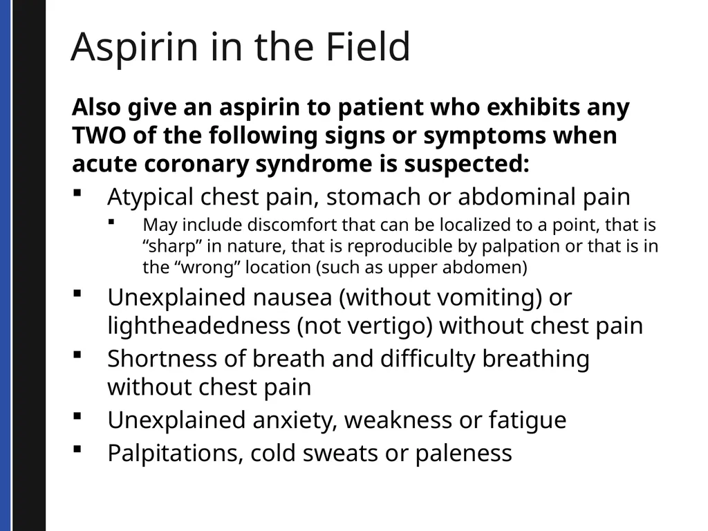
Aspirin in the Field Also give an aspirin to patient who exhibits any TWO of the following signs or symptoms when acute coronary syndrome is suspected:
Atypical chest pain, stomach or abdominal pain
May include discomfort that can be localized to a point, that is “sharp” in nature, that is reproducible by palpation or that is in the “wrong” location (such as upper abdomen)
Unexplained nausea (without vomiting) or lightheadedness (not vertigo) without chest pain
Shortness of breath and difficulty breathing without chest pain
Unexplained anxiety, weakness or fatigue
Palpitations, cold sweats or paleness<br>
Atypical chest pain, stomach or abdominal pain
May include discomfort that can be localized to a point, that is “sharp” in nature, that is reproducible by palpation or that is in the “wrong” location (such as upper abdomen)
Unexplained nausea (without vomiting) or lightheadedness (not vertigo) without chest pain
Shortness of breath and difficulty breathing without chest pain
Unexplained anxiety, weakness or fatigue
Palpitations, cold sweats or paleness<br>
57
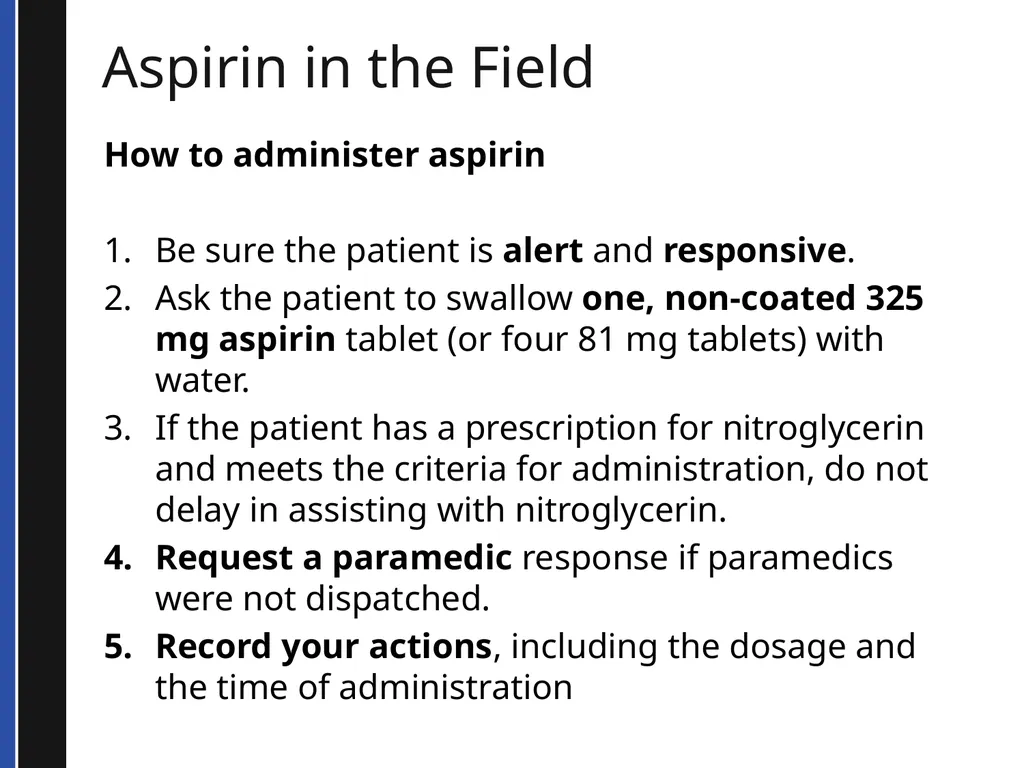
Aspirin in the Field How to administer aspirin
Be sure the patient is alert and responsive.
Ask the patient to swallow one, non-coated 325 mg aspirin tablet (or four 81 mg tablets) with water.
If the patient has a prescription for nitroglycerin and meets the criteria for administration, do not delay in assisting with nitroglycerin.
Request a paramedic response if paramedics were not dispatched.
Record your actions, including the dosage and the time of administration<br>
Be sure the patient is alert and responsive.
Ask the patient to swallow one, non-coated 325 mg aspirin tablet (or four 81 mg tablets) with water.
If the patient has a prescription for nitroglycerin and meets the criteria for administration, do not delay in assisting with nitroglycerin.
Request a paramedic response if paramedics were not dispatched.
Record your actions, including the dosage and the time of administration<br>
58
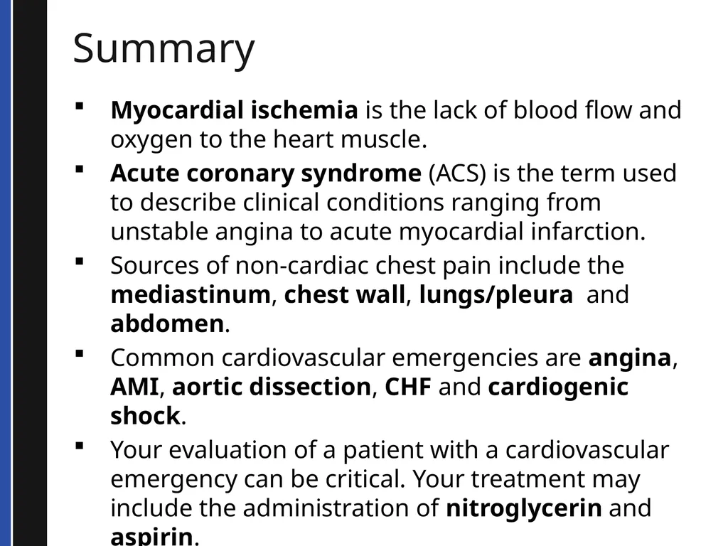
Summary Myocardial ischemia is the lack of blood flow and oxygen to the heart muscle.
Acute coronary syndrome (ACS) is the term used to describe clinical conditions ranging from unstable angina to acute myocardial infarction.
Sources of non-cardiac chest pain include the mediastinum, chest wall, lungs/pleura and abdomen.
Common cardiovascular emergencies are angina, AMI, aortic dissection, CHF and cardiogenic shock.
Your evaluation of a patient with a cardiovascular emergency can be critical. Your treatment may include the administration of nitroglycerin and aspirin.<br>
Acute coronary syndrome (ACS) is the term used to describe clinical conditions ranging from unstable angina to acute myocardial infarction.
Sources of non-cardiac chest pain include the mediastinum, chest wall, lungs/pleura and abdomen.
Common cardiovascular emergencies are angina, AMI, aortic dissection, CHF and cardiogenic shock.
Your evaluation of a patient with a cardiovascular emergency can be critical. Your treatment may include the administration of nitroglycerin and aspirin.<br>