
Chapter 19 Endocrine and Hematologic Emergencies
TD
Published · 140 slides · 0 views

1 / 1






Description
Chapter 19 Endocrine and Hematologic Emergencies National EMS Education Standard Competencies (1 of 3) Medicine Applies fundamental knowledge to provide basic emergency care and transportation based on assessment findings for an acutely ill
Related Topics
Share
Embed code
Download this presentation From Below
"Chapter 19 Endocrine and Hematologic Emergencies" is the property of its rightful owner. Permission is granted to download and print the materials on this website for personal, non-commercial use only, and to display it on your personal computer provided you do not modify the materials and that you retain all copyright notices contained in the materials. By downloading content from our website, you accept the terms of this agreement.
Presentation Transcript
01
Chapter 19
Endocrine and Hematologic Emergencies<br>
Endocrine and Hematologic Emergencies<br>
02
National EMS Education Standard Competencies (1 of 3) Medicine
Applies fundamental knowledge to provide basic emergency care and transportation based on assessment findings for an acutely ill patient.<br>
Applies fundamental knowledge to provide basic emergency care and transportation based on assessment findings for an acutely ill patient.<br>
03
National EMS Education Standard Competencies (2 of 3) Awareness that
Diabetic emergencies cause altered mental status
Anatomy, physiology, pathophysiology, assessment, and management of
Acute diabetic emergencies<br>
Diabetic emergencies cause altered mental status
Anatomy, physiology, pathophysiology, assessment, and management of
Acute diabetic emergencies<br>
04
National EMS Education Standard Competencies (3 of 3) Anatomy, physiology, pathophysiology, assessment, and management of
Sickle cell crisis
Clotting disorders<br>
Sickle cell crisis
Clotting disorders<br>
05
Introduction (1 of 2) Endocrine system influences nearly every:
Cell
Organ
Bodily function
Endocrine disorders can have many signs and symptoms.<br>
Cell
Organ
Bodily function
Endocrine disorders can have many signs and symptoms.<br>
06
Introduction (2 of 2) Hematologic emergencies
Difficult to assess and treat
Your actions may save a life.<br>
Difficult to assess and treat
Your actions may save a life.<br>
07
Anatomy and Physiology (1 of 3) Endocrine system is a communication system that controls functions inside the body.
Glands secrete messenger hormones.
Hormones are chemical substances produced by a gland.
Endocrine disorders are caused by an internal communication problem.<br>
Glands secrete messenger hormones.
Hormones are chemical substances produced by a gland.
Endocrine disorders are caused by an internal communication problem.<br>
08
Anatomy and Physiology (2 of 3) Glucose metabolism
Brain needs glucose and oxygen.
Insulin is necessary for glucose to enter cells.
Without enough insulin, cells do not get fed.<br>
Brain needs glucose and oxygen.
Insulin is necessary for glucose to enter cells.
Without enough insulin, cells do not get fed.<br>
09
Anatomy and Physiology (3 of 3) Glucose metabolism (cont’d)
Pancreas produces and stores glucagon and insulin.
In the pancreas, islets of Langerhans have alpha and beta cells.
Alpha cells produce glucagon.
Beta cells produce insulin.<br>
Pancreas produces and stores glucagon and insulin.
In the pancreas, islets of Langerhans have alpha and beta cells.
Alpha cells produce glucagon.
Beta cells produce insulin.<br>
10
Pathophysiology (1 of 2) Diabetes mellitus impairs body’s ability to use glucose for fuel.
Occurs in about 9.3% of the population
Without treatment, blood glucose levels become too high.
In severe cases, may cause life-threatening illness, or coma and death.
Complications include blindness, cardiovascular disease, and kidney failure.<br>
Occurs in about 9.3% of the population
Without treatment, blood glucose levels become too high.
In severe cases, may cause life-threatening illness, or coma and death.
Complications include blindness, cardiovascular disease, and kidney failure.<br>
11
Pathophysiology (2 of 2) You need to know signs and symptoms of blood glucose that is:
High (hyperglycemia)
Low (hypoglycemia)
Hyperglycemia and hypoglycemia can occur with diabetes mellitus type 1 and type 2.
All hypoglycemic patients require prompt treatment.<br>
High (hyperglycemia)
Low (hypoglycemia)
Hyperglycemia and hypoglycemia can occur with diabetes mellitus type 1 and type 2.
All hypoglycemic patients require prompt treatment.<br>
12
Diabetes Mellitus Type 1 (1 of 8) Autoimmune disorder where the immune system produces antibodies against pancreatic beta cells
Missing the pancreatic hormone insulin
Without insulin, glucose cannot enter the cell, and the cell cannot produce energy.<br>
Missing the pancreatic hormone insulin
Without insulin, glucose cannot enter the cell, and the cell cannot produce energy.<br>
13
Diabetes Mellitus Type 1 (2 of 8) Onset usually happens from early childhood through the fourth decade of life.
Immune system destroys the ability of the pancreas to produce insulin.
Patient must obtain insulin from an external source.
Patients with type 1 diabetes cannot survive without insulin.
Patients who inject insulin often need to check blood glucose levels up to six times a day.<br>
Immune system destroys the ability of the pancreas to produce insulin.
Patient must obtain insulin from an external source.
Patients with type 1 diabetes cannot survive without insulin.
Patients who inject insulin often need to check blood glucose levels up to six times a day.<br>
14
Diabetes Mellitus Type 1 (3 of 8) Many people with type 1 diabetes have an implanted insulin pump.
Continuously measures glucose levels and provides insulin.
Limits the number of times patients have to check their fingerstick glucose level
Can malfunction and diabetic emergencies can develop
Always inquire about the presence of an insulin pump.<br>
Continuously measures glucose levels and provides insulin.
Limits the number of times patients have to check their fingerstick glucose level
Can malfunction and diabetic emergencies can develop
Always inquire about the presence of an insulin pump.<br>
15
Diabetes Mellitus Type 1 (4 of 8) Most common metabolic disease of childhood
New-onset patient will have symptoms related to eating and drinking:
Polyuria
Polydipsia
Polyphagia
Weight loss
Fatigue<br>
New-onset patient will have symptoms related to eating and drinking:
Polyuria
Polydipsia
Polyphagia
Weight loss
Fatigue<br>
16
Diabetes Mellitus Type 1 (5 of 8) When a patient’s blood glucose level is above normal, the kidney’s filtration system becomes overwhelmed and glucose spills into the urine.
Polyuria: frequent urination
Polydipsia: increase in fluid consumption
Polyphagia: severe hunger and increased food intake<br>
Polyuria: frequent urination
Polydipsia: increase in fluid consumption
Polyphagia: severe hunger and increased food intake<br>
17
Diabetes Mellitus Type 1 (6 of 8) When glucose is unavailable to cells, the body turns to burning fat.
This produces acid waste (ketones).
As ketone levels go up in the blood, they spill into the urine.
Kidneys cannot maintain acid–base balance.
Patient breathes faster and deeper
Body attempts to reduce acid level by releasing more carbon dioxide through the lungs.
Known as Kussmaul respirations<br>
This produces acid waste (ketones).
As ketone levels go up in the blood, they spill into the urine.
Kidneys cannot maintain acid–base balance.
Patient breathes faster and deeper
Body attempts to reduce acid level by releasing more carbon dioxide through the lungs.
Known as Kussmaul respirations<br>
18
Diabetes Mellitus Type 1 (7 of 8) If fat metabolism and ketone production continue, diabetic ketoacidosis (DKA) can develop.
May present as generalized illness plus:
Abdominal pain
Body aches
Nausea
Vomiting
Altered mental status or unconsciousness<br>
May present as generalized illness plus:
Abdominal pain
Body aches
Nausea
Vomiting
Altered mental status or unconsciousness<br>
19
Diabetes Mellitus Type 1 (8 of 8) If not recognized and treated, DKA can result in death.
When a patient with DKA has altered mental status, ask family or friends about the patient’s history and presentation.
Obtain a glucose level with a fingerstick using a lancet and a glucometer.
Generally higher than 400 mg/dL<br>
When a patient with DKA has altered mental status, ask family or friends about the patient’s history and presentation.
Obtain a glucose level with a fingerstick using a lancet and a glucometer.
Generally higher than 400 mg/dL<br>
20
Diabetes Mellitus Type 2 (1 of 3) Caused by resistance to the effects of insulin at the cellular level
Association between obesity and increased resistance to the effects of insulin
Pancreas produces more insulin to make up for the increased levels of blood glucose and dysfunction of cellular insulin receptors.
Insulin resistance can sometimes be improved by exercise and dietary modification.<br>
Association between obesity and increased resistance to the effects of insulin
Pancreas produces more insulin to make up for the increased levels of blood glucose and dysfunction of cellular insulin receptors.
Insulin resistance can sometimes be improved by exercise and dietary modification.<br>
21
Diabetes Mellitus Type 2 (2 of 3) Oral medications used to treat type 2 diabetes
Some increase secretion of insulin and pose a high risk of hypoglycemic reaction.
Some stimulate receptors for insulin.
Others decrease the effects of glucagon and decrease the release of glucose stored in the liver.
Injectable medications and insulin are also used for type 2 diabetes.<br>
Some increase secretion of insulin and pose a high risk of hypoglycemic reaction.
Some stimulate receptors for insulin.
Others decrease the effects of glucagon and decrease the release of glucose stored in the liver.
Injectable medications and insulin are also used for type 2 diabetes.<br>
22
Diabetes Mellitus Type 2 (3 of 3) Often diagnosed at a yearly medical examination from complaints related to high blood glucose levels, including:
Recurrent infection
Change in vision
Numbness in the feet<br>
Recurrent infection
Change in vision
Numbness in the feet<br>
23
Symptomatic Hyperglycemia
(1 of 4) Occurs when blood glucose levels are high
Patient is in a state of altered mental status resulting from several combined problems.
In type 1 diabetes, leads to ketoacidosis with dehydration from excessive urination
In type 2 diabetes, leads to a nonketotic hyperosmolar state of dehydration<br>
Patient is in a state of altered mental status resulting from several combined problems.
In type 1 diabetes, leads to ketoacidosis with dehydration from excessive urination
In type 2 diabetes, leads to a nonketotic hyperosmolar state of dehydration<br>
24
Symptomatic Hyperglycemia
(2 of 4) If an individual has hyperglycemia for a protracted length of time, consequences of diabetes may present:
Wounds that do not heal
Numbness in the hands and feet
Blindness
Renal failure
Gastric motility problems<br>
Wounds that do not heal
Numbness in the hands and feet
Blindness
Renal failure
Gastric motility problems<br>
25
Symptomatic Hyperglycemia
(3 of 4) When blood glucose levels are not controlled in diabetes mellitus type 2, HHNS can develop.
Key signs and symptoms of HHNS include:
Hyperglycemia
Altered mental status, drowsiness, lethargy
Severe dehydration, thirst, dark urine
Visual or sensory deficits
Partial paralysis or muscle weakness
Seizures<br>
Key signs and symptoms of HHNS include:
Hyperglycemia
Altered mental status, drowsiness, lethargy
Severe dehydration, thirst, dark urine
Visual or sensory deficits
Partial paralysis or muscle weakness
Seizures<br>
26
Symptomatic Hyperglycemia
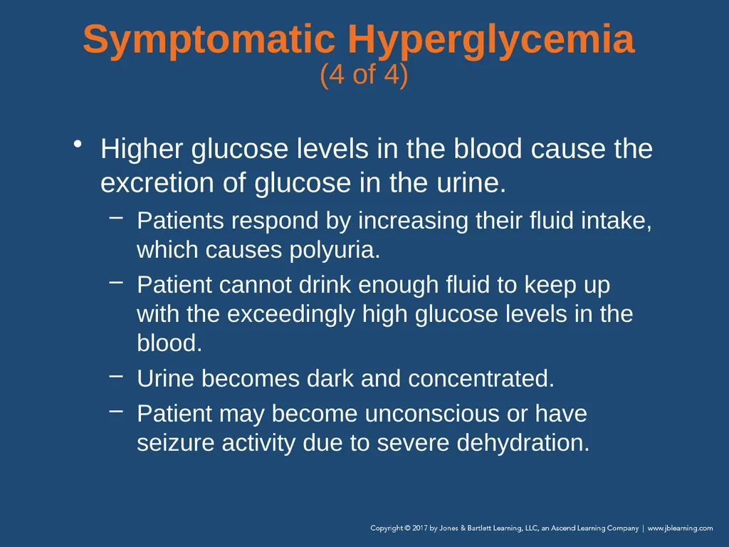
(4 of 4) Higher glucose levels in the blood cause the excretion of glucose in the urine.
Patients respond by increasing their fluid intake, which causes polyuria.
Patient cannot drink enough fluid to keep up with the exceedingly high glucose levels in the blood.
Urine becomes dark and concentrated.
Patient may become unconscious or have seizure activity due to severe dehydration.<br>
Patients respond by increasing their fluid intake, which causes polyuria.
Patient cannot drink enough fluid to keep up with the exceedingly high glucose levels in the blood.
Urine becomes dark and concentrated.
Patient may become unconscious or have seizure activity due to severe dehydration.<br>
27
Symptomatic Hypoglycemia
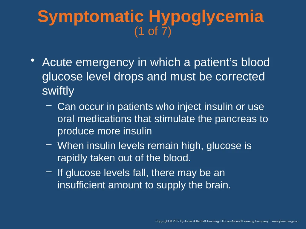
(1 of 7) Acute emergency in which a patient’s blood glucose level drops and must be corrected swiftly
Can occur in patients who inject insulin or use oral medications that stimulate the pancreas to produce more insulin
When insulin levels remain high, glucose is rapidly taken out of the blood.
If glucose levels fall, there may be an insufficient amount to supply the brain.<br>
Can occur in patients who inject insulin or use oral medications that stimulate the pancreas to produce more insulin
When insulin levels remain high, glucose is rapidly taken out of the blood.
If glucose levels fall, there may be an insufficient amount to supply the brain.<br>
28
Symptomatic Hypoglycemia
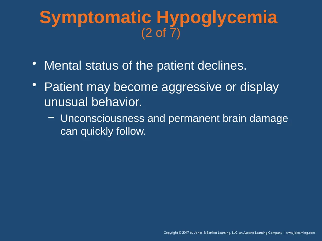
(2 of 7) Mental status of the patient declines.
Patient may become aggressive or display unusual behavior.
Unconsciousness and permanent brain damage can quickly follow.<br>
Patient may become aggressive or display unusual behavior.
Unconsciousness and permanent brain damage can quickly follow.<br>
29
Symptomatic Hypoglycemia
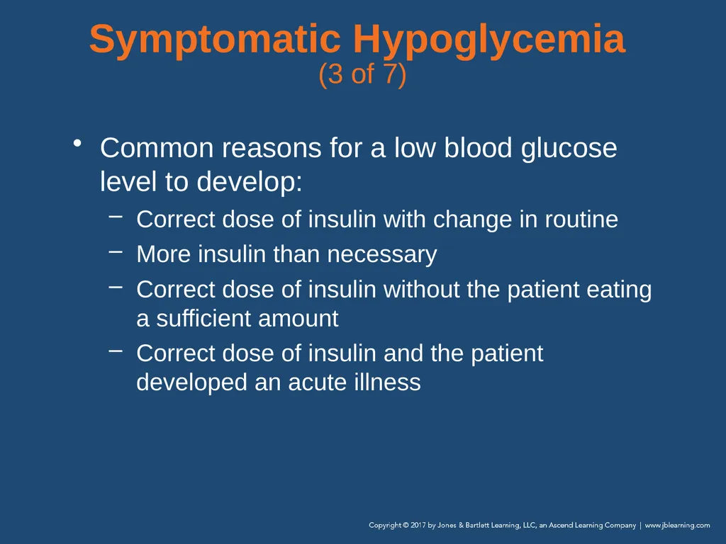
(3 of 7) Common reasons for a low blood glucose level to develop:
Correct dose of insulin with change in routine
More insulin than necessary
Correct dose of insulin without the patient eating a sufficient amount
Correct dose of insulin and the patient developed an acute illness<br>
Correct dose of insulin with change in routine
More insulin than necessary
Correct dose of insulin without the patient eating a sufficient amount
Correct dose of insulin and the patient developed an acute illness<br>
30
Symptomatic Hypoglycemia
(4 of 7) Hypoglycemia develops much more quickly than hyperglycemia.
In some instances, it can occur in a matter of minutes.<br>
In some instances, it can occur in a matter of minutes.<br>
31
Symptomatic Hypoglycemia
(5 of 7) Signs and symptoms of hypoglycemia:
Normal to shallow or rapid respirations
Pale, moist skin
Diaphoresis
Dizziness, headache
Rapid pulse
Normal to low blood pressure<br>
Normal to shallow or rapid respirations
Pale, moist skin
Diaphoresis
Dizziness, headache
Rapid pulse
Normal to low blood pressure<br>
32
Symptomatic Hypoglycemia
(6 of 7) Signs and symptoms of hypoglycemia (cont’d):
Altered mental status
Anxious or combative behavior
Seizure, fainting, or coma
Weakness on one side of the body
Rapid changes in mental status<br>
Altered mental status
Anxious or combative behavior
Seizure, fainting, or coma
Weakness on one side of the body
Rapid changes in mental status<br>
33
Symptomatic Hypoglycemia
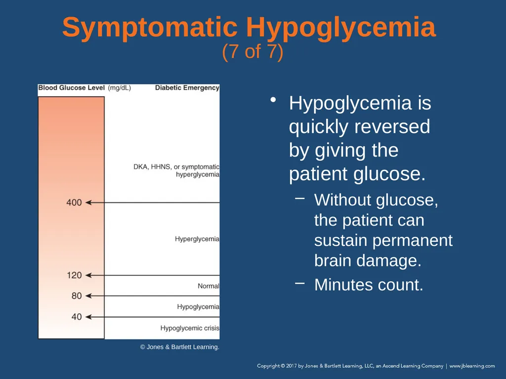
(7 of 7) Hypoglycemia is quickly reversed by giving the patient glucose.
Without glucose, the patient can sustain permanent brain damage.
Minutes count. © Jones & Bartlett Learning.<br>
Without glucose, the patient can sustain permanent brain damage.
Minutes count. © Jones & Bartlett Learning.<br>
34
Scene Size-up Scene safety
Patients with diabetes may use syringes.
Be alert for clues.
Use standard precautions.
Question bystanders on events leading to your arrival.
Keep open the possibility that trauma may have occurred.
Determine MOI/NOI.<br>
Patients with diabetes may use syringes.
Be alert for clues.
Use standard precautions.
Question bystanders on events leading to your arrival.
Keep open the possibility that trauma may have occurred.
Determine MOI/NOI.<br>
35
Primary Assessment (1 of 4) Form a general impression.
Airway and breathing
Patients showing signs of inadequate breathing, a pulse oximetry level less than or equal to 94%, or altered mental status should receive high-flow oxygen (12 to 15 L/min via nonrebreathing mask).<br>
Airway and breathing
Patients showing signs of inadequate breathing, a pulse oximetry level less than or equal to 94%, or altered mental status should receive high-flow oxygen (12 to 15 L/min via nonrebreathing mask).<br>
36
Primary Assessment (2 of 4) Airway and breathing (cont’d)
Hyperglycemic patients may have Kussmaul respirations and sweet, fruity breath.
Hypoglycemic patients will have normal or shallow to rapid respirations.
Manage respiratory distress.<br>
Hyperglycemic patients may have Kussmaul respirations and sweet, fruity breath.
Hypoglycemic patients will have normal or shallow to rapid respirations.
Manage respiratory distress.<br>
37
Primary Assessment (3 of 4) Circulation
Dry, warm skin: hyperglycemia
Moist, pale skin: hypoglycemia
Rapid, weak pulse: symptomatic hypoglycemia<br>
Dry, warm skin: hyperglycemia
Moist, pale skin: hypoglycemia
Rapid, weak pulse: symptomatic hypoglycemia<br>
38
Primary Assessment (4 of 4) Transport decision
Provide prompt transport for patients with altered mental status and inability to swallow.
Further evaluate conscious patients capable of swallowing and able to maintain airway.<br>
Provide prompt transport for patients with altered mental status and inability to swallow.
Further evaluate conscious patients capable of swallowing and able to maintain airway.<br>
39
History Taking (1 of 3) Investigate chief complaint
Obtain history of present illness from responsive patient, family, or bystanders.
If patient has eaten but not taken insulin, hyperglycemia is more likely.<br>
Obtain history of present illness from responsive patient, family, or bystanders.
If patient has eaten but not taken insulin, hyperglycemia is more likely.<br>
40
History Taking (2 of 3) Investigate chief complaint (cont’d)
If patient has taken insulin but not eaten, hypoglycemia is more likely.
Carefully observe signs and symptoms; determine whether hypo- or hyperglycemic.<br>
If patient has taken insulin but not eaten, hypoglycemia is more likely.
Carefully observe signs and symptoms; determine whether hypo- or hyperglycemic.<br>
41
History Taking (3 of 3) SAMPLE history—ask the patient:
Do you take insulin or pills to lower blood sugar?
Do you wear an insulin pump? Is it working properly?
Have you taken your usual insulin dose (or pills) today?
Have you eaten normally today?
Have you had any illnesses, unusual amount of activity, or stress?<br>
Do you take insulin or pills to lower blood sugar?
Do you wear an insulin pump? Is it working properly?
Have you taken your usual insulin dose (or pills) today?
Have you eaten normally today?
Have you had any illnesses, unusual amount of activity, or stress?<br>
42
Secondary Assessment (1 of 3) Physical examination
Assess unresponsive patients from head to toe.
When you suspect a diabetes-related problem, focus on mental status, ability to swallow, and ability to protect airway.<br>
Assess unresponsive patients from head to toe.
When you suspect a diabetes-related problem, focus on mental status, ability to swallow, and ability to protect airway.<br>
43
Secondary Assessment (2 of 3) Vital signs
Use a glucometer, if available and protocols allow.
Hypoglycemia: Respirations are normal to rapid, pulse is weak and rapid, and skin is typically pale and clammy with a low blood pressure
Hyperglycemia: Respirations may be deep and rapid; pulse may be rapid, weak, and thready; and skin may be warm and dry with a normal blood pressure<br>
Use a glucometer, if available and protocols allow.
Hypoglycemia: Respirations are normal to rapid, pulse is weak and rapid, and skin is typically pale and clammy with a low blood pressure
Hyperglycemia: Respirations may be deep and rapid; pulse may be rapid, weak, and thready; and skin may be warm and dry with a normal blood pressure<br>
44
Secondary Assessment (3 of 3) Portable glucometer
Study the operator’s manual for proper use in the field.
Know the upper and lower ranges at which your glucometer functions.
Normal nonfasting adult and child blood glucose level range: 80 to 120 mg/dL; neonates should be above 70 mg/dL<br>
Study the operator’s manual for proper use in the field.
Know the upper and lower ranges at which your glucometer functions.
Normal nonfasting adult and child blood glucose level range: 80 to 120 mg/dL; neonates should be above 70 mg/dL<br>
45
Reassessment (1 of 4) Reassess frequently.
Provide indicated interventions.
Hypoglycemic, conscious, can swallow:
Encourage patient to take glucose tablets or drink juice containing sugar.
Administer highly concentrated sugar gel (if protocols allow).
Provide rapid transport.<br>
Provide indicated interventions.
Hypoglycemic, conscious, can swallow:
Encourage patient to take glucose tablets or drink juice containing sugar.
Administer highly concentrated sugar gel (if protocols allow).
Provide rapid transport.<br>
46
Reassessment (2 of 4) Interventions (cont’d)
Hypoglycemic, unconscious, risk of aspiration:
Patient needs intravenous (IV) glucose or intramuscular (IM or IN) glucagon (beyond EMT competencies).
When in doubt, consult medical control.<br>
Hypoglycemic, unconscious, risk of aspiration:
Patient needs intravenous (IV) glucose or intramuscular (IM or IN) glucagon (beyond EMT competencies).
When in doubt, consult medical control.<br>
47
Reassessment (3 of 4) If unable to test for a blood glucose value:
Perform a thorough assessment.
Contact the hospital to help sort out the signs and symptoms.<br>
Perform a thorough assessment.
Contact the hospital to help sort out the signs and symptoms.<br>
48
Reassessment (4 of 4) Communication and documentation
Coordinate communication and documentation.
Inform receiving hospital about the patient’s history, the present situation, your assessment findings, and your interventions and their results.
Patients who refuse transport after symptoms improve may require even more through documentation.<br>
Coordinate communication and documentation.
Inform receiving hospital about the patient’s history, the present situation, your assessment findings, and your interventions and their results.
Patients who refuse transport after symptoms improve may require even more through documentation.<br>
49
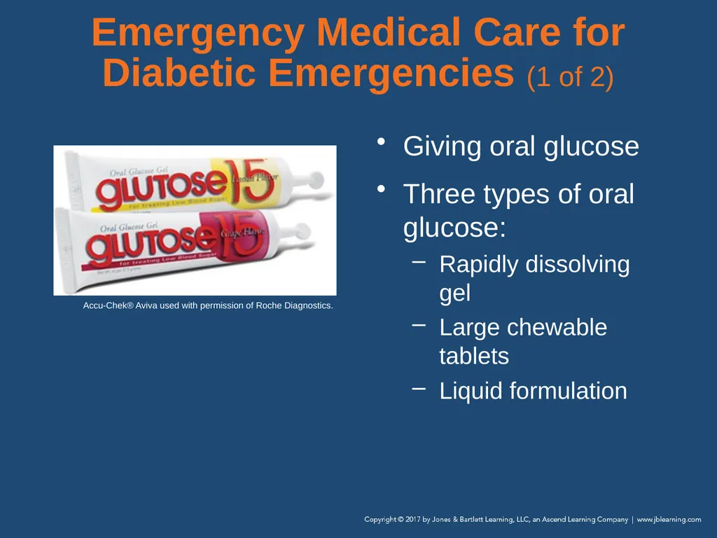
Emergency Medical Care for Diabetic Emergencies (1 of 2) Giving oral glucose
Three types of oral glucose:
Rapidly dissolving gel
Large chewable tablets
Liquid formulation Accu-Chek® Aviva used with permission of Roche Diagnostics.<br>
Three types of oral glucose:
Rapidly dissolving gel
Large chewable tablets
Liquid formulation Accu-Chek® Aviva used with permission of Roche Diagnostics.<br>
50
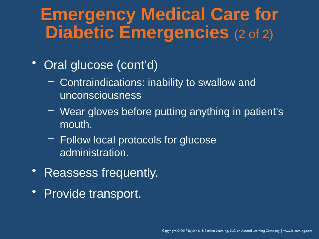
Emergency Medical Care for Diabetic Emergencies (2 of 2) Oral glucose (cont’d)
Contraindications: inability to swallow and unconsciousness
Wear gloves before putting anything in patient’s mouth.
Follow local protocols for glucose administration.
Reassess frequently.
Provide transport.<br>
Contraindications: inability to swallow and unconsciousness
Wear gloves before putting anything in patient’s mouth.
Follow local protocols for glucose administration.
Reassess frequently.
Provide transport.<br>
51
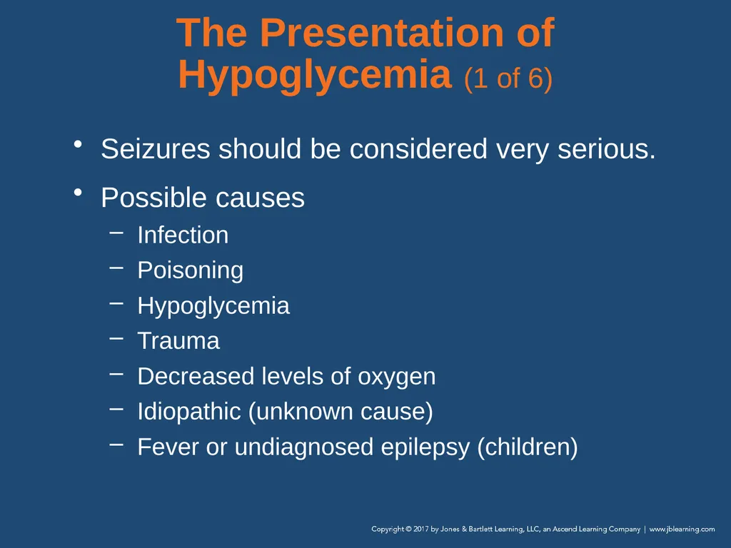
The Presentation of Hypoglycemia (1 of 6) Seizures should be considered very serious.
Possible causes
Infection
Poisoning
Hypoglycemia
Trauma
Decreased levels of oxygen
Idiopathic (unknown cause)
Fever or undiagnosed epilepsy (children)<br>
Possible causes
Infection
Poisoning
Hypoglycemia
Trauma
Decreased levels of oxygen
Idiopathic (unknown cause)
Fever or undiagnosed epilepsy (children)<br>
52
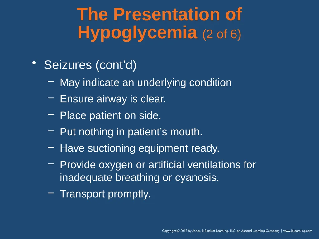
The Presentation of Hypoglycemia (2 of 6) Seizures (cont’d)
May indicate an underlying condition
Ensure airway is clear.
Place patient on side.
Put nothing in patient’s mouth.
Have suctioning equipment ready.
Provide oxygen or artificial ventilations for inadequate breathing or cyanosis.
Transport promptly.<br>
May indicate an underlying condition
Ensure airway is clear.
Place patient on side.
Put nothing in patient’s mouth.
Have suctioning equipment ready.
Provide oxygen or artificial ventilations for inadequate breathing or cyanosis.
Transport promptly.<br>
53
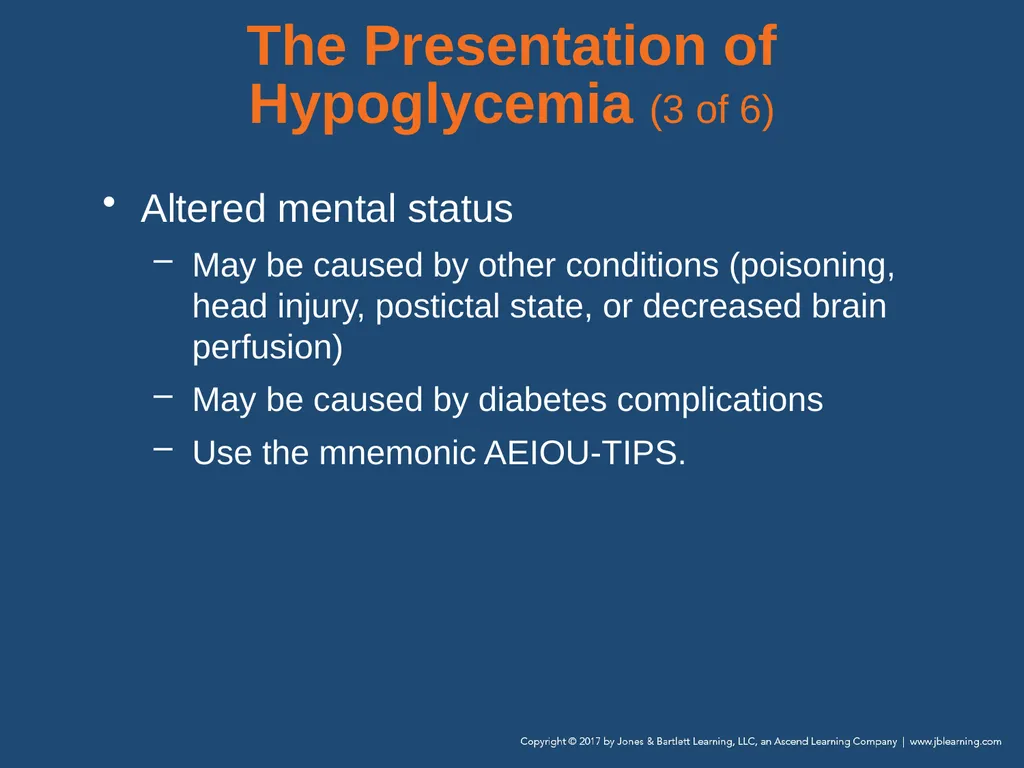
The Presentation of Hypoglycemia (3 of 6) Altered mental status
May be caused by other conditions (poisoning, head injury, postictal state, or decreased brain perfusion)
May be caused by diabetes complications
Use the mnemonic AEIOU-TIPS.<br>
May be caused by other conditions (poisoning, head injury, postictal state, or decreased brain perfusion)
May be caused by diabetes complications
Use the mnemonic AEIOU-TIPS.<br>
54
The Presentation of Hypoglycemia (4 of 6) Altered mental status (cont’d)
Ensure airway is clear.
Be prepared to provide artificial ventilations and suctioning if patient vomits.
Provide prompt transport.
Misdiagnosis of neurologic dysfunction
Symptoms mistaken for intoxication<br>
Ensure airway is clear.
Be prepared to provide artificial ventilations and suctioning if patient vomits.
Provide prompt transport.
Misdiagnosis of neurologic dysfunction
Symptoms mistaken for intoxication<br>
55
The Presentation of Hypoglycemia (5 of 6) Misdiagnosis (cont’d)
A diabetic patient confined by police is at risk.
Look for emergency medical identification bracelet, necklace, or card.
Perform blood glucose test at scene (if protocols allow) or ED.
Diabetes and alcoholism can coexist in a patient.<br>
A diabetic patient confined by police is at risk.
Look for emergency medical identification bracelet, necklace, or card.
Perform blood glucose test at scene (if protocols allow) or ED.
Diabetes and alcoholism can coexist in a patient.<br>
56
The Presentation of Hypoglycemia (6 of 6) Relationship to airway management
Patients with altered mental status can lose gag reflex.
Vomit or tongue may obstruct airway.
Carefully monitor airway.
Place patient in lateral recumbent position.
Make sure suction is available.<br>
Patients with altered mental status can lose gag reflex.
Vomit or tongue may obstruct airway.
Carefully monitor airway.
Place patient in lateral recumbent position.
Make sure suction is available.<br>
57
Hematologic Emergencies Hematology is the study of blood-related diseases.
Three disorders that can create a prehospital emergency:
Sickle cell disease
Hemophilia A
Thrombophilia<br>
Three disorders that can create a prehospital emergency:
Sickle cell disease
Hemophilia A
Thrombophilia<br>
58
Anatomy and Physiology Blood is made up of four components.
Each serves a purpose.
Red blood cells contain hemoglobin, which carries oxygen to the tissues.
White blood cells collect dead cells and provide for their correct disposal.
Platelets are essential for clot formation.
Plasma serves as the transportation medium.<br>
Each serves a purpose.
Red blood cells contain hemoglobin, which carries oxygen to the tissues.
White blood cells collect dead cells and provide for their correct disposal.
Platelets are essential for clot formation.
Plasma serves as the transportation medium.<br>
59
Pathophysiology (1 of 13) Sickle cell disease
Inherited disorder, affects red blood cells
Predominantly in people of African, Caribbean, and South American ancestry
People with sickle cell disease have misshapen RBCs that lead to dysfunction in oxygen binding and unintentional clot formation.
Clots may result in a blockage known as vasoocclusive crisis.
Can result in hypoxia, pain, and organ damage<br>
Inherited disorder, affects red blood cells
Predominantly in people of African, Caribbean, and South American ancestry
People with sickle cell disease have misshapen RBCs that lead to dysfunction in oxygen binding and unintentional clot formation.
Clots may result in a blockage known as vasoocclusive crisis.
Can result in hypoxia, pain, and organ damage<br>
60
Pathophysiology (2 of 13) Sickle cell disease (cont’d)
Sickled cells have a short life span, resulting in more cellular waste products and contributing to sludging of the blood.
Complications include:
Anemia
Gallstones
Jaundice
Splenic dysfunction<br>
Sickled cells have a short life span, resulting in more cellular waste products and contributing to sludging of the blood.
Complications include:
Anemia
Gallstones
Jaundice
Splenic dysfunction<br>
61
Pathophysiology (3 of 13) Sickle cell disease (cont’d)
Vascular occlusion with ischemia:
Acute chest syndrome
Stroke
Joint necrosis
Pain crises
Acute and chronic organ dysfunction/failure
Retinal hemorrhages
Increased risk of infection<br>
Vascular occlusion with ischemia:
Acute chest syndrome
Stroke
Joint necrosis
Pain crises
Acute and chronic organ dysfunction/failure
Retinal hemorrhages
Increased risk of infection<br>
62
Pathophysiology (4 of 13) Sickle cell disease (cont’d)
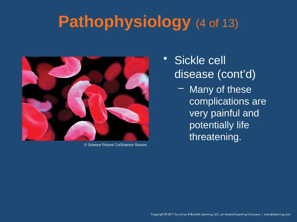
Many of these complications are very painful and potentially life threatening. © Science Picture Co/Science Source.<br>
Many of these complications are very painful and potentially life threatening. © Science Picture Co/Science Source.<br>
63
Pathophysiology (5 of 13) Clotting disorders—hemophilia
Rare: only about 20,000 Americans have the disorder.
Hemophilia A affects mostly males.<br>
Rare: only about 20,000 Americans have the disorder.
Hemophilia A affects mostly males.<br>
64
Pathophysiology (6 of 13) Clotting disorders—hemophilia (cont’d)
Decreased ability to create a clot after an injury, which can be life threatening
Patients typically have intravenous factor VIII replacement infusions, which help the blood clot, either close at hand or with them.<br>
Decreased ability to create a clot after an injury, which can be life threatening
Patients typically have intravenous factor VIII replacement infusions, which help the blood clot, either close at hand or with them.<br>
65
Pathophysiology (7 of 13) Clotting disorders—hemophilia (cont’d)
Common complications of hemophilia A include:
Long-term joint problems that may require a joint replacement
Bleeding in the brain (intracerebral hemorrhage)
Thrombosis due to treatment<br>
Common complications of hemophilia A include:
Long-term joint problems that may require a joint replacement
Bleeding in the brain (intracerebral hemorrhage)
Thrombosis due to treatment<br>
66
Pathophysiology (8 of 13) Clotting disorders—thrombophilia
Disorder in the body’s ability to maintain the smooth flow of blood through the venous and arterial systems
Concentration of particular elements in the blood creates clogging or blockage issues.<br>
Disorder in the body’s ability to maintain the smooth flow of blood through the venous and arterial systems
Concentration of particular elements in the blood creates clogging or blockage issues.<br>
67
Pathophysiology (9 of 13) Clotting disorders—thrombophilia (cont’d)
General term for conditions that result in blood clotting more easily than normal
Inherited (genetic) disorders
Medications or other factors
Patients with cancer
Clots can spontaneously develop in the blood of the patient.<br>
General term for conditions that result in blood clotting more easily than normal
Inherited (genetic) disorders
Medications or other factors
Patients with cancer
Clots can spontaneously develop in the blood of the patient.<br>
68
Pathophysiology (10 of 13) Clotting disorders—deep vein thrombosis (DVT)
Common medical problem in sedentary patients and in patients who have had recent injury or surgery
You may encounter several methods prevent blood clot formation, including:
Blood-thinning medications
Compression stockings
Mechanical devices<br>
Common medical problem in sedentary patients and in patients who have had recent injury or surgery
You may encounter several methods prevent blood clot formation, including:
Blood-thinning medications
Compression stockings
Mechanical devices<br>
69
Pathophysiology (11 of 13) Clotting disorders—DVT (cont’d)
Risk factors include
Recent history of joint replacement who complains of leg swelling
Travelers, truck and long-distance bus drivers
Bedridden nursing home patients<br>
Risk factors include
Recent history of joint replacement who complains of leg swelling
Travelers, truck and long-distance bus drivers
Bedridden nursing home patients<br>
70
Pathophysiology (12 of 13) Clotting disorders—DVT (cont’d)
Treatment
Anticoagulation therapy
Medications are typically administered for at least 3 months after diagnosis of a DVT.
Patients prescribed medications to treat DVT are at increased risk of bleeding complications.<br>
Treatment
Anticoagulation therapy
Medications are typically administered for at least 3 months after diagnosis of a DVT.
Patients prescribed medications to treat DVT are at increased risk of bleeding complications.<br>
71
Pathophysiology (13 of 13) Clotting disorders—DVT (cont’d)
A clot from the DVT can travel from the patient’s lower extremity to the lung, causing a pulmonary embolus.
Pulmonary emboli can cause chest pain, difficulty breathing, or sudden cardiac arrest.<br>
A clot from the DVT can travel from the patient’s lower extremity to the lung, causing a pulmonary embolus.
Pulmonary emboli can cause chest pain, difficulty breathing, or sudden cardiac arrest.<br>
72
Scene Size-up Scene safety
Most sickle cell patients will have had a crisis before.
Wear gloves and eye protection at a minimum.
Consider ALS support.<br>
Most sickle cell patients will have had a crisis before.
Wear gloves and eye protection at a minimum.
Consider ALS support.<br>
73
Primary Assessment (1 of 3) Is the patient in pain and of African American or Mediterranean descent?
Perform cervical spine immobilization, if necessary.
Form a general impression.
Airway and breathing
Inadequate breathing or altered mental status:
High-flow oxygen at 12 to 15 L/min via nonrebreathing mask<br>
Perform cervical spine immobilization, if necessary.
Form a general impression.
Airway and breathing
Inadequate breathing or altered mental status:
High-flow oxygen at 12 to 15 L/min via nonrebreathing mask<br>
74
Primary Assessment (2 of 3) Airway and breathing (cont’d)
Sickle cell crisis patients may have increased respirations or signs of pneumonia.
Manage respiratory distress.<br>
Sickle cell crisis patients may have increased respirations or signs of pneumonia.
Manage respiratory distress.<br>
75
Primary Assessment (3 of 3) Circulation
Sickle cell patients: increased heart rate
Suspected hemophilia patients:
Be alert for signs of acute blood loss.
Note bleeding of unknown origin.
Be alert for signs of hypoxia.
Make a transport decision.
Transport to an ED is recommended for any patient with sickle cell crisis or hemophilia.<br>
Sickle cell patients: increased heart rate
Suspected hemophilia patients:
Be alert for signs of acute blood loss.
Note bleeding of unknown origin.
Be alert for signs of hypoxia.
Make a transport decision.
Transport to an ED is recommended for any patient with sickle cell crisis or hemophilia.<br>
76
History Taking (1 of 3) Investigate chief complaint.
Obtain history of present illness from responsive patients, family, or bystanders.
Physical signs indicating sickle cell crisis:
Swelling of fingers and toes
Priapism
Jaundice<br>
Obtain history of present illness from responsive patients, family, or bystanders.
Physical signs indicating sickle cell crisis:
Swelling of fingers and toes
Priapism
Jaundice<br>
77
History Taking (2 of 3) Ask about:
Single location or felt throughout body?
Visual disturbances?
Nausea, vomiting, or abdominal cramping?
Chest pain or shortness of breath?<br>
Single location or felt throughout body?
Visual disturbances?
Nausea, vomiting, or abdominal cramping?
Chest pain or shortness of breath?<br>
78
History Taking (3 of 3) Obtain SAMPLE history from responsive patient or family member.
Have you had a crisis before?
When was the last time you had a crisis?
How did your last crisis resolve?
Recent illness, unusual amount of activity, or stress?<br>
Have you had a crisis before?
When was the last time you had a crisis?
How did your last crisis resolve?
Recent illness, unusual amount of activity, or stress?<br>
79
Secondary Assessment Physical examination
Focus on major joints.
Evaluate and document mental status using (AVPU).
Vital signs
Obtain complete set of vital signs.
Look for signs of sickle cell crisis.
Use pulse oximeter, if available.<br>
Focus on major joints.
Evaluate and document mental status using (AVPU).
Vital signs
Obtain complete set of vital signs.
Look for signs of sickle cell crisis.
Use pulse oximeter, if available.<br>
80
Reassessment (1 of 2) Reassess vital signs frequently.
Evaluate interventions.
Adjust or change the interventions as needed.
Document each assessment.
Administer supplemental oxygen.
Hospital care for sickle cell crisis:
Analgesics, penicillin, IV fluid, blood transfusion<br>
Evaluate interventions.
Adjust or change the interventions as needed.
Document each assessment.
Administer supplemental oxygen.
Hospital care for sickle cell crisis:
Analgesics, penicillin, IV fluid, blood transfusion<br>
81
Reassessment (2 of 2) Hospital care for hemophilia:
IV therapy (for hypotension)
Transfusion of plasma
Communicate with hospital staff for continuity of care and document clearly.<br>
IV therapy (for hypotension)
Transfusion of plasma
Communicate with hospital staff for continuity of care and document clearly.<br>
82
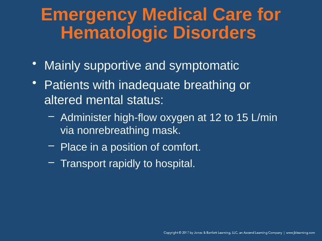
Emergency Medical Care for Hematologic Disorders Mainly supportive and symptomatic
Patients with inadequate breathing or altered mental status:
Administer high-flow oxygen at 12 to 15 L/min via nonrebreathing mask.
Place in a position of comfort.
Transport rapidly to hospital.<br>
Patients with inadequate breathing or altered mental status:
Administer high-flow oxygen at 12 to 15 L/min via nonrebreathing mask.
Place in a position of comfort.
Transport rapidly to hospital.<br>
83
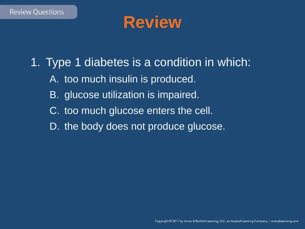
Review Type 1 diabetes is a condition in which:
too much insulin is produced.
glucose utilization is impaired.
too much glucose enters the cell.
the body does not produce glucose.<br>
too much insulin is produced.
glucose utilization is impaired.
too much glucose enters the cell.
the body does not produce glucose.<br>
84
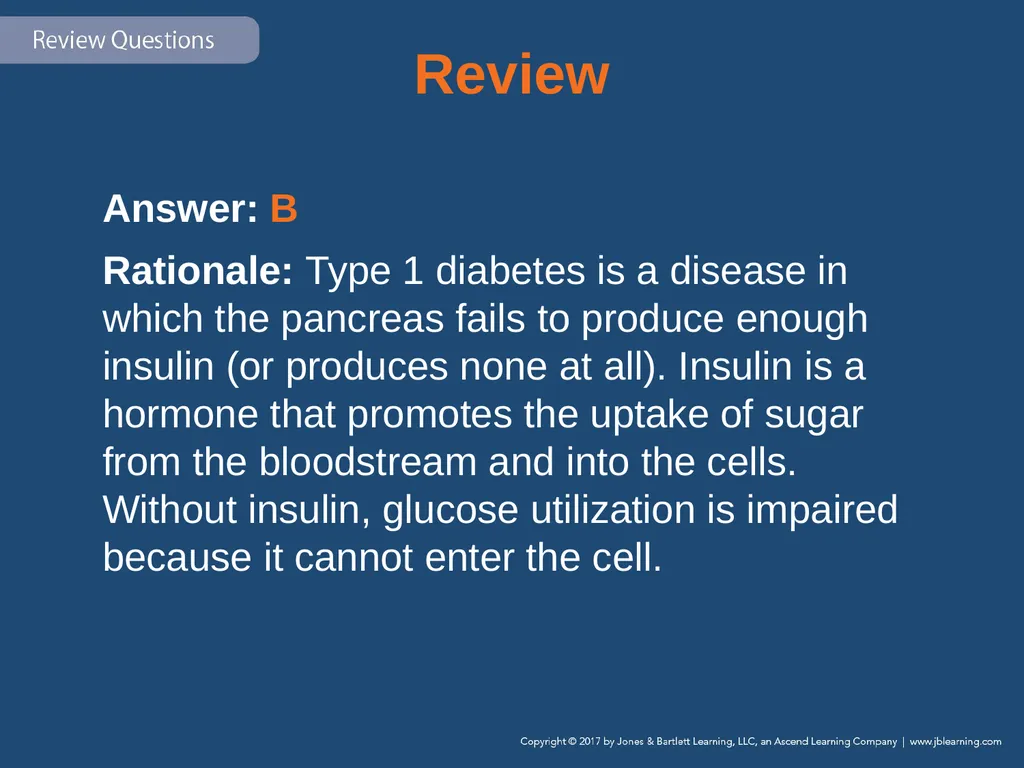
Review Answer: B
Rationale: Type 1 diabetes is a disease in which the pancreas fails to produce enough insulin (or produces none at all). Insulin is a hormone that promotes the uptake of sugar from the bloodstream and into the cells. Without insulin, glucose utilization is impaired because it cannot enter the cell.<br>
Rationale: Type 1 diabetes is a disease in which the pancreas fails to produce enough insulin (or produces none at all). Insulin is a hormone that promotes the uptake of sugar from the bloodstream and into the cells. Without insulin, glucose utilization is impaired because it cannot enter the cell.<br>
85
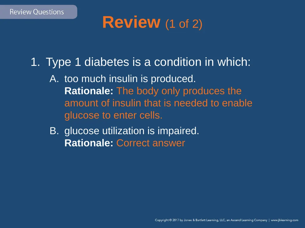
Review (1 of 2) Type 1 diabetes is a condition in which:
too much insulin is produced. Rationale: The body only produces the amount of insulin that is needed to enable glucose to enter cells.
glucose utilization is impaired. Rationale: Correct answer<br>
too much insulin is produced. Rationale: The body only produces the amount of insulin that is needed to enable glucose to enter cells.
glucose utilization is impaired. Rationale: Correct answer<br>
86
Review (2 of 2) Type 1 diabetes is a condition in which:
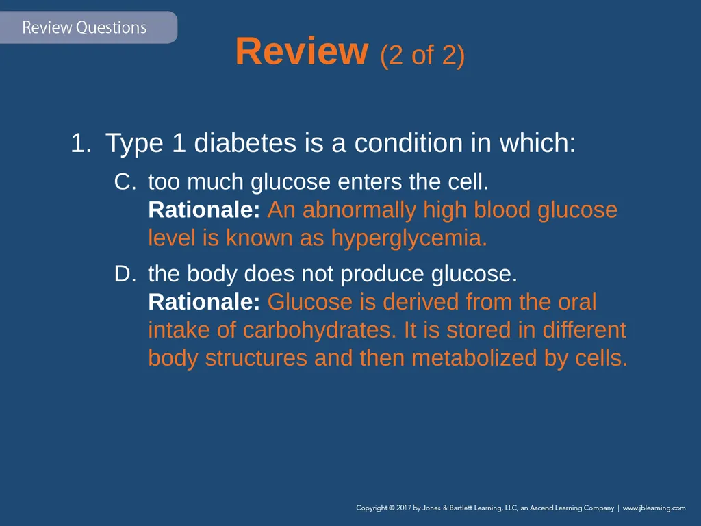
too much glucose enters the cell. Rationale: An abnormally high blood glucose level is known as hyperglycemia.
the body does not produce glucose. Rationale: Glucose is derived from the oral intake of carbohydrates. It is stored in different body structures and then metabolized by cells.<br>
too much glucose enters the cell. Rationale: An abnormally high blood glucose level is known as hyperglycemia.
the body does not produce glucose. Rationale: Glucose is derived from the oral intake of carbohydrates. It is stored in different body structures and then metabolized by cells.<br>
87
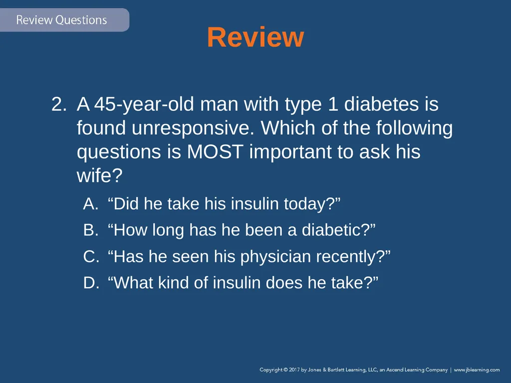
Review A 45-year-old man with type 1 diabetes is found unresponsive. Which of the following questions is MOST important to ask his wife?
“Did he take his insulin today?”
“How long has he been a diabetic?”
“Has he seen his physician recently?”
“What kind of insulin does he take?”<br>
“Did he take his insulin today?”
“How long has he been a diabetic?”
“Has he seen his physician recently?”
“What kind of insulin does he take?”<br>
88
Review Answer: A
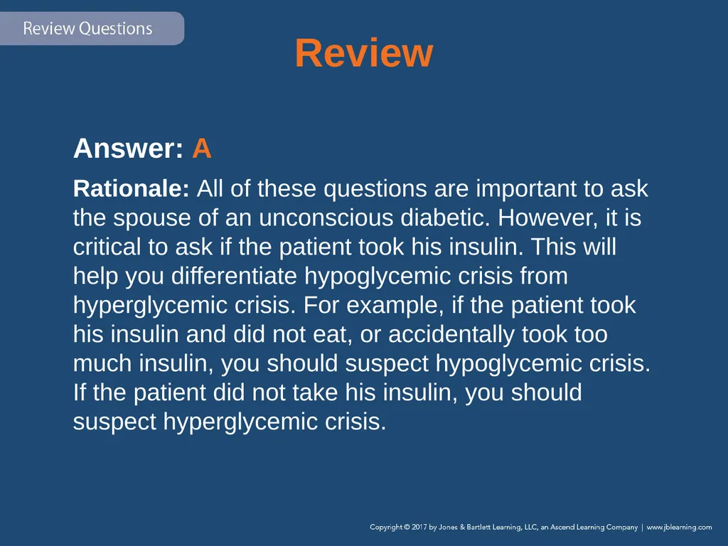
Rationale: All of these questions are important to ask the spouse of an unconscious diabetic. However, it is critical to ask if the patient took his insulin. This will help you differentiate hypoglycemic crisis from hyperglycemic crisis. For example, if the patient took his insulin and did not eat, or accidentally took too much insulin, you should suspect hypoglycemic crisis. If the patient did not take his insulin, you should suspect hyperglycemic crisis.<br>
Rationale: All of these questions are important to ask the spouse of an unconscious diabetic. However, it is critical to ask if the patient took his insulin. This will help you differentiate hypoglycemic crisis from hyperglycemic crisis. For example, if the patient took his insulin and did not eat, or accidentally took too much insulin, you should suspect hypoglycemic crisis. If the patient did not take his insulin, you should suspect hyperglycemic crisis.<br>
89
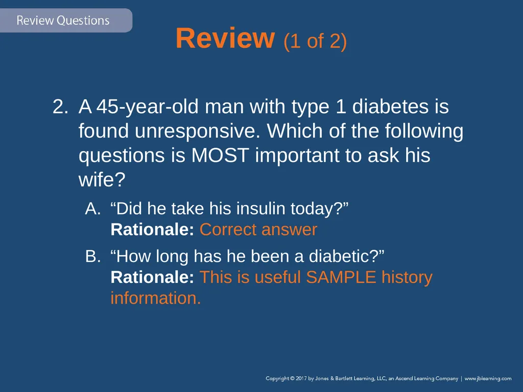
Review (1 of 2) A 45-year-old man with type 1 diabetes is found unresponsive. Which of the following questions is MOST important to ask his wife?
“Did he take his insulin today?” Rationale: Correct answer
“How long has he been a diabetic?” Rationale: This is useful SAMPLE history information.<br>
“Did he take his insulin today?” Rationale: Correct answer
“How long has he been a diabetic?” Rationale: This is useful SAMPLE history information.<br>
90
Review (2 of 2) A 45-year-old man with type 1 diabetes is found unresponsive. Which of the following questions is MOST important to ask his wife?
“Has he seen his physician recently?” Rationale: This is also important SAMPLE history information.
“What kind of insulin does he take?” Rationale: This provides important information about a patient’s medications.<br>
“Has he seen his physician recently?” Rationale: This is also important SAMPLE history information.
“What kind of insulin does he take?” Rationale: This provides important information about a patient’s medications.<br>
91
Review A diabetic patient presents with a blood glucose level of 310 mg/dL and severe dehydration. The patient’s dehydration is the result of:
excretion of glucose and water from the kidneys.
a deficiency of insulin that causes internal fluid loss.
an infection that often accompanies hyperglycemia.
an inability to produce energy because of insulin depletion.<br>
excretion of glucose and water from the kidneys.
a deficiency of insulin that causes internal fluid loss.
an infection that often accompanies hyperglycemia.
an inability to produce energy because of insulin depletion.<br>
92
Review Answer: A
Rationale: In severe hyperglycemia, the kidneys excrete excess glucose from the body. This process requires a large amount of water to accomplish; therefore, water is excreted with the glucose, resulting in dehydration.<br>
Rationale: In severe hyperglycemia, the kidneys excrete excess glucose from the body. This process requires a large amount of water to accomplish; therefore, water is excreted with the glucose, resulting in dehydration.<br>
93
Review (1 of 4) A diabetic patient presents with a blood glucose level of 310 mg/dL and severe dehydration. The patient’s dehydration is the result of:
excretion of glucose and water from the kidneys. Rationale: Correct answer<br>
excretion of glucose and water from the kidneys. Rationale: Correct answer<br>
94
Review (2 of 4) A diabetic patient presents with a blood glucose level of 310 mg/dL and severe dehydration. The patient’s dehydration is the result of:
a deficiency of insulin that causes internal fluid loss. Rationale: A lack of insulin will cause the glucose level to rise, and it is the glucose that causes the fluid loss.<br>
a deficiency of insulin that causes internal fluid loss. Rationale: A lack of insulin will cause the glucose level to rise, and it is the glucose that causes the fluid loss.<br>
95
Review (3 of 4) A diabetic patient presents with a blood glucose level of 310 mg/dL and severe dehydration. The patient’s dehydration is the result of:
an infection that often accompanies hyperglycemia. Rationale: An infection is an invasion of the body by an organism—glucose is not a foreign element.<br>
an infection that often accompanies hyperglycemia. Rationale: An infection is an invasion of the body by an organism—glucose is not a foreign element.<br>
96
Review (4 of 4) A diabetic patient presents with a blood glucose level of 310 mg/dL and severe dehydration. The patient’s dehydration is the result of:
an inability to produce energy because of insulin depletion. Rationale: A body’s inability to metabolize glucose does not cause a fever.<br>
an inability to produce energy because of insulin depletion. Rationale: A body’s inability to metabolize glucose does not cause a fever.<br>
97
Review Which combination of factors would MOST likely cause a hypoglycemic crisis in a diabetic patient?
Eating a meal and taking insulin
Skipping a meal and taking insulin
Eating a meal and not taking insulin
Skipping a meal and not taking insulin<br>
Eating a meal and taking insulin
Skipping a meal and taking insulin
Eating a meal and not taking insulin
Skipping a meal and not taking insulin<br>
98
Review Answer: B
Rationale: The combination that would most likely cause a hypoglycemic crisis is skipping a meal and taking insulin. The patient will use up all available glucose in the bloodstream and become hypoglycemic. Left untreated, hypoglycemic crisis may cause permanent brain damage or even death.<br>
Rationale: The combination that would most likely cause a hypoglycemic crisis is skipping a meal and taking insulin. The patient will use up all available glucose in the bloodstream and become hypoglycemic. Left untreated, hypoglycemic crisis may cause permanent brain damage or even death.<br>
99
Review (1 of 2) Which combination of factors would MOST likely cause a hypoglycemic crisis in a diabetic patient?
Eating a meal and taking insulin Rationale: This process will maintain the body’s glucose level.
Skipping a meal and taking insulin Rationale: Correct answer<br>
Eating a meal and taking insulin Rationale: This process will maintain the body’s glucose level.
Skipping a meal and taking insulin Rationale: Correct answer<br>
100
Review (2 of 2) Which combination of factors would MOST likely cause a hypoglycemic crisis in a diabetic patient?
Eating a meal and not taking insulin Rationale: Eating will cause the glucose levels to rise.
Skipping a meal and not taking insulin Rationale: Glucose levels should remain the same but may be influenced by the patient’s metabolic rate or physical activities. This does not cause a hypoglycemic crisis.<br>
Eating a meal and not taking insulin Rationale: Eating will cause the glucose levels to rise.
Skipping a meal and not taking insulin Rationale: Glucose levels should remain the same but may be influenced by the patient’s metabolic rate or physical activities. This does not cause a hypoglycemic crisis.<br>
101
Review A 19-year-old diabetic male was found unresponsive on the couch by his roommate. After confirming that the patient is unresponsive, you should:
suction his oropharynx.
manually open his airway.
administer high-flow oxygen.
begin assisting his ventilations.<br>
suction his oropharynx.
manually open his airway.
administer high-flow oxygen.
begin assisting his ventilations.<br>
102
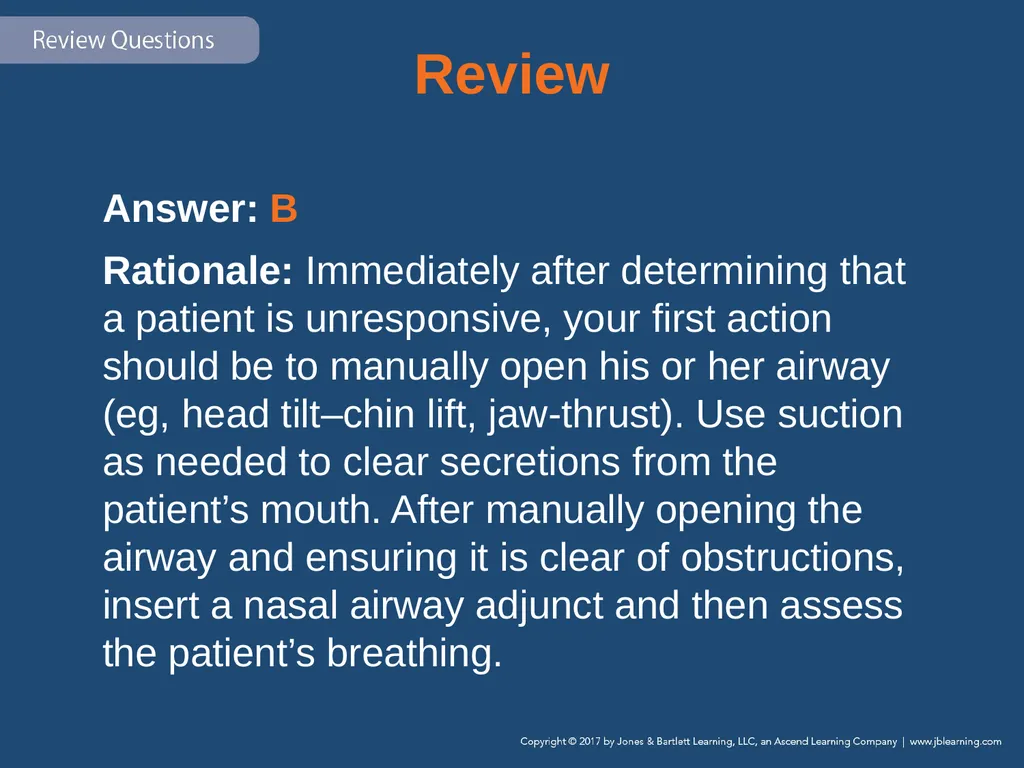
Review Answer: B
Rationale: Immediately after determining that a patient is unresponsive, your first action should be to manually open his or her airway (eg, head tilt–chin lift, jaw-thrust). Use suction as needed to clear secretions from the patient’s mouth. After manually opening the airway and ensuring it is clear of obstructions, insert a nasal airway adjunct and then assess the patient’s breathing.<br>
Rationale: Immediately after determining that a patient is unresponsive, your first action should be to manually open his or her airway (eg, head tilt–chin lift, jaw-thrust). Use suction as needed to clear secretions from the patient’s mouth. After manually opening the airway and ensuring it is clear of obstructions, insert a nasal airway adjunct and then assess the patient’s breathing.<br>
103
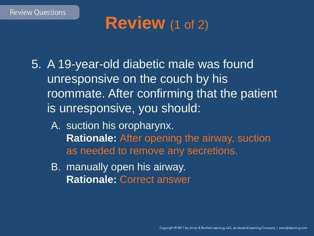
Review (1 of 2) A 19-year-old diabetic male was found unresponsive on the couch by his roommate. After confirming that the patient is unresponsive, you should:
suction his oropharynx. Rationale: After opening the airway, suction as needed to remove any secretions.
manually open his airway. Rationale: Correct answer<br>
suction his oropharynx. Rationale: After opening the airway, suction as needed to remove any secretions.
manually open his airway. Rationale: Correct answer<br>
104
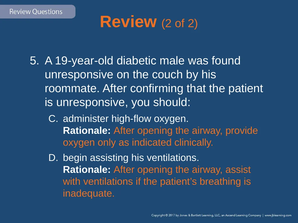
Review (2 of 2) A 19-year-old diabetic male was found unresponsive on the couch by his roommate. After confirming that the patient is unresponsive, you should:
administer high-flow oxygen. Rationale: After opening the airway, provide oxygen only as indicated clinically.
begin assisting his ventilations. Rationale: After opening the airway, assist with ventilations if the patient’s breathing is inadequate.<br>
administer high-flow oxygen. Rationale: After opening the airway, provide oxygen only as indicated clinically.
begin assisting his ventilations. Rationale: After opening the airway, assist with ventilations if the patient’s breathing is inadequate.<br>
105
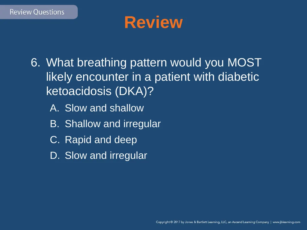
Review What breathing pattern would you MOST likely encounter in a patient with diabetic ketoacidosis (DKA)?
Slow and shallow
Shallow and irregular
Rapid and deep
Slow and irregular<br>
Slow and shallow
Shallow and irregular
Rapid and deep
Slow and irregular<br>
106
Review Answer: C
Rationale: Kussmaul respirations—a rapid and deep breathing pattern seen in patients with DKA—indicates that the body is attempting to eliminate ketones via the respiratory system. A fruity or acetone breath odor is usually present in patients with Kussmaul respirations.<br>
Rationale: Kussmaul respirations—a rapid and deep breathing pattern seen in patients with DKA—indicates that the body is attempting to eliminate ketones via the respiratory system. A fruity or acetone breath odor is usually present in patients with Kussmaul respirations.<br>
107
Review (1 of 2) What breathing pattern would you MOST likely encounter in a patient with diabetic ketoacidosis (DKA)?
Slow and shallow Rationale: Agonal respirations are seen with cerebral anoxia and may have an occasional gasp.
Shallow and irregular Rationale: Agonal respirations are seen with cerebral anoxia and may have an occasional gasp.<br>
Slow and shallow Rationale: Agonal respirations are seen with cerebral anoxia and may have an occasional gasp.
Shallow and irregular Rationale: Agonal respirations are seen with cerebral anoxia and may have an occasional gasp.<br>
108
Review (2 of 2) What breathing pattern would you MOST likely encounter in a patient with diabetic ketoacidosis (DKA)?
Rapid and deep Rationale: Correct answer
Slow and irregular Rationale: Slow and irregular respiration results from increased intracranial pressure and can also have periods of apnea.<br>
Rapid and deep Rationale: Correct answer
Slow and irregular Rationale: Slow and irregular respiration results from increased intracranial pressure and can also have periods of apnea.<br>
109
Review A woman called EMS because her 12-year-old son, who had been experiencing excessive urination, thirst, and hunger for the past 36 hours, has an altered mental status and is breathing fast. You should be MOST suspicious for:
low blood sugar.
hypoglycemia.
hypoglycemic crisis.
hyperglycemic crisis.<br>
low blood sugar.
hypoglycemia.
hypoglycemic crisis.
hyperglycemic crisis.<br>
110
Review Answer: D
Rationale: The child is experiencing a hyperglycemic crisis secondary to severe hyperglycemia. Hyperglycemic crisis is characterized by a slow onset and excessive urination (polyuria), thirst (polydipsia), and hunger (polyphagia). Other signs include rapid, deep breathing with a fruity or acetone breath odor (Kussmaul respirations); a rapid, thready pulse; and an altered mental status.<br>
Rationale: The child is experiencing a hyperglycemic crisis secondary to severe hyperglycemia. Hyperglycemic crisis is characterized by a slow onset and excessive urination (polyuria), thirst (polydipsia), and hunger (polyphagia). Other signs include rapid, deep breathing with a fruity or acetone breath odor (Kussmaul respirations); a rapid, thready pulse; and an altered mental status.<br>
111
Review (1 of 2) A woman called EMS because her 12-year-old son, who had been experiencing excessive urination, thirst, and hunger for the past
36 hours, has an altered mental status and is breathing fast. You should be MOST suspicious for:
low blood sugar. Rationale: Low blood sugar does not cause frequent urination.
hypoglycemia. Rationale: Hypoglycemia is low blood sugar.<br>
low blood sugar. Rationale: Low blood sugar does not cause frequent urination.
hypoglycemia. Rationale: Hypoglycemia is low blood sugar.<br>
112
Review (2 of 2) A woman called EMS because her 12-year-old son, who had been experiencing excessive urination, thirst, and hunger for the past
36 hours, has an altered mental status and is breathing fast. You should be MOST suspicious for:
hypoglycemic crisis. Rationale: Hypoglycemic crisis does not produce any of these symptoms.
hyperglycemic crisis. Rationale: Correct answer<br>
hypoglycemic crisis. Rationale: Hypoglycemic crisis does not produce any of these symptoms.
hyperglycemic crisis. Rationale: Correct answer<br>
113
Review If the cells do not receive glucose, they will begin to metabolize:
fat.
acid.
sugar.
ketones.<br>
fat.
acid.
sugar.
ketones.<br>
114
Review Answer: A
Rationale: If the body’s cells do not receive glucose, they will begin to metabolize the next most readily available substance—fat. Fat metabolism results in the production of ketoacids, which are released into the bloodstream (hence the term “ketoacidosis”).<br>
Rationale: If the body’s cells do not receive glucose, they will begin to metabolize the next most readily available substance—fat. Fat metabolism results in the production of ketoacids, which are released into the bloodstream (hence the term “ketoacidosis”).<br>
115
Review (1 of 2) If the cells do not receive glucose, they will begin to metabolize:
fat. Rationale: Correct answer
acid. Rationale: Fatty acids are a by-product (waste product) of the metabolism of fat.<br>
fat. Rationale: Correct answer
acid. Rationale: Fatty acids are a by-product (waste product) of the metabolism of fat.<br>
116
Review (2 of 2) If the cells do not receive glucose, they will begin to metabolize:
sugar. Rationale: Sugar is glucose.
ketones. Rationale: Ketones are a by-product (waste product) of the metabolism of fat.<br>
sugar. Rationale: Sugar is glucose.
ketones. Rationale: Ketones are a by-product (waste product) of the metabolism of fat.<br>
117
Review In contrast to a hyperglycemic crisis, a hypoglycemic crisis:
rarely presents with seizures.
presents over a period of hours to days.
should not routinely be treated with glucose.
usually responds immediately after treatment.<br>
rarely presents with seizures.
presents over a period of hours to days.
should not routinely be treated with glucose.
usually responds immediately after treatment.<br>
118
Review Answer: D
Rationale: Hypoglycemic crisis usually responds immediately following treatment with glucose. Patients with hyperglycemic crisis generally respond to treatment gradually, within 6–12 hours following the appropriate treatment. Seizures can occur with both hyperglycemic crisis and hypoglycemic crisis, but are more common in patients with hypoglycemic crisis.<br>
Rationale: Hypoglycemic crisis usually responds immediately following treatment with glucose. Patients with hyperglycemic crisis generally respond to treatment gradually, within 6–12 hours following the appropriate treatment. Seizures can occur with both hyperglycemic crisis and hypoglycemic crisis, but are more common in patients with hypoglycemic crisis.<br>
119
Review (1 of 2) In contrast to a hyperglycemic crisis, a hypoglycemic crisis:
rarely presents with seizures. Rationale: Hypoglycemic crisis can produce seizures.
presents over a period of hours to days. Rationale: Hypoglycemic crisis has a rapid onset of symptoms (possible minutes).<br>
rarely presents with seizures. Rationale: Hypoglycemic crisis can produce seizures.
presents over a period of hours to days. Rationale: Hypoglycemic crisis has a rapid onset of symptoms (possible minutes).<br>
120
Review (2 of 2) In contrast to a hyperglycemic crisis, a hypoglycemic crisis:
should not routinely be treated with glucose. Rationale: Hypoglycemic crisis is always treated with glucose.
usually responds immediately after treatment. Rationale: Correct answer<br>
should not routinely be treated with glucose. Rationale: Hypoglycemic crisis is always treated with glucose.
usually responds immediately after treatment. Rationale: Correct answer<br>
121
Review Patients with diabetic ketoacidosis experience polydipsia because:
they are dehydrated secondary to excessive urination.
the cells of the body are starved due to a lack of glucose.
fatty acids are being metabolized at the cellular level.
hyperglycemia usually causes severe internal water loss.<br>
they are dehydrated secondary to excessive urination.
the cells of the body are starved due to a lack of glucose.
fatty acids are being metabolized at the cellular level.
hyperglycemia usually causes severe internal water loss.<br>
122
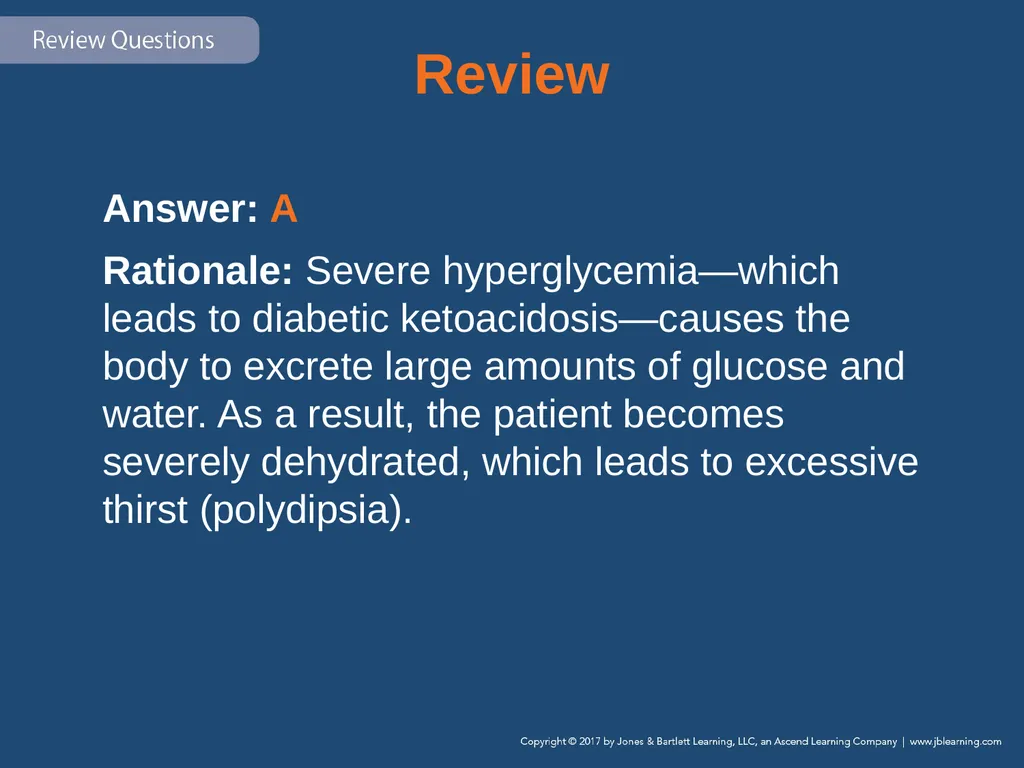
Review Answer: A
Rationale: Severe hyperglycemia—which leads to diabetic ketoacidosis—causes the body to excrete large amounts of glucose and water. As a result, the patient becomes severely dehydrated, which leads to excessive thirst (polydipsia).<br>
Rationale: Severe hyperglycemia—which leads to diabetic ketoacidosis—causes the body to excrete large amounts of glucose and water. As a result, the patient becomes severely dehydrated, which leads to excessive thirst (polydipsia).<br>
123
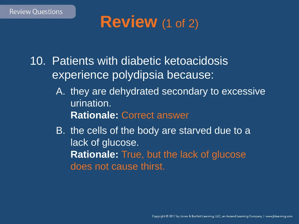
Review (1 of 2) Patients with diabetic ketoacidosis experience polydipsia because:
they are dehydrated secondary to excessive urination. Rationale: Correct answer
the cells of the body are starved due to a lack of glucose. Rationale: True, but the lack of glucose does not cause thirst.<br>
they are dehydrated secondary to excessive urination. Rationale: Correct answer
the cells of the body are starved due to a lack of glucose. Rationale: True, but the lack of glucose does not cause thirst.<br>
124
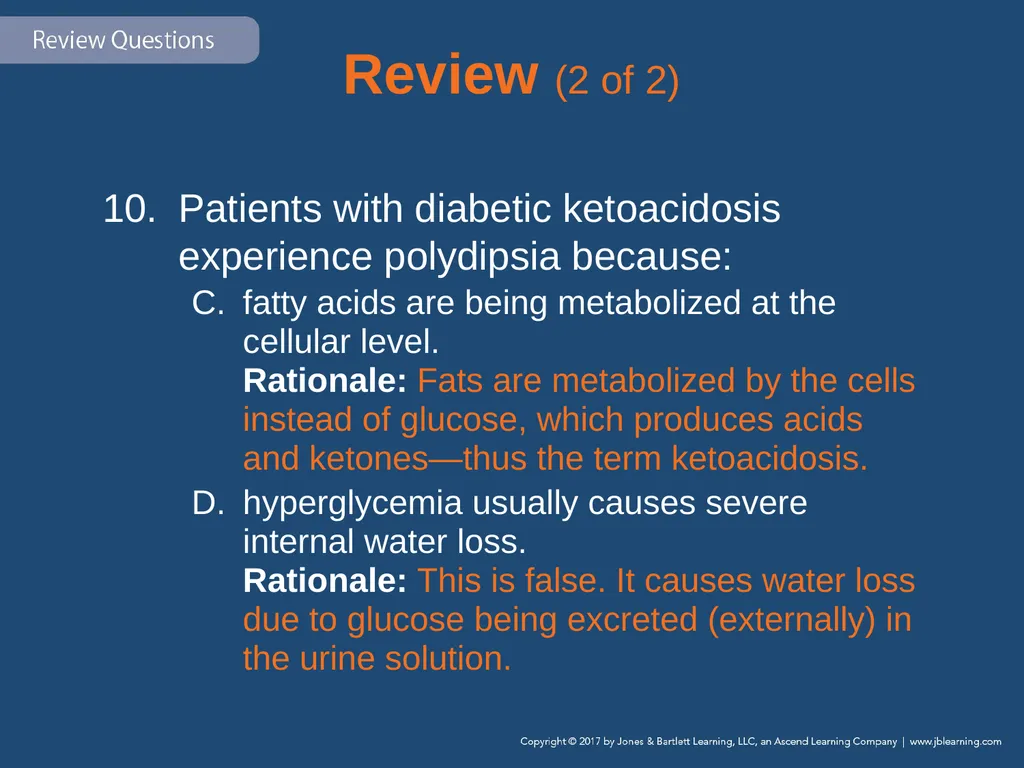
Review (2 of 2) Patients with diabetic ketoacidosis experience polydipsia because:
fatty acids are being metabolized at the cellular level. Rationale: Fats are metabolized by the cells instead of glucose, which produces acids and ketones—thus the term ketoacidosis.
hyperglycemia usually causes severe internal water loss. Rationale: This is false. It causes water loss due to glucose being excreted (externally) in the urine solution.<br>
fatty acids are being metabolized at the cellular level. Rationale: Fats are metabolized by the cells instead of glucose, which produces acids and ketones—thus the term ketoacidosis.
hyperglycemia usually causes severe internal water loss. Rationale: This is false. It causes water loss due to glucose being excreted (externally) in the urine solution.<br>
125
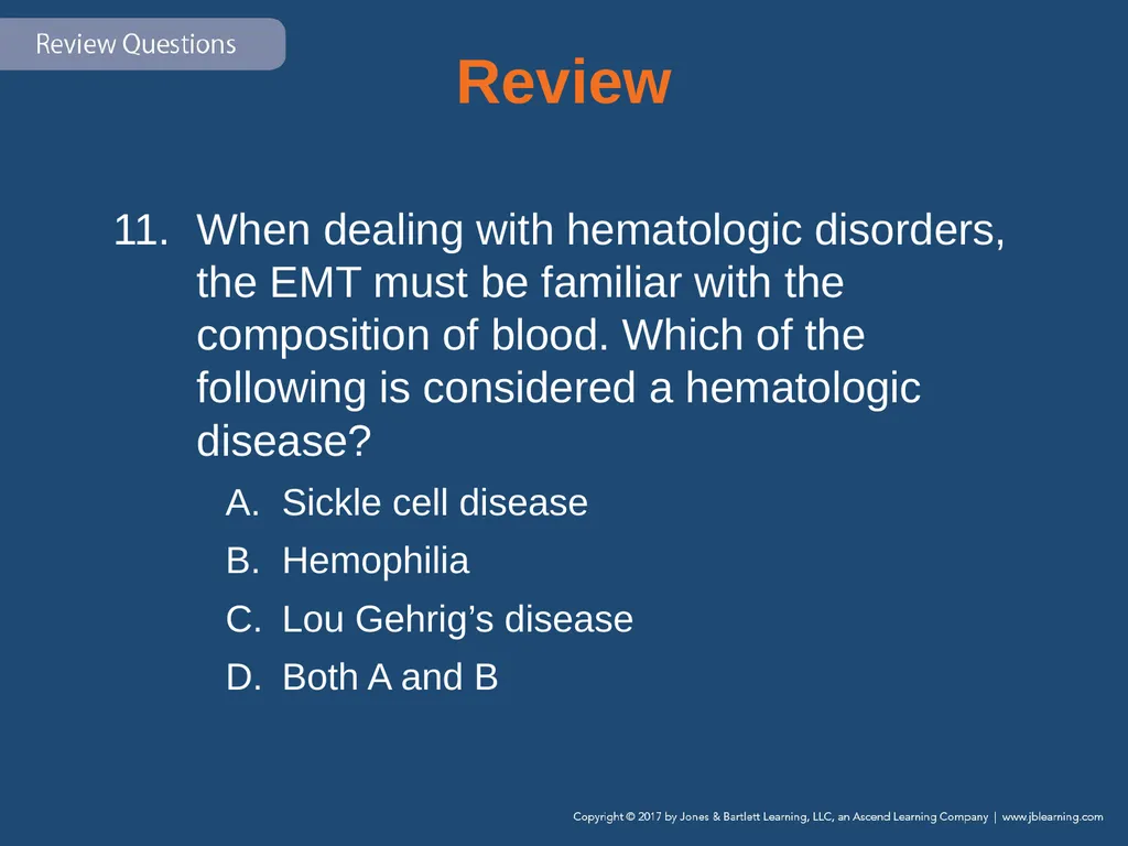
Review When dealing with hematologic disorders, the EMT must be familiar with the composition of blood. Which of the following is considered a hematologic disease?
Sickle cell disease
Hemophilia
Lou Gehrig’s disease
Both A and B<br>
Sickle cell disease
Hemophilia
Lou Gehrig’s disease
Both A and B<br>
126
Review Answer: D
Rationale: Hematology is the study and prevention of blood-related diseases, such as sickle cell disease and hemophilia.<br>
Rationale: Hematology is the study and prevention of blood-related diseases, such as sickle cell disease and hemophilia.<br>
127
Review (1 of 2) When dealing with hematologic disorders, the EMT must be familiar with the composition of blood. Which of the following is considered a hematologic disease?
Sickle cell disease Rationale: Sickle cell disease is a hematologic disorder affecting the red blood cells.
Hemophilia Rationale: Hemophilia is a hematologic disorder affecting the blood’s ability to clot.<br>
Sickle cell disease Rationale: Sickle cell disease is a hematologic disorder affecting the red blood cells.
Hemophilia Rationale: Hemophilia is a hematologic disorder affecting the blood’s ability to clot.<br>
128
Review (2 of 2) When dealing with hematologic disorders, the EMT must be familiar with the composition of blood. Which of the following is considered a hematologic disease?
Lou Gehrig’s disease Rationale: Lou Gehrig’s disease affects the nerve cells in the brain and spinal cord.
Both A and B Rationale: Correct answer<br>
Lou Gehrig’s disease Rationale: Lou Gehrig’s disease affects the nerve cells in the brain and spinal cord.
Both A and B Rationale: Correct answer<br>
129
Review What are the two main components of blood?
Erythrocytes and hemoglobin
Cells and plasma
Leukocytes and white blood cells
Platelets and neutrophils<br>
Erythrocytes and hemoglobin
Cells and plasma
Leukocytes and white blood cells
Platelets and neutrophils<br>
130
Review Answer: B
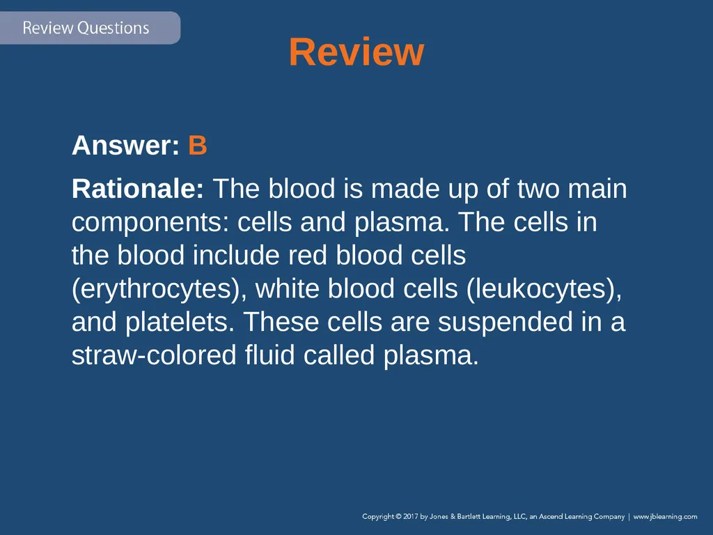
Rationale: The blood is made up of two main components: cells and plasma. The cells in the blood include red blood cells (erythrocytes), white blood cells (leukocytes), and platelets. These cells are suspended in a straw-colored fluid called plasma.<br>
Rationale: The blood is made up of two main components: cells and plasma. The cells in the blood include red blood cells (erythrocytes), white blood cells (leukocytes), and platelets. These cells are suspended in a straw-colored fluid called plasma.<br>
131
Review (1 of 2) What are the two main components of blood?
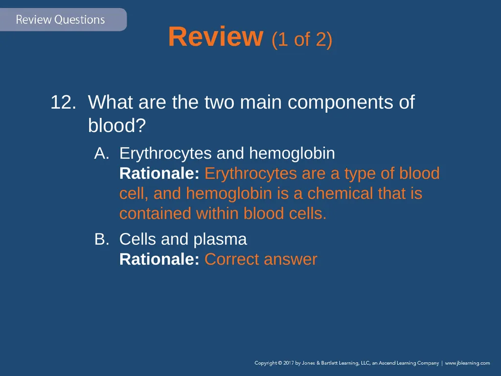
Erythrocytes and hemoglobin Rationale: Erythrocytes are a type of blood cell, and hemoglobin is a chemical that is contained within blood cells.
Cells and plasma Rationale: Correct answer<br>
Erythrocytes and hemoglobin Rationale: Erythrocytes are a type of blood cell, and hemoglobin is a chemical that is contained within blood cells.
Cells and plasma Rationale: Correct answer<br>
132
Review (2 of 2) What are the two main components of blood?
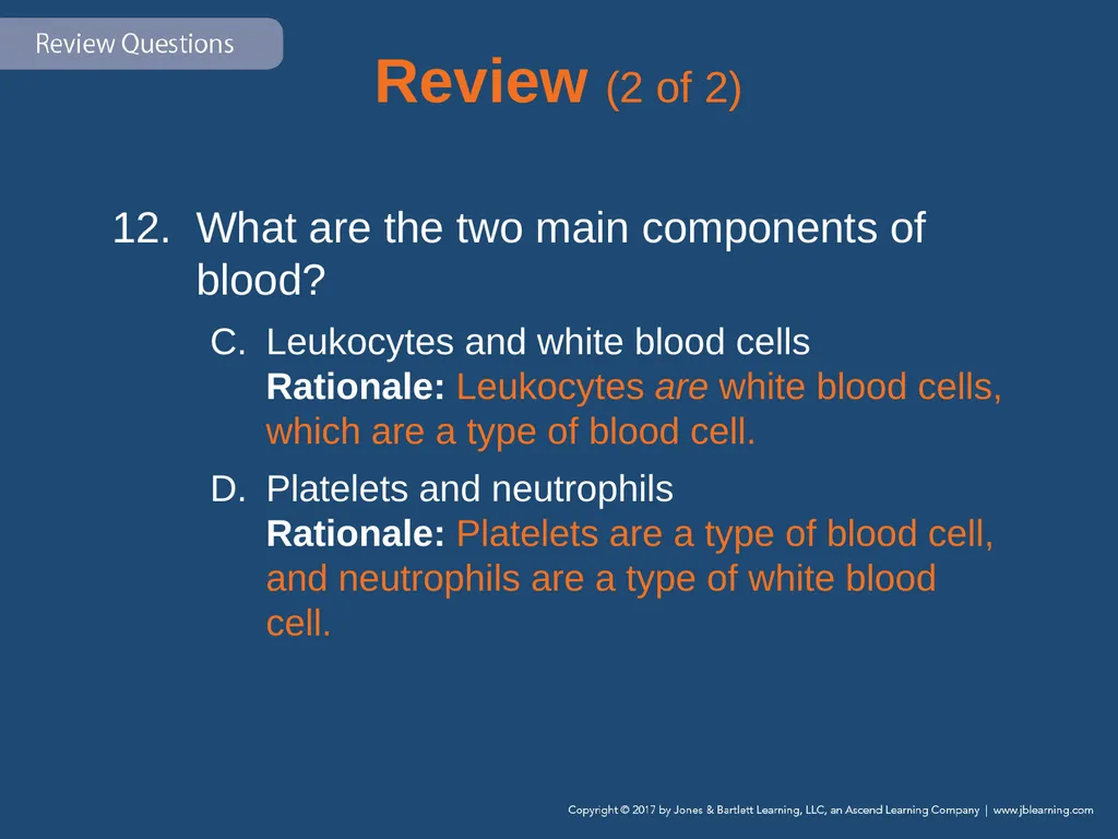
Leukocytes and white blood cells Rationale: Leukocytes are white blood cells, which are a type of blood cell.
Platelets and neutrophils Rationale: Platelets are a type of blood cell, and neutrophils are a type of white blood cell.<br>
Leukocytes and white blood cells Rationale: Leukocytes are white blood cells, which are a type of blood cell.
Platelets and neutrophils Rationale: Platelets are a type of blood cell, and neutrophils are a type of white blood cell.<br>
133
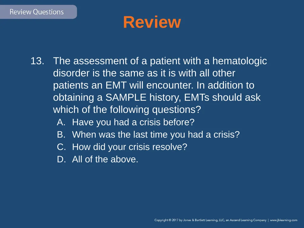
Review The assessment of a patient with a hematologic disorder is the same as it is with all other patients an EMT will encounter. In addition to obtaining a SAMPLE history, EMTs should ask which of the following questions?
Have you had a crisis before?
When was the last time you had a crisis?
How did your crisis resolve?
All of the above.<br>
Have you had a crisis before?
When was the last time you had a crisis?
How did your crisis resolve?
All of the above.<br>
134
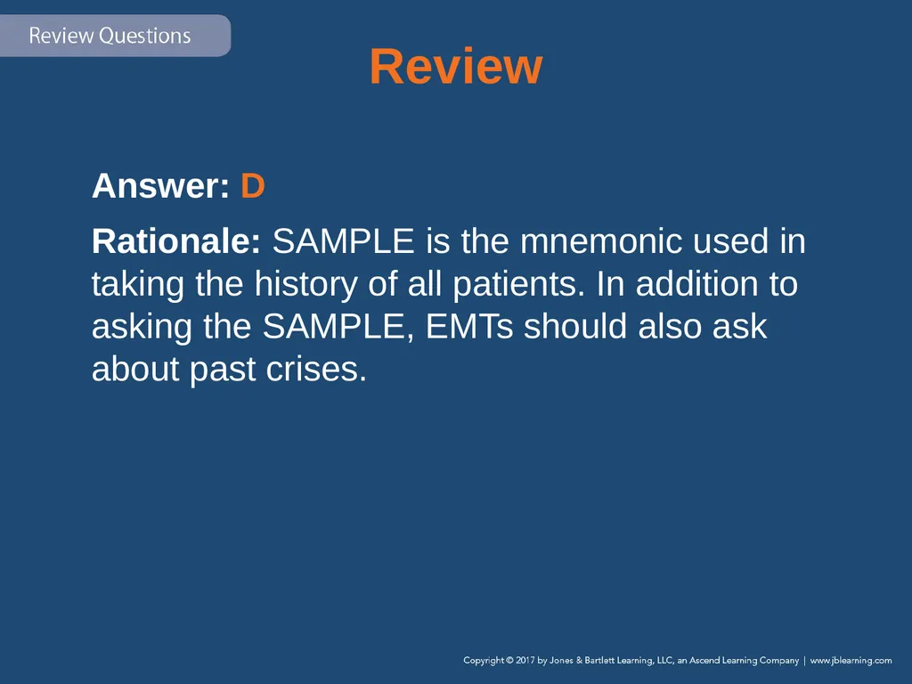
Review Answer: D
Rationale: SAMPLE is the mnemonic used in taking the history of all patients. In addition to asking the SAMPLE, EMTs should also ask about past crises.<br>
Rationale: SAMPLE is the mnemonic used in taking the history of all patients. In addition to asking the SAMPLE, EMTs should also ask about past crises.<br>
135
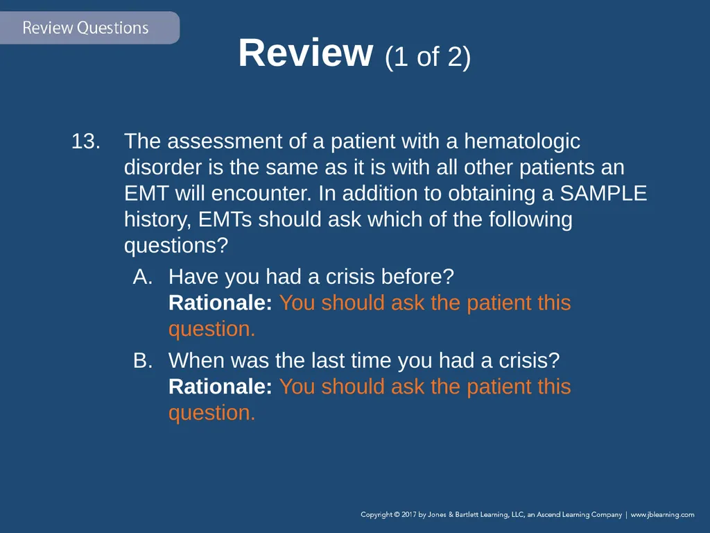
Review (1 of 2) The assessment of a patient with a hematologic disorder is the same as it is with all other patients an EMT will encounter. In addition to obtaining a SAMPLE history, EMTs should ask which of the following questions?
Have you had a crisis before? Rationale: You should ask the patient this question.
When was the last time you had a crisis? Rationale: You should ask the patient this question.<br>
Have you had a crisis before? Rationale: You should ask the patient this question.
When was the last time you had a crisis? Rationale: You should ask the patient this question.<br>
136
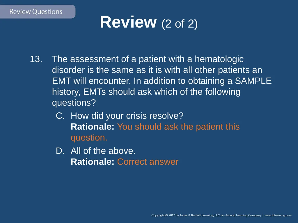
Review (2 of 2) The assessment of a patient with a hematologic disorder is the same as it is with all other patients an EMT will encounter. In addition to obtaining a SAMPLE history, EMTs should ask which of the following questions?
How did your crisis resolve? Rationale: You should ask the patient this question.
All of the above. Rationale: Correct answer<br>
How did your crisis resolve? Rationale: You should ask the patient this question.
All of the above. Rationale: Correct answer<br>
137
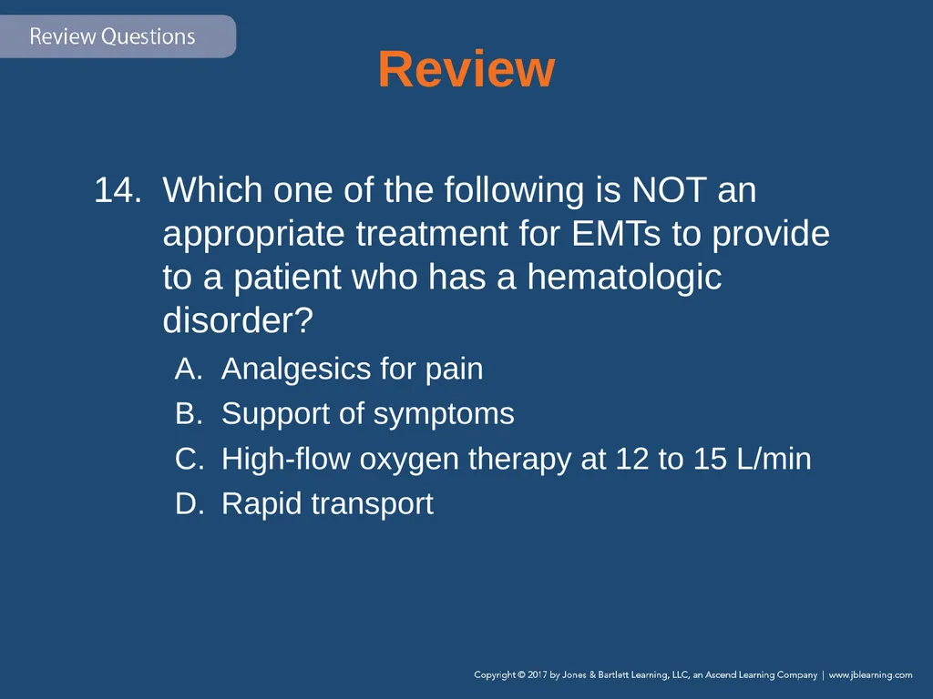
Review Which one of the following is NOT an appropriate treatment for EMTs to provide to a patient who has a hematologic disorder?
Analgesics for pain
Support of symptoms
High-flow oxygen therapy at 12 to 15 L/min
Rapid transport<br>
Analgesics for pain
Support of symptoms
High-flow oxygen therapy at 12 to 15 L/min
Rapid transport<br>
138
Review Answer: A
Rationale: Although analgesics would benefit a patient suffering from a hematologic disorder, the administration of such medications is not in the scope of practice for the EMT. ALS providers would have to be present to provide this emergency care.<br>
Rationale: Although analgesics would benefit a patient suffering from a hematologic disorder, the administration of such medications is not in the scope of practice for the EMT. ALS providers would have to be present to provide this emergency care.<br>
139
Review (1 of 2) Which one of the following is NOT an appropriate treatment for EMTs to provide to a patient who has a hematologic disorder?
Analgesics for pain Rationale: Correct answer
Support of symptoms Rationale: This is an appropriate treatment.<br>
Analgesics for pain Rationale: Correct answer
Support of symptoms Rationale: This is an appropriate treatment.<br>
140
Review (2 of 2) Which one of the following is NOT an appropriate treatment for EMTs to provide to a patient who has a hematologic disorder?
High-flow oxygen therapy at 12 to 15 L/min Rationale: This is an appropriate treatment.
Rapid transport Rationale: This is an appropriate treatment.<br>
High-flow oxygen therapy at 12 to 15 L/min Rationale: This is an appropriate treatment.
Rapid transport Rationale: This is an appropriate treatment.<br>