
Chapter 29 Genitourinary and Renal Disorders
FD
Published · 52 slides · 0 views

1 / 1










Description
Chapter 29 Genitourinary and Renal Disorders Comprehensive Lecture National EMS Education Standard Competencies Medicine Integrates assessment findings with principles of epidemiology and pathophysiology to formulate a field impression and
Related Topics
Share
Embed code
Download this presentation From Below
"Chapter 29 Genitourinary and Renal Disorders" is the property of its rightful owner. Permission is granted to download and print the materials on this website for personal, non-commercial use only, and to display it on your personal computer provided you do not modify the materials and that you retain all copyright notices contained in the materials. By downloading content from our website, you accept the terms of this agreement.
Presentation Transcript
01
Chapter 29
Genitourinary and Renal Disorders
Comprehensive Lecture<br>
Comprehensive Lecture<br>
02
National EMS Education Standard Competencies Medicine
Integrates assessment findings with principles of epidemiology and pathophysiology to formulate a field impression and implement a comprehensive treatment/disposition plan for a patient with a medical complaint.<br>
Integrates assessment findings with principles of epidemiology and pathophysiology to formulate a field impression and implement a comprehensive treatment/disposition plan for a patient with a medical complaint.<br>
03
Anatomy and Physiology Review
(1 of 4) Genitourinary System
Genito – refers to the genital organs and the male and female reproductive system
Urinary – system responsible for the following processes:
Regulation of water and electrolytes
Regulation of the acid base balance
Excretion of waste products and foreign chemicals
Regulation of the arterial blood pressure
Production of red blood cells
Stimulation of glucose production<br>
Genito – refers to the genital organs and the male and female reproductive system
Urinary – system responsible for the following processes:
Regulation of water and electrolytes
Regulation of the acid base balance
Excretion of waste products and foreign chemicals
Regulation of the arterial blood pressure
Production of red blood cells
Stimulation of glucose production<br>
04
Anatomy and Physiology Review
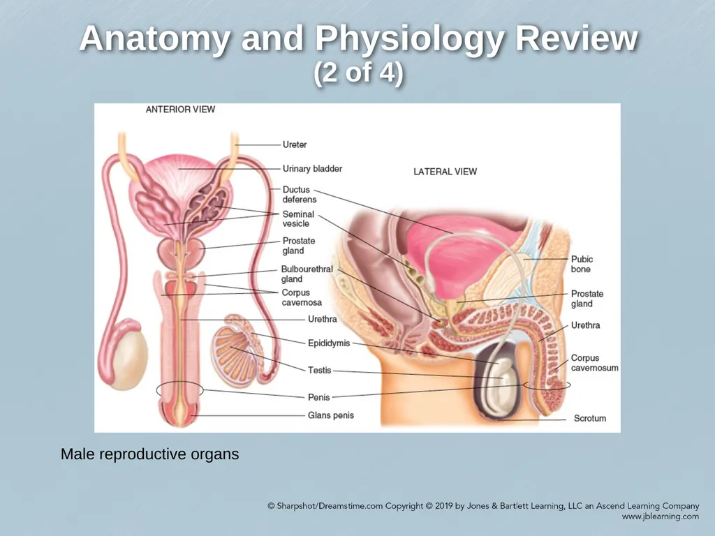
(2 of 4) Male reproductive organs<br>
05
Anatomy and Physiology Review
(3 of 4) Female reproductive organs<br>
06
Anatomy and Physiology Review
(4 of 4) Components of the urinary system<br>
07
Renal Diseases (1 of 25) Kidneys: two bean shaped organs
Basic functional unit of the kidney is the nephron
Filter blood
Remove waste products
Produce urine<br>
Basic functional unit of the kidney is the nephron
Filter blood
Remove waste products
Produce urine<br>
08
Renal Diseases (2 of 25) Acute/Chronic Renal Failure
Prerenal – related to events outside the kidney
Intrarenal – disease or damage within the kidney
Postrenal – disease that blocks the system that collects urine
Classification depends on duration of renal failure and reversibility potential<br>
Prerenal – related to events outside the kidney
Intrarenal – disease or damage within the kidney
Postrenal – disease that blocks the system that collects urine
Classification depends on duration of renal failure and reversibility potential<br>
09
Renal Diseases (3 of 25) Acute Kidney Injury (AKI)
Life-threatening
Occurs when kidneys cannot excrete the daily load of toxins in the urine
Patients divided into two groups:
Oliguric – excrete less than 500 mL/d
Nonoliguric – excrete greater than 500 mL/day
Mortality rate of hospitalized patients is 62%, but may be reversible if caught early and treated
Causes – trauma, shock, infection, urinary obstruction, and multisystem diseases.<br>
Life-threatening
Occurs when kidneys cannot excrete the daily load of toxins in the urine
Patients divided into two groups:
Oliguric – excrete less than 500 mL/d
Nonoliguric – excrete greater than 500 mL/day
Mortality rate of hospitalized patients is 62%, but may be reversible if caught early and treated
Causes – trauma, shock, infection, urinary obstruction, and multisystem diseases.<br>
10
Renal Diseases (4 of 25) Acute Kidney Injury (AKI) (cont.)
Quick onset; deteriorating function includes decrease or cessation of urine output, which can cause uremia.
Uremia may be associated with the following findings:
Generalized edema
Acidosis
High concentrations of nonprotein nitrogens, or other products of renal excretion
If not recognized early and treated, can lead to heart failure volume overload, hyperkalemia, metabolic acidosis<br>
Quick onset; deteriorating function includes decrease or cessation of urine output, which can cause uremia.
Uremia may be associated with the following findings:
Generalized edema
Acidosis
High concentrations of nonprotein nitrogens, or other products of renal excretion
If not recognized early and treated, can lead to heart failure volume overload, hyperkalemia, metabolic acidosis<br>
11
Renal Diseases (5 of 25) Acute Kidney Injury (AKI) (cont.)
Prerenal AKI
Inadequate perfusion of the kidneys
Obstruction to renal arteries results in decreased blood flow to the kidneys and causes an increase in renal vascular resistance
Patients are critically ill and may have preexisting medical conditions.
Signs and symptoms: dizziness, dry mouth, thirst, hypotension.
Treatment –improve kidney perfusion and function by treating the underlying condition.<br>
Prerenal AKI
Inadequate perfusion of the kidneys
Obstruction to renal arteries results in decreased blood flow to the kidneys and causes an increase in renal vascular resistance
Patients are critically ill and may have preexisting medical conditions.
Signs and symptoms: dizziness, dry mouth, thirst, hypotension.
Treatment –improve kidney perfusion and function by treating the underlying condition.<br>
12
Renal Diseases (6 of 25) Acute Kidney Injury (AKI) (cont.)
Intrarenal AKI
Results from conditions that damage or injure one or both kidneys (e.g., tubular diseases)
90% of cases are caused by ischemia or toxins, and can lead to acute tubular necrosis.
Associated with hypertension, autoimmune diseases and pyelonephritis
Signs and symptoms – fever, flank and joint pain, headache, and hypertension; in severe cases renal dialysis or kidney transplantation may be necessary.<br>
Intrarenal AKI
Results from conditions that damage or injure one or both kidneys (e.g., tubular diseases)
90% of cases are caused by ischemia or toxins, and can lead to acute tubular necrosis.
Associated with hypertension, autoimmune diseases and pyelonephritis
Signs and symptoms – fever, flank and joint pain, headache, and hypertension; in severe cases renal dialysis or kidney transplantation may be necessary.<br>
13
Renal Diseases (7 of 25) Acute Kidney Injury (AKI) (cont.)
Postrenal AKI
Caused by ureteral and urethral obstruction- obstructs the urine flow from both kidneys.
Blockage of urine causes pressure to build in the renal nephrons and may shut down the nephrons.
Signs and symptoms – urine retention, distended bladder, gross hematuria, lower back pain, pain in abdomen, groin or genitalia.
Can be reversed by removing the obstruction to urine flow, or diverting the flow of urine.
Treatment – nephrostomy tubes are placed through the patient’s back into the kidney, and connected to an external urinary collection bag.<br>
Postrenal AKI
Caused by ureteral and urethral obstruction- obstructs the urine flow from both kidneys.
Blockage of urine causes pressure to build in the renal nephrons and may shut down the nephrons.
Signs and symptoms – urine retention, distended bladder, gross hematuria, lower back pain, pain in abdomen, groin or genitalia.
Can be reversed by removing the obstruction to urine flow, or diverting the flow of urine.
Treatment – nephrostomy tubes are placed through the patient’s back into the kidney, and connected to an external urinary collection bag.<br>
14
Renal Diseases (8 of 25) Chronic Kidney Failure
Progressive, irreversible systemic disease
Develops over time; renal function declines, leads to end-stage renal disease requiring dialysis or kidney transplant.
Possible cause - congenital disorders or prolonged pyelonephritis
Kidneys compensate for renal damage by hyperfiltration causing further damage and loss of kidney function.
Buildup of fluid and waste causes azotemia and uremia, and most body systems are affected.
Complications include hypertension, heart failure, or anemia; once diagnosed treatments begin to delay or stop the progressive loss of kidney function.<br>
Progressive, irreversible systemic disease
Develops over time; renal function declines, leads to end-stage renal disease requiring dialysis or kidney transplant.
Possible cause - congenital disorders or prolonged pyelonephritis
Kidneys compensate for renal damage by hyperfiltration causing further damage and loss of kidney function.
Buildup of fluid and waste causes azotemia and uremia, and most body systems are affected.
Complications include hypertension, heart failure, or anemia; once diagnosed treatments begin to delay or stop the progressive loss of kidney function.<br>
15
Renal Diseases (9 of 25) Chronic Kidney Failure (cont.)
End stage requires dialysis or transplantation
Six systemic manifestations:
Gastrointestinal – anorexia, vomiting, metallic taste Cardiopulmonary – hypertension, pericarditis,
Nervous system – anxiety, delirium, hallucinations Metabolic or endocrine – glucose intolerance, anemia
Personality changes – fatigue, mental dullness,
Signs of uremia – uremic frost, pasty, yellow skin
Treatment options – kidney replacement therapy, peritoneal dialysis, hemodialysis, supportive care.<br>
End stage requires dialysis or transplantation
Six systemic manifestations:
Gastrointestinal – anorexia, vomiting, metallic taste Cardiopulmonary – hypertension, pericarditis,
Nervous system – anxiety, delirium, hallucinations Metabolic or endocrine – glucose intolerance, anemia
Personality changes – fatigue, mental dullness,
Signs of uremia – uremic frost, pasty, yellow skin
Treatment options – kidney replacement therapy, peritoneal dialysis, hemodialysis, supportive care.<br>
16
Renal Diseases (10 of 25) Transplantation
Can provide the best survival rate and quality of life
Most transplanted kidneys do not function forever
10 years post-surgery, approximately 60% of living donor kidneys and just under half of deceased donor kidneys will still be functioning.<br>
Can provide the best survival rate and quality of life
Most transplanted kidneys do not function forever
10 years post-surgery, approximately 60% of living donor kidneys and just under half of deceased donor kidneys will still be functioning.<br>
17
Renal Diseases (11 of 25) Renal Dialysis
Technique used to normalize blood chemistry and remove excess fluid in patients with acute CRF
Two types:
Hemodialysis
Peritoneal dialysis
Transfer depends on difference in concentrations of solutions, molecular size, and how long the blood and dialysate remain in contact with membrane<br>
Technique used to normalize blood chemistry and remove excess fluid in patients with acute CRF
Two types:
Hemodialysis
Peritoneal dialysis
Transfer depends on difference in concentrations of solutions, molecular size, and how long the blood and dialysate remain in contact with membrane<br>
18
Renal Diseases (12 of 25) Renal Dialysis (cont.)
Hemodialysis
Patient’s heparinized blood is pumped through a surgically constructed arteriovenous fistula or graft
Blood is taken from the arm and flows through a dialyzer machine that filters the blood through tiny filaments in a dialysis solution; blood is returned to the body.
Performed 3 or more times per week at an outpatient dialysis center or hospital
Each session lasts 3-4 hours.<br>
Hemodialysis
Patient’s heparinized blood is pumped through a surgically constructed arteriovenous fistula or graft
Blood is taken from the arm and flows through a dialyzer machine that filters the blood through tiny filaments in a dialysis solution; blood is returned to the body.
Performed 3 or more times per week at an outpatient dialysis center or hospital
Each session lasts 3-4 hours.<br>
19
Renal Diseases (13 of 25) Arteriovenous shunt<br>
20
Renal Diseases (14 of 25) Renal Dialysis (cont.)
Peritoneal Dialysis
Dialysis membrane is the patient’s peritoneum; using an implanted catheter, 2 L of dialysate are infused into the peritoneal cavity over 10 minutes.
Fluids move by osmosis and solutes diffuse from the blood in the peritoneal capillaries into the dialysate during the dwell period.
Equilibration occurs after several hours
Two systems are used:
Continuous ambulatory peritoneal dialysis
Automated peritoneal dialysis<br>
Peritoneal Dialysis
Dialysis membrane is the patient’s peritoneum; using an implanted catheter, 2 L of dialysate are infused into the peritoneal cavity over 10 minutes.
Fluids move by osmosis and solutes diffuse from the blood in the peritoneal capillaries into the dialysate during the dwell period.
Equilibration occurs after several hours
Two systems are used:
Continuous ambulatory peritoneal dialysis
Automated peritoneal dialysis<br>
21
Renal Diseases (15 of 25) Renal Dialysis (cont.)
Peritoneal Dialysis (cont.)
Slower than hemodialysis
Complication – peritonitis, caused by an infection when proper aseptic technique is not used.
Signs and symptoms – cloudy drainage, exit-site redness, tenderness, swelling, or pus
May be performed regularly in the home by patient or family caregiver.<br>
Peritoneal Dialysis (cont.)
Slower than hemodialysis
Complication – peritonitis, caused by an infection when proper aseptic technique is not used.
Signs and symptoms – cloudy drainage, exit-site redness, tenderness, swelling, or pus
May be performed regularly in the home by patient or family caregiver.<br>
22
Renal Diseases (16 of 25) Peritoneal dialysis<br>
23
Renal Diseases (17 of 25) Renal Dialysis (cont.)
Dialysis Emergencies
May be due to the disease process or from complications of the dialysis
Vascular access problems
Bleeding at the site of puncture
Bleeding from graft or fistula is minimal but excessive pressure can cause thrombosis.
If severe external hemorrhage occurs and cannot be controlled, apply a tourniquet.<br>
Dialysis Emergencies
May be due to the disease process or from complications of the dialysis
Vascular access problems
Bleeding at the site of puncture
Bleeding from graft or fistula is minimal but excessive pressure can cause thrombosis.
If severe external hemorrhage occurs and cannot be controlled, apply a tourniquet.<br>
24
Renal Diseases (18 of 25) Renal Dialysis (cont.)
Dialysis Emergencies (cont.)
Vascular access problems
Pseudoaneurysm – dilation resembling an aneurysm occurring at the graft site
Occluded fistulas and grafts due to thrombus formation
Infection results from a puncture made during dialysis
Signs of infection include unexplained fever or malaise.<br>
Dialysis Emergencies (cont.)
Vascular access problems
Pseudoaneurysm – dilation resembling an aneurysm occurring at the graft site
Occluded fistulas and grafts due to thrombus formation
Infection results from a puncture made during dialysis
Signs of infection include unexplained fever or malaise.<br>
25
Renal Diseases (19 of 25) Renal Dialysis (cont.)
Dialysis Emergencies (cont.)
Hemorrhage
Dialysis patients are at higher risk because of their exposure to anticoagulants during hemodialysis and decreased platelet function.
Patients with hemorrhage from trauma or medical condition should be monitored closely for signs of hypovolemia.
Anemia (common with dialysis) lowers patients’ ability to compensate for blood loss. Significant blood loss may produce dyspnea or angina.<br>
Dialysis Emergencies (cont.)
Hemorrhage
Dialysis patients are at higher risk because of their exposure to anticoagulants during hemodialysis and decreased platelet function.
Patients with hemorrhage from trauma or medical condition should be monitored closely for signs of hypovolemia.
Anemia (common with dialysis) lowers patients’ ability to compensate for blood loss. Significant blood loss may produce dyspnea or angina.<br>
26
Renal Diseases (20 of 25) Renal Dialysis (cont.)
Dialysis Emergencies (cont.)
Hypotension
Occurs with hemodialysis
Patient may not be able to maintain normal blood pressure and must be managed cautiously with the administration of volume-expanding fluids
Avoid fluid overload which can cause hypertension and signs of heart failure. If patients do not respond, consider other serious causes.<br>
Dialysis Emergencies (cont.)
Hypotension
Occurs with hemodialysis
Patient may not be able to maintain normal blood pressure and must be managed cautiously with the administration of volume-expanding fluids
Avoid fluid overload which can cause hypertension and signs of heart failure. If patients do not respond, consider other serious causes.<br>
27
Renal Diseases (21 of 25) Renal Dialysis (cont.)
Dialysis Emergencies (cont.)
Chest pain
Results from episodes of hypotension and mild hypoxemia during dialysis
Symptoms are relieved by administration of oxygen, fluid replacement, and antianginal medications.
Dysrhythmias due to myocardial ischemia may be associated with dialysis<br>
Dialysis Emergencies (cont.)
Chest pain
Results from episodes of hypotension and mild hypoxemia during dialysis
Symptoms are relieved by administration of oxygen, fluid replacement, and antianginal medications.
Dysrhythmias due to myocardial ischemia may be associated with dialysis<br>
28
Renal Diseases (22 of 25) Renal Dialysis (cont.)
Dialysis Emergencies (cont.)
Severe hyperkalemia
Life-threatening emergency resulting from poor dietary regulation and missed dialysis.
ECG shows a tall or tented T-wave
Any patient with renal failure in cardiac arrest should be suspected of having severe hyperkalemia.
Treatment – physician may recommend separate administration of calcium and sodium bicarbonate during resuscitation.<br>
Dialysis Emergencies (cont.)
Severe hyperkalemia
Life-threatening emergency resulting from poor dietary regulation and missed dialysis.
ECG shows a tall or tented T-wave
Any patient with renal failure in cardiac arrest should be suspected of having severe hyperkalemia.
Treatment – physician may recommend separate administration of calcium and sodium bicarbonate during resuscitation.<br>
29
Renal Diseases (23 of 25) Renal Dialysis (cont.)
Dialysis Emergencies (cont.)
Disequilibrium syndrome
Neurological findings that occur during or immediately following dialysis.
Mild symptoms (headache, fatigue)
Severe symptoms (confusion, coma)
Results from disproportionate decrease in the osmolality of the extracellular fluid compared with that of the intracellular compartment in the brain or cerebrospinal fluid.<br>
Dialysis Emergencies (cont.)
Disequilibrium syndrome
Neurological findings that occur during or immediately following dialysis.
Mild symptoms (headache, fatigue)
Severe symptoms (confusion, coma)
Results from disproportionate decrease in the osmolality of the extracellular fluid compared with that of the intracellular compartment in the brain or cerebrospinal fluid.<br>
30
Renal Diseases (24 of 25) Renal Dialysis (cont.)
Dialysis Emergencies (cont.)
Air embolism
May enter the patient’s blood stream if there is negative pressure on the venous side of dialysis tubing or the machine malfunctions
May travel to the right ventricle of the heart where it can block passage of the blood to the left myocardium.
Patient requires a high concentration oxygen, infusion of IV fluid, and rapid transport to a medical facility.<br>
Dialysis Emergencies (cont.)
Air embolism
May enter the patient’s blood stream if there is negative pressure on the venous side of dialysis tubing or the machine malfunctions
May travel to the right ventricle of the heart where it can block passage of the blood to the left myocardium.
Patient requires a high concentration oxygen, infusion of IV fluid, and rapid transport to a medical facility.<br>
31
Renal Diseases (25 of 25) Renal Dialysis (cont.)
Dialysis Emergencies (cont.)
Management
Patients with chronic or acute renal failure:
Provide airway and ventilatory support with supplemental high-concentration oxygen if hypoxic
Vascular access for fluid replacement, medication therapy, or fluid resuscitation if needed
ECG and other vital sign monitoring
Rapid transport to an appropriate medical facility with hyperbaric chamber<br>
Dialysis Emergencies (cont.)
Management
Patients with chronic or acute renal failure:
Provide airway and ventilatory support with supplemental high-concentration oxygen if hypoxic
Vascular access for fluid replacement, medication therapy, or fluid resuscitation if needed
ECG and other vital sign monitoring
Rapid transport to an appropriate medical facility with hyperbaric chamber<br>
32
Urinary System Conditions (1 of 4) Urinary Retention
May cause acute pain
Signs and symptoms
Severe abdominal pain associated with an urgent need (but inability) to urinate
Distended bladder
Treatment – once in the ER, a urethral catheter is inserted to empty the bladder; supportive care is given.
Some EMS systems may permit urinary catheterization in the prehospital setting.<br>
May cause acute pain
Signs and symptoms
Severe abdominal pain associated with an urgent need (but inability) to urinate
Distended bladder
Treatment – once in the ER, a urethral catheter is inserted to empty the bladder; supportive care is given.
Some EMS systems may permit urinary catheterization in the prehospital setting.<br>
33
Urinary System Conditions (2 of 4) Urinary Tract Infection
First develops in the lower urinary tract, may progress to upper tract if left untreated
Common sites of lower UTI – urethra and bladder
Causes – contraceptive devices, unsafe sexual practices, presence of renal stones, suppressed immune system
Signs and symptoms – dysuria, urinary frequency, cloudy or rust-colored urine, fever, chills, and malaise
Diagnoses –urinalysis and microscopic examination for blood cells, sediment and bacteria.
Treatment – antibiotics<br>
First develops in the lower urinary tract, may progress to upper tract if left untreated
Common sites of lower UTI – urethra and bladder
Causes – contraceptive devices, unsafe sexual practices, presence of renal stones, suppressed immune system
Signs and symptoms – dysuria, urinary frequency, cloudy or rust-colored urine, fever, chills, and malaise
Diagnoses –urinalysis and microscopic examination for blood cells, sediment and bacteria.
Treatment – antibiotics<br>
34
Urinary System Conditions (3 of 4) Pyelonephritis
Inflammation of the kidney parenchyma occurring as a result of a lower UTI infection.
Bacterial infection
Signs and symptoms – usually abrupt, fever, chills, flank pain, nausea, vomiting
Treatment – antibiotics, fluid replacement, sometimes hospitalization<br>
Inflammation of the kidney parenchyma occurring as a result of a lower UTI infection.
Bacterial infection
Signs and symptoms – usually abrupt, fever, chills, flank pain, nausea, vomiting
Treatment – antibiotics, fluid replacement, sometimes hospitalization<br>
35
Urinary System Conditions (4 of 4) Urinary Calculus
Kidney stones
Result from supersaturation of the urine with insoluble salts..
Risk factors – dehydration, drug use, and surgery
Chemical composition – calcium, uric acid, struvite, and cystine
Signs and symptoms –acute, excruciating pain accompanied by restlessness, nausea, and vomiting, etc.
Prehospital care – IV fluids, transport in a comfortable position, antiemetics, and pain management
Treatment – if the stone does not pass spontaneously, lithotripsy or surgical intervention may be required.<br>
Kidney stones
Result from supersaturation of the urine with insoluble salts..
Risk factors – dehydration, drug use, and surgery
Chemical composition – calcium, uric acid, struvite, and cystine
Signs and symptoms –acute, excruciating pain accompanied by restlessness, nausea, and vomiting, etc.
Prehospital care – IV fluids, transport in a comfortable position, antiemetics, and pain management
Treatment – if the stone does not pass spontaneously, lithotripsy or surgical intervention may be required.<br>
36
Male Genital Tract Disorders
(1 of 15) Epididymitis
Inflammation of the epididymitis
Cause – bacterial infection associated with other structures of the genitourinary tract
Signs and symptoms – gradual onset of unilateral scrotum pain radiating to spermatic cord. Tender swelling of the scrotum and testicle, producing inflammation of one or both testes. Fever, malaise, urethral discharge
Treatment – antibiotics, bed rest, analgesics, elevation of the scrotum<br>
Inflammation of the epididymitis
Cause – bacterial infection associated with other structures of the genitourinary tract
Signs and symptoms – gradual onset of unilateral scrotum pain radiating to spermatic cord. Tender swelling of the scrotum and testicle, producing inflammation of one or both testes. Fever, malaise, urethral discharge
Treatment – antibiotics, bed rest, analgesics, elevation of the scrotum<br>
37
Male Genital Tract Disorders
(2 of 15) Fournier Gangrene
Bacterial infection of the skin affecting the genitals and perineum
Urologic surgical emergency resulting from an infected wound or abrasion
Fatal if infection enters the bloodstream
Most common in men between the ages of 60 and 80 years
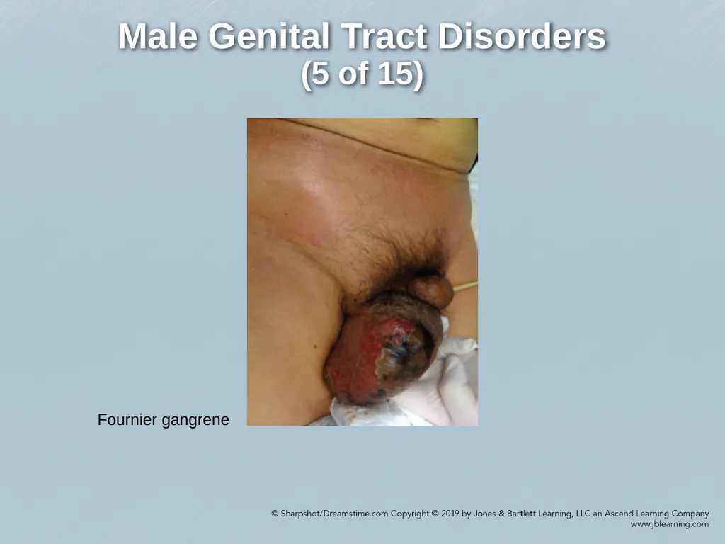
Signs and symptoms – intense pain and tenderness in the genitalia, crepitus of the skin, drainage of pus, and fever.<br>
Bacterial infection of the skin affecting the genitals and perineum
Urologic surgical emergency resulting from an infected wound or abrasion
Fatal if infection enters the bloodstream
Most common in men between the ages of 60 and 80 years
Signs and symptoms – intense pain and tenderness in the genitalia, crepitus of the skin, drainage of pus, and fever.<br>
38
Male Genital Tract Disorders
(3 of 15) Fournier Gangrene (cont.)
5 Stages:
Fever and lethargy (2 to 7 days)
Intense genital pain and tenderness
Increasing genital pain and tenderness with progressive erythema of the overlying skin
Dusky appearance of the overlying skin
Obvious gangrene of a portion of the genitalia; purulent drainage from wounds<br>
5 Stages:
Fever and lethargy (2 to 7 days)
Intense genital pain and tenderness
Increasing genital pain and tenderness with progressive erythema of the overlying skin
Dusky appearance of the overlying skin
Obvious gangrene of a portion of the genitalia; purulent drainage from wounds<br>
39
Male Genital Tract Disorders
(4 of 15) Fournier Gangrene (cont.)
Treatment
Prehospital – ranges from emotional support and rapid transport to full resuscitative measures to manage shock; following stabilization- antibiotics, hyperbaric oxygen therapy, and surgery.<br>
Treatment
Prehospital – ranges from emotional support and rapid transport to full resuscitative measures to manage shock; following stabilization- antibiotics, hyperbaric oxygen therapy, and surgery.<br>
40
Male Genital Tract Disorders
(5 of 15) Fournier gangrene<br>
41
Male Genital Tract Disorders
(6 of 15) Phimosis
Tightness of the prepuce of the penis. Prevents retraction of the foreskin over the glans.
Causes – failure of the foreskin to loosen during growth, infection, genital disease and trauma.
Usually painless
Paraphimosis is the inability to pull the retracted foreskin back over the head of the penis; can restrict blood flow and requires emergency care.<br>
Tightness of the prepuce of the penis. Prevents retraction of the foreskin over the glans.
Causes – failure of the foreskin to loosen during growth, infection, genital disease and trauma.
Usually painless
Paraphimosis is the inability to pull the retracted foreskin back over the head of the penis; can restrict blood flow and requires emergency care.<br>
42
Male Genital Tract Disorders
(7 of 15) Priapism
Persistent and painful erection lasting 4 hours or longer without sexual stimulation
Develops when blood in the penis becomes trapped and unable to drain
Categorized into two groups:
Low flow
High flow<br>
Persistent and painful erection lasting 4 hours or longer without sexual stimulation
Develops when blood in the penis becomes trapped and unable to drain
Categorized into two groups:
Low flow
High flow<br>
43
Male Genital Tract Disorders
(8 of 15) Priapism (cont.)
Causes –antidepressants; trauma to spinal cord or genital area; black widow spider bites, carbon monoxide poisoning; illicit drug use
All patients should be transported and evaluated by a physician<br>
Causes –antidepressants; trauma to spinal cord or genital area; black widow spider bites, carbon monoxide poisoning; illicit drug use
All patients should be transported and evaluated by a physician<br>
44
Male Genital Tract Disorders
(9 of 15) Benign Prostatic Hypertrophy
Enlargement of the prostate gland.
Signs and symptoms – not all men are symptomatic; some patients report urinary frequency, weak urine stream, or difficulty starting and stopping urination
Treatment – depends on severity of signs and symptoms and impact on daily life. Options include medications, surgery.<br>
Enlargement of the prostate gland.
Signs and symptoms – not all men are symptomatic; some patients report urinary frequency, weak urine stream, or difficulty starting and stopping urination
Treatment – depends on severity of signs and symptoms and impact on daily life. Options include medications, surgery.<br>
45
Male Genital Tract Disorders
(10 of 15) Testicular Masses
Enlargement or growth on one or both testicles; most are benign.
Most common masses:
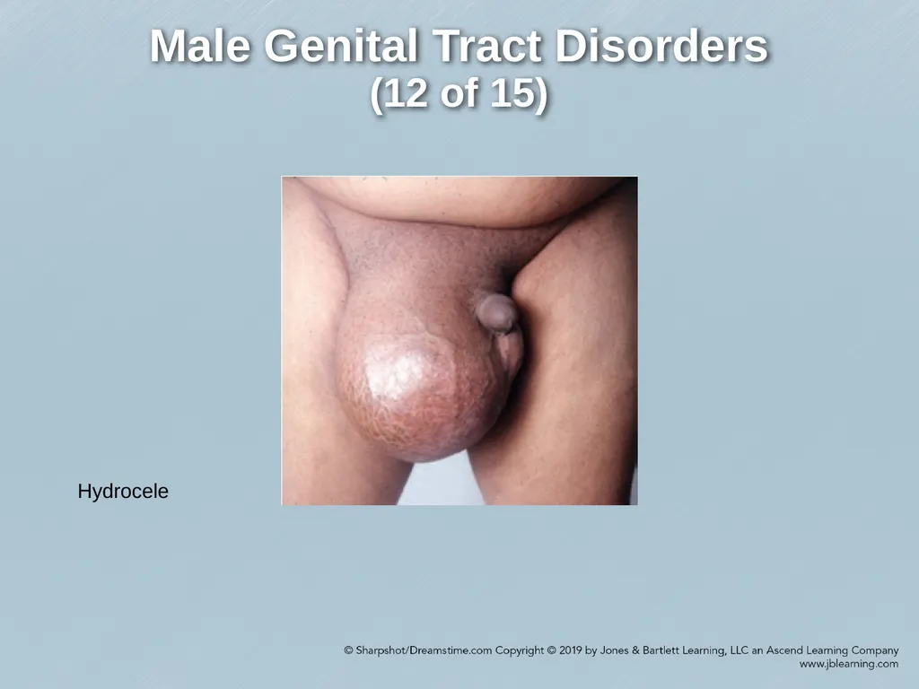
Hydroceles – fluid-filled sac along the spermatic cord in the scrotum
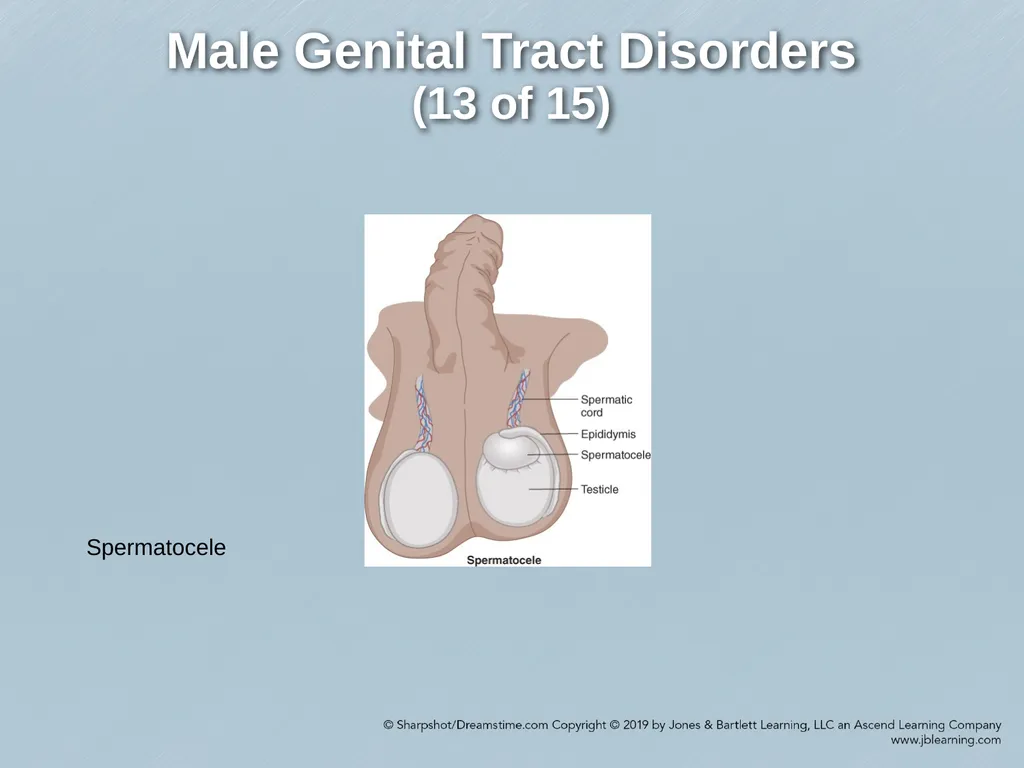
Spermatocele – benign cystic accumulation of sperm that arises from the head of the epididymis
Varicocele – enlargement of the veins that drain the testicles; May cause a heavy sensation or dull ache in the genital area.<br>
Enlargement or growth on one or both testicles; most are benign.
Most common masses:
Hydroceles – fluid-filled sac along the spermatic cord in the scrotum
Spermatocele – benign cystic accumulation of sperm that arises from the head of the epididymis
Varicocele – enlargement of the veins that drain the testicles; May cause a heavy sensation or dull ache in the genital area.<br>
46
Male Genital Tract Disorders
(11 of 15) Testicular Masses (cont.)
Soft scrotal swellings often more prominent when standing or exercising.
Diagnosis – ultrasonographic and transillumination techniques
Treatment – none; however, surgery may be needed to drain fluid or tie off the affected veins.
Testicular cancer may present as a testicular mass with or without pain<br>
Soft scrotal swellings often more prominent when standing or exercising.
Diagnosis – ultrasonographic and transillumination techniques
Treatment – none; however, surgery may be needed to drain fluid or tie off the affected veins.
Testicular cancer may present as a testicular mass with or without pain<br>
47
Male Genital Tract Disorders
(12 of 15) Hydrocele<br>
48
Male Genital Tract Disorders
(13 of 15) Spermatocele<br>
49
Male Genital Tract Disorders
(14 of 15) Testicular Torsion
Urologic emergency; a testicle twists on its spermatic cord, disrupting blood supply.
Causes –blunt trauma to the scrotal area; spontaneous
Signs and symptoms – tender epididymis and painful swelling of the scrotal sac; associated with nausea and vomiting<br>
Urologic emergency; a testicle twists on its spermatic cord, disrupting blood supply.
Causes –blunt trauma to the scrotal area; spontaneous
Signs and symptoms – tender epididymis and painful swelling of the scrotal sac; associated with nausea and vomiting<br>
50
Male Genital Tract Disorders
(15 of 15) Testicular Torsion (cont.)
Diagnoses – diagnose and treat within 6 hours to prevent loss of testis.
Treatment – apply ice packs to the scrotum, manual; surgical repair is necessary within 4-6 hours of onset; rapid transport to ER is critical.<br>
Diagnoses – diagnose and treat within 6 hours to prevent loss of testis.
Treatment – apply ice packs to the scrotum, manual; surgical repair is necessary within 4-6 hours of onset; rapid transport to ER is critical.<br>
51
Physical Examination of Patients with Genitourinary or Renal Disorders (1 of 2) Patient’s privacy is priority
Proper draping
Same sex paramedics should perform the exam or a chaperone should be present.
Exam should include the following:
Primary assessment
Focused history
Physical examination<br>
Proper draping
Same sex paramedics should perform the exam or a chaperone should be present.
Exam should include the following:
Primary assessment
Focused history
Physical examination<br>
52
Physical Examination of Patients with Genitourinary or Renal Disorders (2 of 2) Management and Treatment Plan
Address life threats, monitor ECG and vital signs, and transport for evaluation.
Patients should not be allowed to eat or drink because surgery may be indicated.<br>
Address life threats, monitor ECG and vital signs, and transport for evaluation.
Patients should not be allowed to eat or drink because surgery may be indicated.<br>