
Chapter 35 Shock Comprehensive Lecture National
LO
Published · 59 slides · 0 views
1 / 1

Description
Chapter 35 Shock Comprehensive Lecture National EMS Education Standard Competencies Shock and Resuscitation Integrates a comprehensive knowledge of causes and pathophysiology of shock, respiratory failure, or arrest into the management of
Related Topics
Share
Embed code
Download this presentation From Below
"Chapter 35 Shock Comprehensive Lecture National" is the property of its rightful owner. Permission is granted to download and print the materials on this website for personal, non-commercial use only, and to display it on your personal computer provided you do not modify the materials and that you retain all copyright notices contained in the materials. By downloading content from our website, you accept the terms of this agreement.
Presentation Transcript
01
Chapter 35
Shock
Comprehensive Lecture<br>
Comprehensive Lecture<br>
02
National EMS Education Standard Competencies Shock and Resuscitation
Integrates a comprehensive knowledge of causes and pathophysiology of shock, respiratory failure, or arrest into the management of these conditions, with an emphasis on early intervention to prevent arrest.<br>
Integrates a comprehensive knowledge of causes and pathophysiology of shock, respiratory failure, or arrest into the management of these conditions, with an emphasis on early intervention to prevent arrest.<br>
03
Shock Shock is not a single event, and it does not have a specific cause and treatment.
It is a complex group of physiologic abnormalities that can result from a variety of disease states and injuries.
There are many complexities involved in shock: It is not adequately defined by pulse rate, blood pressure, or cardiac function.
At its fundamental core, shock is hypoxia at the cellular level.<br>
It is a complex group of physiologic abnormalities that can result from a variety of disease states and injuries.
There are many complexities involved in shock: It is not adequately defined by pulse rate, blood pressure, or cardiac function.
At its fundamental core, shock is hypoxia at the cellular level.<br>
04
Oxygen Delivery to the Tissues (1 of 11) Cells require oxygen for the production of energy by aerobic metabolism. Systemic oxygen delivery to the tissues depends on two factors:
Adequate delivery of blood through the capillary bed in tissues (perfusion)
Adequate oxygen content of the blood<br>
Adequate delivery of blood through the capillary bed in tissues (perfusion)
Adequate oxygen content of the blood<br>
05
Oxygen Delivery to the Tissues (2 of 11) Heart
Systemic oxygen delivery is the product of cardiac output (CO) and the arterial oxygen content.
CO depends on several factors, including the strength of the heart’s contraction (inotropy), the rate of contraction (chronotropy), and the amount of venous return available to the ventricle (preload).<br>
Systemic oxygen delivery is the product of cardiac output (CO) and the arterial oxygen content.
CO depends on several factors, including the strength of the heart’s contraction (inotropy), the rate of contraction (chronotropy), and the amount of venous return available to the ventricle (preload).<br>
06
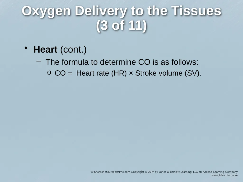
Oxygen Delivery to the Tissues (3 of 11) Heart (cont.)
The formula to determine CO is as follows:
CO = Heart rate (HR) × Stroke volume (SV).<br>
The formula to determine CO is as follows:
CO = Heart rate (HR) × Stroke volume (SV).<br>
07
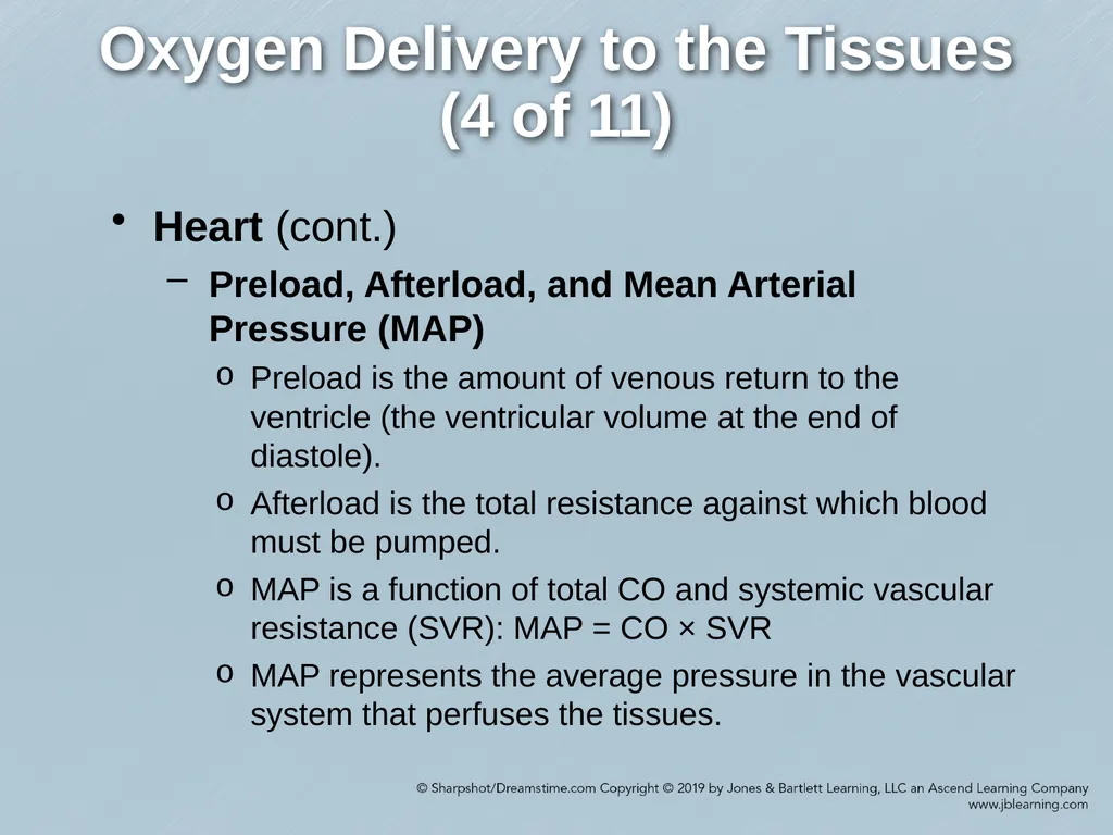
Oxygen Delivery to the Tissues (4 of 11) Heart (cont.)
Preload, Afterload, and Mean Arterial Pressure (MAP)
Preload is the amount of venous return to the ventricle (the ventricular volume at the end of diastole).
Afterload is the total resistance against which blood must be pumped.
MAP is a function of total CO and systemic vascular resistance (SVR): MAP = CO × SVR
MAP represents the average pressure in the vascular system that perfuses the tissues.<br>
Preload, Afterload, and Mean Arterial Pressure (MAP)
Preload is the amount of venous return to the ventricle (the ventricular volume at the end of diastole).
Afterload is the total resistance against which blood must be pumped.
MAP is a function of total CO and systemic vascular resistance (SVR): MAP = CO × SVR
MAP represents the average pressure in the vascular system that perfuses the tissues.<br>
08
Oxygen Delivery to the Tissues (5 of 11) Heart (cont.)
Ejection Fraction
Ejection fraction is a measurement of the percentage of blood volume pumped out of the left ventricle with each contraction.
Normally, 5070% of the blood is ejected out of the left ventricle. A borderline ejection fraction is 4149%.<br>
Ejection Fraction
Ejection fraction is a measurement of the percentage of blood volume pumped out of the left ventricle with each contraction.
Normally, 5070% of the blood is ejected out of the left ventricle. A borderline ejection fraction is 4149%.<br>
09
Oxygen Delivery to the Tissues (6 of 11) Vasculature
The entire vascular system of the body is lined with smooth, low-friction endothelial cells. All vessels larger than capillaries have layers of tissues surrounding the endothelium, known as tunicae.<br>
The entire vascular system of the body is lined with smooth, low-friction endothelial cells. All vessels larger than capillaries have layers of tissues surrounding the endothelium, known as tunicae.<br>
10
Oxygen Delivery to the Tissues (7 of 11) Vasculature (cont.)
SVR
Blood flow to an organ is dependent on three factors: arterial pressure, venous pressure, and vascular resistance.
Flow = [Arterial pressure – Venous pressure]/Vascular resistance.
Changes in resistance are the primary means by which blood flow is regulated within organs.
On a systemic level, afterload (SVR) is the total resistance against which blood must be pumped.<br>
SVR
Blood flow to an organ is dependent on three factors: arterial pressure, venous pressure, and vascular resistance.
Flow = [Arterial pressure – Venous pressure]/Vascular resistance.
Changes in resistance are the primary means by which blood flow is regulated within organs.
On a systemic level, afterload (SVR) is the total resistance against which blood must be pumped.<br>
11
Oxygen Delivery to the Tissues (8 of 11) Vasculature (cont.)
SVR (cont.)
SVR is a measurement of friction between the vessel walls and fluid, and between the molecules of the fluid themselves, both of which oppose flow.
Fluid flows through any kind of tube—including blood vessels—in response to pressure gradients between the two ends of the tube.
Systemic pressure (left-sided pressure) and pulmonic pressure (right-sided pressure) are the measurements of pressure in the vascular system. Both types of pressures have two phases: systolic and diastolic.
The difference between these two pressures, known as the pulse pressure, reflects the tone of the arterial system.<br>
SVR (cont.)
SVR is a measurement of friction between the vessel walls and fluid, and between the molecules of the fluid themselves, both of which oppose flow.
Fluid flows through any kind of tube—including blood vessels—in response to pressure gradients between the two ends of the tube.
Systemic pressure (left-sided pressure) and pulmonic pressure (right-sided pressure) are the measurements of pressure in the vascular system. Both types of pressures have two phases: systolic and diastolic.
The difference between these two pressures, known as the pulse pressure, reflects the tone of the arterial system.<br>
12
Oxygen Delivery to the Tissues (9 of 11) Vasculature (cont.)
Microcirculation
Microcirculation refers to circulation of blood through the smallest vessels in the circulatory system (arterioles, venules, shunts, and capillaries). Microcirculation exists in all tissues and organs except the cartilage, epithelia, cornea, and lens of the eye.<br>
Microcirculation
Microcirculation refers to circulation of blood through the smallest vessels in the circulatory system (arterioles, venules, shunts, and capillaries). Microcirculation exists in all tissues and organs except the cartilage, epithelia, cornea, and lens of the eye.<br>
13
Oxygen Delivery to the Tissues (10 of 11) Vasculature (cont.)
Microcirculation (cont.)
The following mechanisms control blood flow to the tissues:
Local control of blood flow by the tissues
Nervous control of blood flow
Baroreceptor reflexes
Chemoreceptor reflexes
Central nervous system ischemia response
Hormonal mechanisms
Adrenal–medullary mechanism
Renin–angiotensin–aldosterone mechanism
Vasopressin mechanism
Reabsorption of tissue fluid<br>
Microcirculation (cont.)
The following mechanisms control blood flow to the tissues:
Local control of blood flow by the tissues
Nervous control of blood flow
Baroreceptor reflexes
Chemoreceptor reflexes
Central nervous system ischemia response
Hormonal mechanisms
Adrenal–medullary mechanism
Renin–angiotensin–aldosterone mechanism
Vasopressin mechanism
Reabsorption of tissue fluid<br>
14
Oxygen Delivery to the Tissues (11 of 11) Lungs
Tissue cells require adequate oxygen to function.
To ensure that they obtain the needed oxygen, adequate oxygen must be available to be picked up by the red blood cells as they pass through the capillary membranes in the lungs.
The high partial pressure of oxygen in inspired air, adequate depth and rate of ventilation, and matching of pulmonary ventilation and perfusion make adequate oxygenation possible.<br>
Tissue cells require adequate oxygen to function.
To ensure that they obtain the needed oxygen, adequate oxygen must be available to be picked up by the red blood cells as they pass through the capillary membranes in the lungs.
The high partial pressure of oxygen in inspired air, adequate depth and rate of ventilation, and matching of pulmonary ventilation and perfusion make adequate oxygenation possible.<br>
15
The Body as a Container
(1 of 2) The healthy body can be viewed as a smooth-flowing fluid delivery system inside a container.
This container must be filled to achieve adequate preload and tissue oxygenation.
If the patient is hypovolemic and the 5-L container remains the same size despite the 3-L volume, the patient becomes hypotensive or loses pressure in the container.<br>
This container must be filled to achieve adequate preload and tissue oxygenation.
If the patient is hypovolemic and the 5-L container remains the same size despite the 3-L volume, the patient becomes hypotensive or loses pressure in the container.<br>
16
The Body as a Container
(2 of 2) If fluid is adequate for a 5-L container but the container size has been enlarged to 7 L by illness or injury that results in vasodilation, the 5 L of fluid does not provide adequate preload for the container.
Factors that occasionally are responsible for vasodilation include cardiac and blood pressure medications, allergic reaction, heat- and cold-related injuries, and alcohol or other drug use.<br>
Factors that occasionally are responsible for vasodilation include cardiac and blood pressure medications, allergic reaction, heat- and cold-related injuries, and alcohol or other drug use.<br>
17
Capillary-Cellular Relationship in Shock (1 of 2) No matter what the cause of shock, this condition is characterized by a homeostatic vasoconstrictor response with arteriolar narrowing, a reduction in the number of open capillaries, and a decrease in venous return and CO.
Without treatment, anaerobic metabolism develops, leading to an increase in lactic acid and metabolic acidosis.<br>
Without treatment, anaerobic metabolism develops, leading to an increase in lactic acid and metabolic acidosis.<br>
18
Capillary-Cellular Relationship in Shock (2 of 2) Severe microcirculatory failure cannot be remedied with fluid replacement.
Eventual outcomes can include heart failure, gastrointestinal bleeding, sepsis, severe pancreatitis, pulmonary thrombosis, respiratory failure, and death.<br>
Eventual outcomes can include heart failure, gastrointestinal bleeding, sepsis, severe pancreatitis, pulmonary thrombosis, respiratory failure, and death.<br>
19
Classifications of Shock
(1 of 8) Hypovolemic Shock
Due to an inadequate circulating blood volume
Most common causes are hemorrhage and dehydration
Illnesses and injuries that can lead to hypovolemic shock include trauma, gastrointestinal bleeding, burns, diarrhea, vomiting, endocrine disorders, and internal third-space loss, as in peritonitis.<br>
Due to an inadequate circulating blood volume
Most common causes are hemorrhage and dehydration
Illnesses and injuries that can lead to hypovolemic shock include trauma, gastrointestinal bleeding, burns, diarrhea, vomiting, endocrine disorders, and internal third-space loss, as in peritonitis.<br>
20
Classifications of Shock
(2 of 8) Cardiogenic Shock
Cardiogenic shock is the result of a severe compromise in CO such that inadequate tissue perfusion occurs despite an adequate amount of circulating blood volume.
Cardiogenic shock can result from pump failure, depression of cardiac contractility, mechanical obstruction to forward blood flow, or regurgitation of the left ventricular output.
Myocardial infarction is the most common cause of cardiogenic shock.<br>
Cardiogenic shock is the result of a severe compromise in CO such that inadequate tissue perfusion occurs despite an adequate amount of circulating blood volume.
Cardiogenic shock can result from pump failure, depression of cardiac contractility, mechanical obstruction to forward blood flow, or regurgitation of the left ventricular output.
Myocardial infarction is the most common cause of cardiogenic shock.<br>
21
Classifications of Shock
(3 of 8) Obstructive Shock
Obstructive shock is a form of shock associated with the inability to produce adequate CO despite normal intravascular volume and myocardial function.
Causes of obstructive shock include pericardial tamponade, tension pneumothorax, and pulmonary embolism.
Pericardial tamponade directly impairs diastolic filling of the right ventricle; tension pneumothorax indirectly impairs right ventricular filling by obstructing venous return; and pulmonary embolism may obstruct lobar arteries and cause impaired systolic contraction.<br>
Obstructive shock is a form of shock associated with the inability to produce adequate CO despite normal intravascular volume and myocardial function.
Causes of obstructive shock include pericardial tamponade, tension pneumothorax, and pulmonary embolism.
Pericardial tamponade directly impairs diastolic filling of the right ventricle; tension pneumothorax indirectly impairs right ventricular filling by obstructing venous return; and pulmonary embolism may obstruct lobar arteries and cause impaired systolic contraction.<br>
22
Classifications of Shock
(4 of 8) Distributive Shock
Distributive shock occurs when peripheral vasodilation causes a decrease in SVR.
The most common causes of distributive shock are neurogenic shock, anaphylactic shock, and septic shock.
In contrast to hypovolemic and cardiogenic shock, distributive shock is a “warm shock.”<br>
Distributive shock occurs when peripheral vasodilation causes a decrease in SVR.
The most common causes of distributive shock are neurogenic shock, anaphylactic shock, and septic shock.
In contrast to hypovolemic and cardiogenic shock, distributive shock is a “warm shock.”<br>
23
Classifications of Shock
(5 of 8) Distributive Shock (cont.)
Neurogenic Shock
Neurogenic shock (also known as vasogenic shock) results from loss of normal vasomotor tone due to unopposed parasympathetic response after disruption of the spinal cord at midthoracic levels and above.<br>
Neurogenic Shock
Neurogenic shock (also known as vasogenic shock) results from loss of normal vasomotor tone due to unopposed parasympathetic response after disruption of the spinal cord at midthoracic levels and above.<br>
24
Classifications of Shock
(6 of 8) Distributive Shock (cont.)
Anaphylactic Shock
Anaphylactic shock is the most extreme form of anaphylaxis.
Anaphylaxis may occur when the body is exposed to an antigen that produces a severe allergic reaction.
The body responds by releasing histamine and other mediators that act on receptors in the systemic and pulmonary microcirculation and produce an effect on bronchial smooth muscle.<br>
Anaphylactic Shock
Anaphylactic shock is the most extreme form of anaphylaxis.
Anaphylaxis may occur when the body is exposed to an antigen that produces a severe allergic reaction.
The body responds by releasing histamine and other mediators that act on receptors in the systemic and pulmonary microcirculation and produce an effect on bronchial smooth muscle.<br>
25
Classifications of Shock
(7 of 8) Distributive Shock (cont.)
Sepsis and Septic Shock
Sepsis is a systemic inflammatory response syndrome (SIRS) to a known or presumed infection; it can lead to severe organ dysfunction and death.
This syndrome involves altered function in major organs and body processes.
Septic shock is a subset of sepsis and a more severe syndrome in which profound circulatory, cellular, and metabolic abnormalities cause a greater risk of mortality than does sepsis alone.<br>
Sepsis and Septic Shock
Sepsis is a systemic inflammatory response syndrome (SIRS) to a known or presumed infection; it can lead to severe organ dysfunction and death.
This syndrome involves altered function in major organs and body processes.
Septic shock is a subset of sepsis and a more severe syndrome in which profound circulatory, cellular, and metabolic abnormalities cause a greater risk of mortality than does sepsis alone.<br>
26
Classifications of Shock
(8 of 8) Distributive Shock (cont.)
Sepsis and Septic Shock (cont.)
A number of screening criteria have been developed to support prehospital sepsis identification. The basis of most of these tools is identification of suspected infection, an inflammatory response, and evidence of poor end-organ perfusion.
The most common sites of infection include the lungs, urinary tract, and skin.
Signs of inflammatory response include the SIRS criteria.<br>
Sepsis and Septic Shock (cont.)
A number of screening criteria have been developed to support prehospital sepsis identification. The basis of most of these tools is identification of suspected infection, an inflammatory response, and evidence of poor end-organ perfusion.
The most common sites of infection include the lungs, urinary tract, and skin.
Signs of inflammatory response include the SIRS criteria.<br>
27
Progression of Shock (1 of 8) Compensated Shock
Associated with decreased blood flow and perfusion to the tissues
Compensatory responses of the body can overcome the decrease in available fluid
An increase in catecholamine production maintains CO and a normal systolic blood pressure
If the underlying cause of shock is untreated, the compensatory mechanisms will eventually collapse.<br>
Associated with decreased blood flow and perfusion to the tissues
Compensatory responses of the body can overcome the decrease in available fluid
An increase in catecholamine production maintains CO and a normal systolic blood pressure
If the underlying cause of shock is untreated, the compensatory mechanisms will eventually collapse.<br>
28
Progression of Shock (2 of 8) Uncompensated Shock
Occurs when the body is no longer able to maintain systemic blood pressure
The systolic pressure usually drops before the diastolic pressure
Systolic pressure depends more on blood volume
Diastolic pressure may rise at first because of vasoconstriction<br>
Occurs when the body is no longer able to maintain systemic blood pressure
The systolic pressure usually drops before the diastolic pressure
Systolic pressure depends more on blood volume
Diastolic pressure may rise at first because of vasoconstriction<br>
29
Progression of Shock (3 of 8) Uncompensated Shock (cont.)
The decrease in systolic pressure, along with maintained or increased diastolic pressure, can lead to a narrow pulse pressure—so narrowed, in fact, that the pulse pressure is not detectable with a blood pressure cuff.
Effects on the cardiovascular system include a decreased preload and an increased rate of contraction caused by catecholamine stimulation.
Myocardial contractions initially can be stronger as a result of catecholamine release.<br>
The decrease in systolic pressure, along with maintained or increased diastolic pressure, can lead to a narrow pulse pressure—so narrowed, in fact, that the pulse pressure is not detectable with a blood pressure cuff.
Effects on the cardiovascular system include a decreased preload and an increased rate of contraction caused by catecholamine stimulation.
Myocardial contractions initially can be stronger as a result of catecholamine release.<br>
30
Progression of Shock (4 of 8) Uncompensated Shock (cont.)
In the latter phases of uncompensated shock, myocardial strength may decrease as a result of the following factors:
Ischemia can result from a reduction in circulating red blood cells, a lower pO2 level, and decreased coronary perfusion because of hypotension (especially diastolic hypotension).
Cardiodepressant substances (e.g., myocardial toxin released from the ischemic pancreas) can depress heart function in late shock.<br>
In the latter phases of uncompensated shock, myocardial strength may decrease as a result of the following factors:
Ischemia can result from a reduction in circulating red blood cells, a lower pO2 level, and decreased coronary perfusion because of hypotension (especially diastolic hypotension).
Cardiodepressant substances (e.g., myocardial toxin released from the ischemic pancreas) can depress heart function in late shock.<br>
31
Progression of Shock (5 of 8) Uncompensated Shock (cont.)
In the latter phases of uncompensated shock, myocardial strength may decrease as a result of the following factors: (cont.)
Injury of the myocardium (essentially simulating myocardial infarction) can result from associated ischemia.
Decreased preload can lead to decreased contractility.
Acidosis can lead to decreased contractility.
Cardiac rhythm disturbances can result from hypoxia.<br>
In the latter phases of uncompensated shock, myocardial strength may decrease as a result of the following factors: (cont.)
Injury of the myocardium (essentially simulating myocardial infarction) can result from associated ischemia.
Decreased preload can lead to decreased contractility.
Acidosis can lead to decreased contractility.
Cardiac rhythm disturbances can result from hypoxia.<br>
32
Progression of Shock (6 of 8) Irreversible Shock
The progression of cellular ischemia and necrosis and subsequent organ death, even with oxygenation and perfusion restored, indicate irreversible shockthe third phase of shock.
Despite a return to normal perfusion, patients with irreversible shock as a result of massive cellular damage do not survive.
Cells and the vital organs begin to die from the lack of energy, the membrane pumps fail, and the various organelles in the cells sequentially break down.<br>
The progression of cellular ischemia and necrosis and subsequent organ death, even with oxygenation and perfusion restored, indicate irreversible shockthe third phase of shock.
Despite a return to normal perfusion, patients with irreversible shock as a result of massive cellular damage do not survive.
Cells and the vital organs begin to die from the lack of energy, the membrane pumps fail, and the various organelles in the cells sequentially break down.<br>
33
Progression of Shock (7 of 8) Variations in Physiologic Response to Shock
Determining factors include the following:
Age
Older adults are less able to compensate.
Children compensate for a longer period but deteriorate faster.
General Health
Preexisting disease
Other injuries
Pregnancy<br>
Determining factors include the following:
Age
Older adults are less able to compensate.
Children compensate for a longer period but deteriorate faster.
General Health
Preexisting disease
Other injuries
Pregnancy<br>
34
Progression of Shock (8 of 8) Variations in Physiologic Response to Shock (cont.)
Determining factors include the following: (cont.)
Ability to activate compensatory mechanisms
Medications, some of which can interfere with compensatory mechanisms
Specific organ system(s) affected<br>
Determining factors include the following: (cont.)
Ability to activate compensatory mechanisms
Medications, some of which can interfere with compensatory mechanisms
Specific organ system(s) affected<br>
35
Assessment and Management of the Patient in Shock (1 of 10) Primary Survey
Exsanguinating Hemorrhage
Address any exsanguinating hemorrhage with wound packing for truncal injuries or tourniquet application for extremity wounds.
In a trauma patient, in whom hemorrhagic shock is the most common type of shock to develop, circulatory assessment begins with identification of uncontrolled hemorrhage.
In cases of external hemorrhage, applying direct pressure often controls bleeding.<br>
Exsanguinating Hemorrhage
Address any exsanguinating hemorrhage with wound packing for truncal injuries or tourniquet application for extremity wounds.
In a trauma patient, in whom hemorrhagic shock is the most common type of shock to develop, circulatory assessment begins with identification of uncontrolled hemorrhage.
In cases of external hemorrhage, applying direct pressure often controls bleeding.<br>
36
Assessment and Management of the Patient in Shock (2 of 10) Primary Survey (cont.)
Airway
Confirm that the airway is open and patent to ensure adequate air movement.
Breathing
The patient’s respiratory pattern often reflects the adequacy of ventilation, so it can offer clues to the presence of shock.<br>
Airway
Confirm that the airway is open and patent to ensure adequate air movement.
Breathing
The patient’s respiratory pattern often reflects the adequacy of ventilation, so it can offer clues to the presence of shock.<br>
37
Assessment and Management of the Patient in Shock (3 of 10) Primary Survey (cont.)
Circulation
The paramedic should evaluate the rate, character, and location of the patient’s pulses as part of the circulatory assessment.
Patients with neurogenic shock or cardiogenic shock caused by heart block may be bradycardic, while pulse rates increase fairly early in patients with hypovolemic shock.
The increased HR helps to maintain an adequate CO.<br>
Circulation
The paramedic should evaluate the rate, character, and location of the patient’s pulses as part of the circulatory assessment.
Patients with neurogenic shock or cardiogenic shock caused by heart block may be bradycardic, while pulse rates increase fairly early in patients with hypovolemic shock.
The increased HR helps to maintain an adequate CO.<br>
38
Assessment and Management of the Patient in Shock (4 of 10) Primary Survey (cont.)
Circulation (cont.)
The character of the pulse can be strong or weak.
Tissue perfusion sometimes can be estimated by evaluating the color, moisture, and temperature of the skin.
The capillary refill test can offer useful details on the pediatric patient’s tissue perfusion.
If the paramedic suspects internal bleeding, rapid transport to a proper facility while ensuring adequate ventilation is the highest priority.<br>
Circulation (cont.)
The character of the pulse can be strong or weak.
Tissue perfusion sometimes can be estimated by evaluating the color, moisture, and temperature of the skin.
The capillary refill test can offer useful details on the pediatric patient’s tissue perfusion.
If the paramedic suspects internal bleeding, rapid transport to a proper facility while ensuring adequate ventilation is the highest priority.<br>
39
Assessment and Management of the Patient in Shock (5 of 10) Primary Survey (cont.)
Disability
The evaluation of the patient’s level of consciousness is crucial in assessing cerebral oxygenation. The patient can become restless, agitated, and confused as cerebral ischemia develops.<br>
Disability
The evaluation of the patient’s level of consciousness is crucial in assessing cerebral oxygenation. The patient can become restless, agitated, and confused as cerebral ischemia develops.<br>
40
Assessment and Management of the Patient in Shock (6 of 10) Primary Survey (cont.)
Exposure of the Body Surfaces
The paramedic should expose the body surfaces in the primary survey as indicated by scenario or mechanism of injury.
A visual inspection can reveal conditions that may be life threatening but that are hidden by clothing.<br>
Exposure of the Body Surfaces
The paramedic should expose the body surfaces in the primary survey as indicated by scenario or mechanism of injury.
A visual inspection can reveal conditions that may be life threatening but that are hidden by clothing.<br>
41
Assessment and Management of the Patient in Shock (7 of 10) Secondary Assessment
A systematic approach offers a way to evaluate potentially life-threatening conditions and allows the paramedic to further assess the patient’s perfusion status.
Serial blood pressure assessments should be obtained, trended, and evaluated in the context of the patient’s normal state.
The systolic pressure falls when the heart can no longer pump enough blood to fill the container at the end of cardiac contraction.<br>
A systematic approach offers a way to evaluate potentially life-threatening conditions and allows the paramedic to further assess the patient’s perfusion status.
Serial blood pressure assessments should be obtained, trended, and evaluated in the context of the patient’s normal state.
The systolic pressure falls when the heart can no longer pump enough blood to fill the container at the end of cardiac contraction.<br>
42
Assessment and Management of the Patient in Shock (8 of 10) Secondary Assessment (cont.)
Pulse pressure should be noted.
The paramedic should consider evaluation of orthostatic vital signs in conscious patients suspected of having lost circulating blood volume.
A fluid deficit can persist even after the systolic pressure returns to normal following fluid replacement.
If available, serum lactate can be measured using an approved testing device.<br>
Pulse pressure should be noted.
The paramedic should consider evaluation of orthostatic vital signs in conscious patients suspected of having lost circulating blood volume.
A fluid deficit can persist even after the systolic pressure returns to normal following fluid replacement.
If available, serum lactate can be measured using an approved testing device.<br>
43
Assessment and Management of the Patient in Shock (9 of 10) Differential Shock Assessment
Cardiogenic Shock
The patient with cardiogenic shock often has a chief complaint of chest pain, dyspnea, or extreme HRs.
Obstructive Shock
Patients with this type of shock often are the victims of a major chest injury or they have a history that is consistent with pulmonary embolism.<br>
Cardiogenic Shock
The patient with cardiogenic shock often has a chief complaint of chest pain, dyspnea, or extreme HRs.
Obstructive Shock
Patients with this type of shock often are the victims of a major chest injury or they have a history that is consistent with pulmonary embolism.<br>
44
Assessment and Management of the Patient in Shock (10 of 10) Differential Shock Assessment (cont.)
Distributive Shock
When the patient has some type of distributive shock (neurogenic shock, anaphylactic shock, and septic shock), the history or scene assessment may reveal a mechanism that suggests vasodilation as a cause of the shock state.
Signs and symptoms of distributive shock that are unusual in the presence of hypovolemic shock include warm and flushed skin (especially in dependent areas).<br>
Distributive Shock
When the patient has some type of distributive shock (neurogenic shock, anaphylactic shock, and septic shock), the history or scene assessment may reveal a mechanism that suggests vasodilation as a cause of the shock state.
Signs and symptoms of distributive shock that are unusual in the presence of hypovolemic shock include warm and flushed skin (especially in dependent areas).<br>
45
Resuscitation (1 of 8) Red Blood Cell Oxygenation
Adequate oxygenation of red blood cells is required for adequate tissue oxygenation, which in turn means the patient must have a patent airway.
Ventilation also must be supported with oxygen to maintain an SpO2 level of 94% or greater.<br>
Adequate oxygenation of red blood cells is required for adequate tissue oxygenation, which in turn means the patient must have a patent airway.
Ventilation also must be supported with oxygen to maintain an SpO2 level of 94% or greater.<br>
46
Resuscitation (2 of 8) Ratio of Volume to Container Size
The second requirement in maintaining adequate oxygen-carrying capacity is that the container be full of fluid.
To achieve this condition, the paramedic can decrease the size of the container, especially when the shock state is not associated with hemorrhage.<br>
The second requirement in maintaining adequate oxygen-carrying capacity is that the container be full of fluid.
To achieve this condition, the paramedic can decrease the size of the container, especially when the shock state is not associated with hemorrhage.<br>
47
Resuscitation (3 of 8) Fluid Resuscitation in Shock
Almost every patient in shock requires volume expanders as part of resuscitation. The selection of IV fluids for initial volume replacement varies according to the medical direction.
In prehospital care, the most common emergency requiring fluid replacement is loss of volume caused by hemorrhage or dehydration.<br>
Almost every patient in shock requires volume expanders as part of resuscitation. The selection of IV fluids for initial volume replacement varies according to the medical direction.
In prehospital care, the most common emergency requiring fluid replacement is loss of volume caused by hemorrhage or dehydration.<br>
48
Resuscitation (4 of 8) Fluid Resuscitation in Shock (cont.)
Crystalloids
Crystalloid solutions are created by dissolving crystals such as salts and sugars in water.
Hypertonic solutions have higher osmotic pressure than do body cells (greater than 300 mOsm/L).
Lactated Ringer solution is a well-balanced solution that contains many of the chemicals found in human blood.<br>
Crystalloids
Crystalloid solutions are created by dissolving crystals such as salts and sugars in water.
Hypertonic solutions have higher osmotic pressure than do body cells (greater than 300 mOsm/L).
Lactated Ringer solution is a well-balanced solution that contains many of the chemicals found in human blood.<br>
49
Resuscitation (5 of 8) Fluid Resuscitation in Shock (cont.)
Colloids
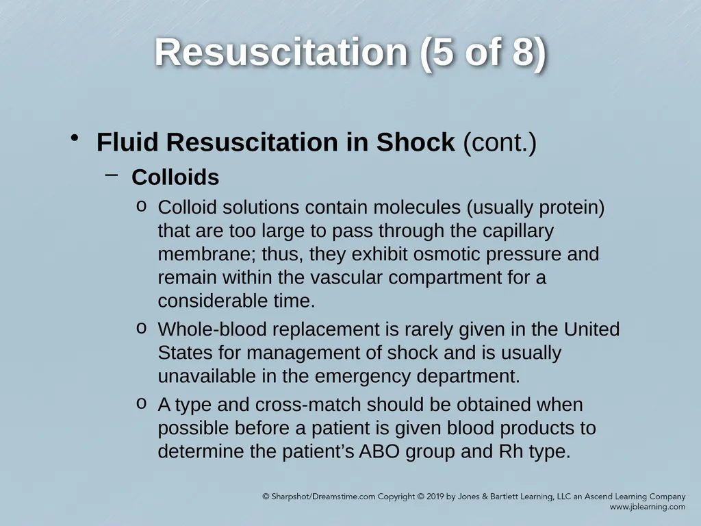
Colloid solutions contain molecules (usually protein) that are too large to pass through the capillary membrane; thus, they exhibit osmotic pressure and remain within the vascular compartment for a considerable time.
Whole-blood replacement is rarely given in the United States for management of shock and is usually unavailable in the emergency department.
A type and cross-match should be obtained when possible before a patient is given blood products to determine the patient’s ABO group and Rh type.<br>
Colloids
Colloid solutions contain molecules (usually protein) that are too large to pass through the capillary membrane; thus, they exhibit osmotic pressure and remain within the vascular compartment for a considerable time.
Whole-blood replacement is rarely given in the United States for management of shock and is usually unavailable in the emergency department.
A type and cross-match should be obtained when possible before a patient is given blood products to determine the patient’s ABO group and Rh type.<br>
50
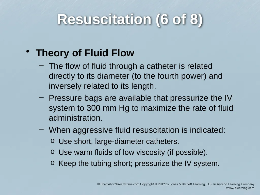
Resuscitation (6 of 8) Theory of Fluid Flow
The flow of fluid through a catheter is related directly to its diameter (to the fourth power) and inversely related to its length.
Pressure bags are available that pressurize the IV system to 300 mm Hg to maximize the rate of fluid administration.
When aggressive fluid resuscitation is indicated:
Use short, large-diameter catheters.
Use warm fluids of low viscosity (if possible).
Keep the tubing short; pressurize the IV system.<br>
The flow of fluid through a catheter is related directly to its diameter (to the fourth power) and inversely related to its length.
Pressure bags are available that pressurize the IV system to 300 mm Hg to maximize the rate of fluid administration.
When aggressive fluid resuscitation is indicated:
Use short, large-diameter catheters.
Use warm fluids of low viscosity (if possible).
Keep the tubing short; pressurize the IV system.<br>
51
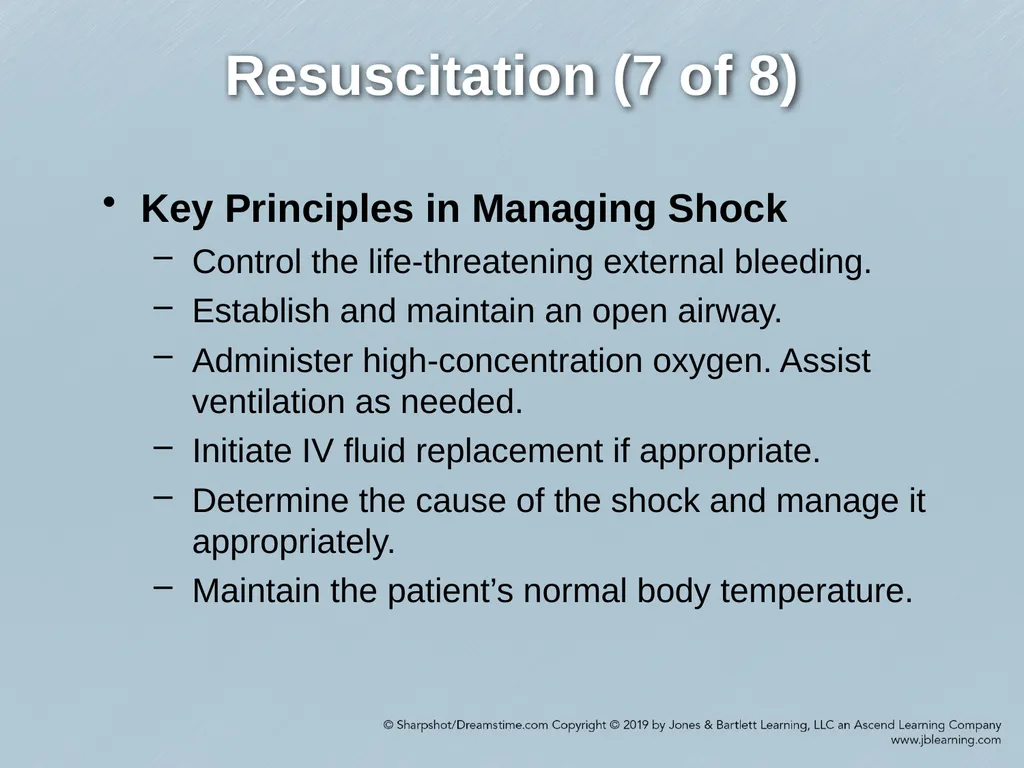
Resuscitation (7 of 8) Key Principles in Managing Shock
Control the life-threatening external bleeding.
Establish and maintain an open airway.
Administer high-concentration oxygen. Assist ventilation as needed.
Initiate IV fluid replacement if appropriate.
Determine the cause of the shock and manage it appropriately.
Maintain the patient’s normal body temperature.<br>
Control the life-threatening external bleeding.
Establish and maintain an open airway.
Administer high-concentration oxygen. Assist ventilation as needed.
Initiate IV fluid replacement if appropriate.
Determine the cause of the shock and manage it appropriately.
Maintain the patient’s normal body temperature.<br>
52
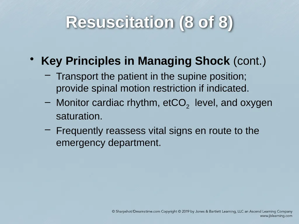
Resuscitation (8 of 8) Key Principles in Managing Shock (cont.)
Transport the patient in the supine position; provide spinal motion restriction if indicated.
Monitor cardiac rhythm, etCO2 level, and oxygen saturation.
Frequently reassess vital signs en route to the emergency department.<br>
Transport the patient in the supine position; provide spinal motion restriction if indicated.
Monitor cardiac rhythm, etCO2 level, and oxygen saturation.
Frequently reassess vital signs en route to the emergency department.<br>
53
Management of Specific Forms of Shock (1 of 5) Hypovolemic Shock
The management of hypovolemic shock is not considered complete until the volume is replaced and the cause or causes of shock are corrected.
Crystalloid fluid replacement is done in cases of simple dehydration or volume replacement because of hemorrhage, definitive surgery, critical care support, and postoperative rehabilitation.
In nontraumatic hypovolemic shock, infuse crystalloid fluids beginning with 30 mL/kg to a maximum of 1 L over less than 15 min.<br>
The management of hypovolemic shock is not considered complete until the volume is replaced and the cause or causes of shock are corrected.
Crystalloid fluid replacement is done in cases of simple dehydration or volume replacement because of hemorrhage, definitive surgery, critical care support, and postoperative rehabilitation.
In nontraumatic hypovolemic shock, infuse crystalloid fluids beginning with 30 mL/kg to a maximum of 1 L over less than 15 min.<br>
54
Management of Specific Forms of Shock (2 of 5) Cardiogenic Shock
The management of cardiogenic shock focuses on improving the pumping action of the heart and on managing cardiac rhythm irregularities.
Drug therapy for cardiogenic shock varies according to the cause of the shock. It can include vasopressors (norepinephrine, epinephrine, and dopamine), inotropic drugs, and antidysrhythmics (usually after fluid infusion).<br>
The management of cardiogenic shock focuses on improving the pumping action of the heart and on managing cardiac rhythm irregularities.
Drug therapy for cardiogenic shock varies according to the cause of the shock. It can include vasopressors (norepinephrine, epinephrine, and dopamine), inotropic drugs, and antidysrhythmics (usually after fluid infusion).<br>
55
Management of Specific Forms of Shock (3 of 5) Obstructive Shock
Common causes of obstructive shock include pericardial tamponade, tension pneumothorax, and pulmonary embolism.
The goals of prehospital care are to identify and relieve the obstruction to flow.
Emergency care for pericardial tamponade includes aggressive fluid replacement to maintain adequate preload.<br>
Common causes of obstructive shock include pericardial tamponade, tension pneumothorax, and pulmonary embolism.
The goals of prehospital care are to identify and relieve the obstruction to flow.
Emergency care for pericardial tamponade includes aggressive fluid replacement to maintain adequate preload.<br>
56
Management of Specific Forms of Shock (4 of 5) Distributive Shock
Neurogenic Shock
The management of neurogenic shock is similar to the management of hypovolemia.
Anaphylactic Shock
IM administration of epinephrine is the treatment of choice for patients with acute anaphylactic reactions.
Crystalloids may compensate for the increased container size caused by vasodilation resulting from histamine release during an anaphylactic reaction.<br>
Neurogenic Shock
The management of neurogenic shock is similar to the management of hypovolemia.
Anaphylactic Shock
IM administration of epinephrine is the treatment of choice for patients with acute anaphylactic reactions.
Crystalloids may compensate for the increased container size caused by vasodilation resulting from histamine release during an anaphylactic reaction.<br>
57
Management of Specific Forms of Shock (5 of 5) Distributive Shock (cont.)
Septic Shock
The management of septic shock in the prehospital setting can include the management of hypovolemia (if present) and the correction of metabolic acid–base imbalance.
Patients with adrenal insufficiency associated with sepsis may not respond to fluid resuscitation or sympathomimetic drugs. In such a case, a corticosteroid is administered as part of the shock resuscitation.<br>
Septic Shock
The management of septic shock in the prehospital setting can include the management of hypovolemia (if present) and the correction of metabolic acid–base imbalance.
Patients with adrenal insufficiency associated with sepsis may not respond to fluid resuscitation or sympathomimetic drugs. In such a case, a corticosteroid is administered as part of the shock resuscitation.<br>
58
Integration of Patient Assessment and the Treatment Plan (1 of 2) Many complications of shock are possible.
The goals of prehospital care for the patient with severe hemorrhage or shock include:
Rapid recognition of the event
Initiation of treatment and prevention of additional injury
Rapid transport to an appropriate medical facility by ground or air ambulance
Advanced notification of the receiving facility<br>
The goals of prehospital care for the patient with severe hemorrhage or shock include:
Rapid recognition of the event
Initiation of treatment and prevention of additional injury
Rapid transport to an appropriate medical facility by ground or air ambulance
Advanced notification of the receiving facility<br>
59
Integration of Patient Assessment and the Treatment Plan (2 of 2) The paramedic should follow guidelines established by the local protocol and medical direction in determining the appropriate prehospital level of care for patients and in identifying the appropriate medical facility for patient transport.<br>