
CHAPTER 6 Proteins and Heme Derivatives Learning
PC
Published · 27 slides · 0 views
1 / 1

Description
CHAPTER 6 Proteins and Heme Derivatives Learning Objectives (1 of 2) Define key terms associated with amino acids, proteins, and heme derivatives. Describe the basic structure and general properties of amino acids. Differentiate between
Related Topics
Share
Embed code
Download this presentation From Below
"CHAPTER 6 Proteins and Heme Derivatives Learning" is the property of its rightful owner. Permission is granted to download and print the materials on this website for personal, non-commercial use only, and to display it on your personal computer provided you do not modify the materials and that you retain all copyright notices contained in the materials. By downloading content from our website, you accept the terms of this agreement.
Presentation Transcript
01
CHAPTER 6 Proteins and Heme Derivatives<br>
02
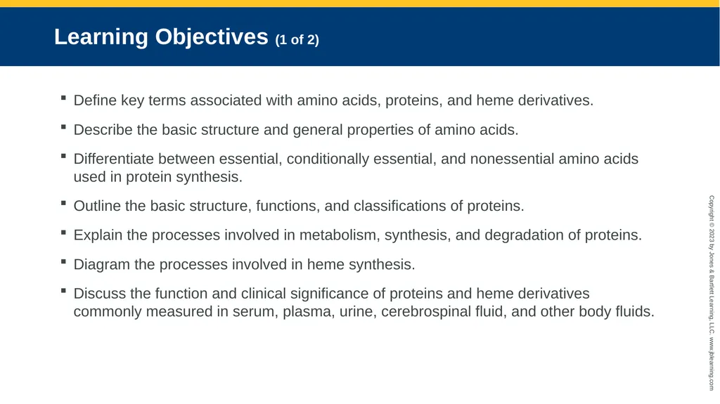
Learning Objectives (1 of 2) Define key terms associated with amino acids, proteins, and heme derivatives.
Describe the basic structure and general properties of amino acids.
Differentiate between essential, conditionally essential, and nonessential amino acids used in protein synthesis.
Outline the basic structure, functions, and classifications of proteins.
Explain the processes involved in metabolism, synthesis, and degradation of proteins.
Diagram the processes involved in heme synthesis.
Discuss the function and clinical significance of proteins and heme derivatives commonly measured in serum, plasma, urine, cerebrospinal fluid, and other body fluids.<br>
Describe the basic structure and general properties of amino acids.
Differentiate between essential, conditionally essential, and nonessential amino acids used in protein synthesis.
Outline the basic structure, functions, and classifications of proteins.
Explain the processes involved in metabolism, synthesis, and degradation of proteins.
Diagram the processes involved in heme synthesis.
Discuss the function and clinical significance of proteins and heme derivatives commonly measured in serum, plasma, urine, cerebrospinal fluid, and other body fluids.<br>
03
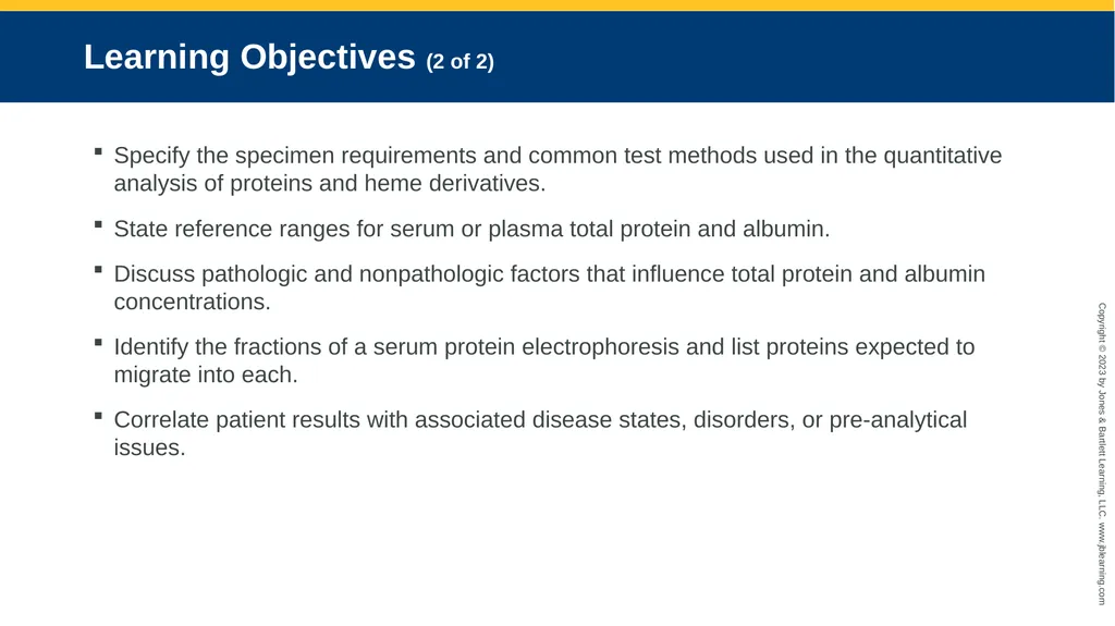
Learning Objectives (2 of 2) Specify the specimen requirements and common test methods used in the quantitative analysis of proteins and heme derivatives.
State reference ranges for serum or plasma total protein and albumin.
Discuss pathologic and nonpathologic factors that influence total protein and albumin concentrations.
Identify the fractions of a serum protein electrophoresis and list proteins expected to migrate into each.
Correlate patient results with associated disease states, disorders, or pre-analytical issues.<br>
State reference ranges for serum or plasma total protein and albumin.
Discuss pathologic and nonpathologic factors that influence total protein and albumin concentrations.
Identify the fractions of a serum protein electrophoresis and list proteins expected to migrate into each.
Correlate patient results with associated disease states, disorders, or pre-analytical issues.<br>
04
Amino Acids (1 of 3) Basic Structure
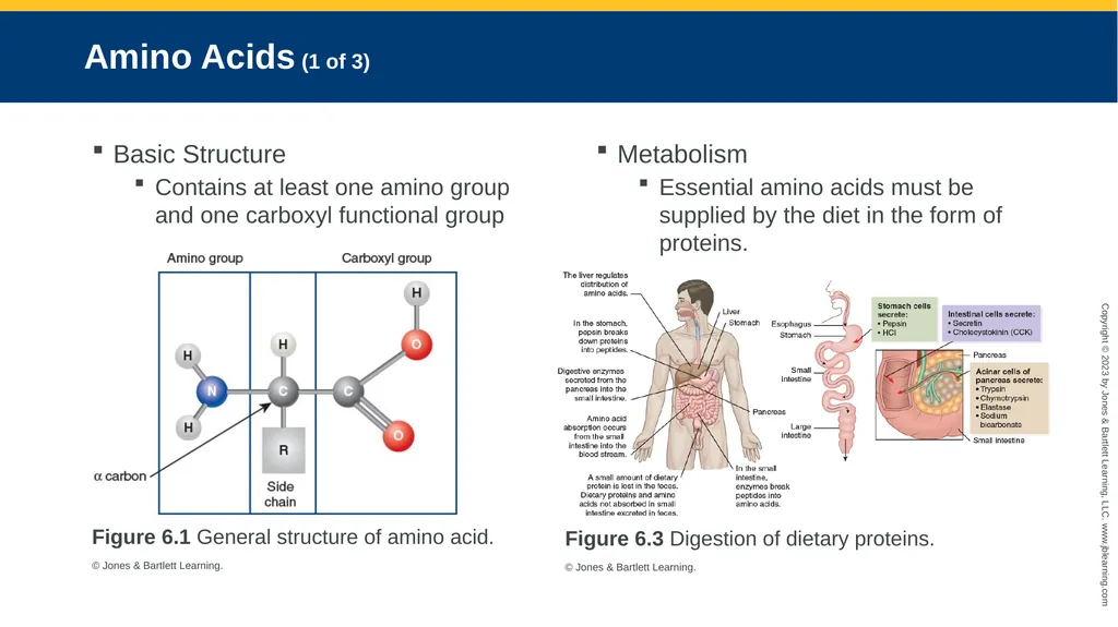
Contains at least one amino group and one carboxyl functional group Figure 6.1 General structure of amino acid.
© Jones & Bartlett Learning. Metabolism
Essential amino acids must be supplied by the diet in the form of proteins. Figure 6.3 Digestion of dietary proteins.
© Jones & Bartlett Learning.<br>
Contains at least one amino group and one carboxyl functional group Figure 6.1 General structure of amino acid.
© Jones & Bartlett Learning. Metabolism
Essential amino acids must be supplied by the diet in the form of proteins. Figure 6.3 Digestion of dietary proteins.
© Jones & Bartlett Learning.<br>
05
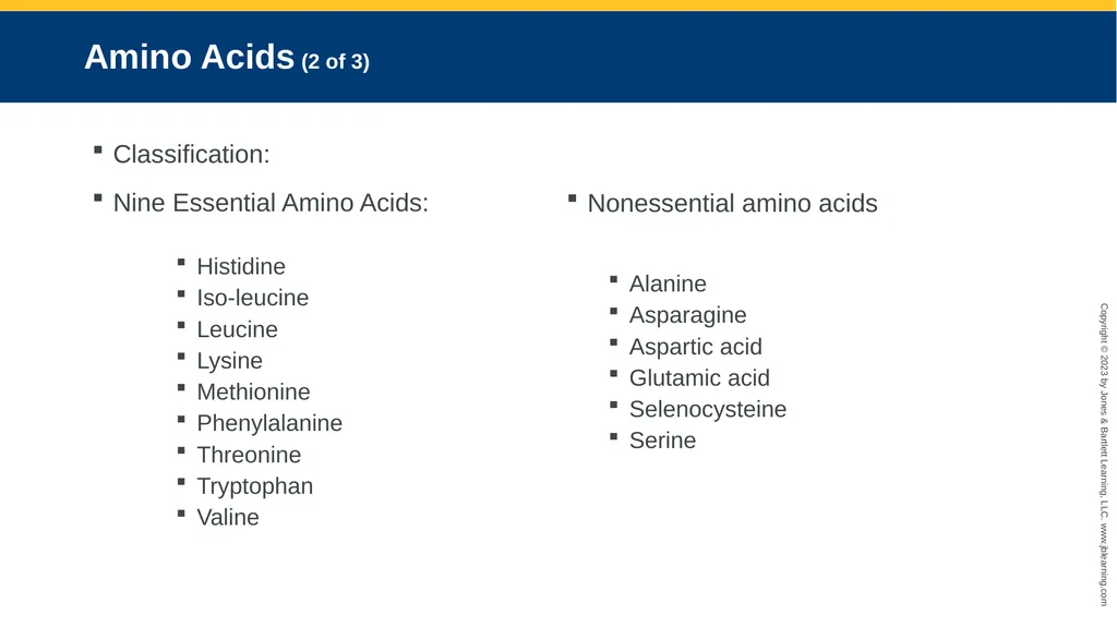
Amino Acids (2 of 3) Classification:
Nine Essential Amino Acids:
Histidine
Iso-leucine
Leucine
Lysine
Methionine
Phenylalanine
Threonine
Tryptophan
Valine Nonessential amino acids
Alanine
Asparagine
Aspartic acid
Glutamic acid
Selenocysteine
Serine<br>
Nine Essential Amino Acids:
Histidine
Iso-leucine
Leucine
Lysine
Methionine
Phenylalanine
Threonine
Tryptophan
Valine Nonessential amino acids
Alanine
Asparagine
Aspartic acid
Glutamic acid
Selenocysteine
Serine<br>
06
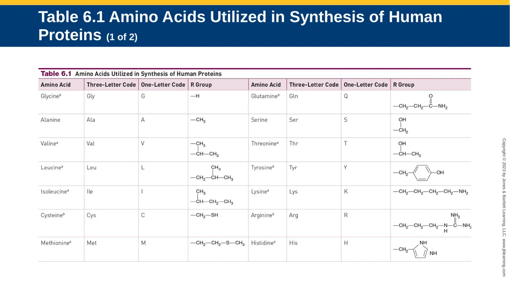
Table 6.1 Amino Acids Utilized in Synthesis of Human Proteins (1 of 2)<br>
07
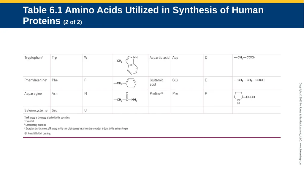
Table 6.1 Amino Acids Utilized in Synthesis of Human Proteins (2 of 2)<br>
08
Amino Acids (3 of 3) Conditionally Essential Amino Acids
Arginine
Cysteine
Glutamine
Glycine
Proline
Tyrosine Aminoacidopathies
A class of inborn errors of metabolism in which an enzyme defect inhibits the body’s ability to metabolize certain amino acids
Can cause severe medical complications, such as brain damage, due to the accumulation of toxic amino acids or their by-products in the blood and tissues<br>
Arginine
Cysteine
Glutamine
Glycine
Proline
Tyrosine Aminoacidopathies
A class of inborn errors of metabolism in which an enzyme defect inhibits the body’s ability to metabolize certain amino acids
Can cause severe medical complications, such as brain damage, due to the accumulation of toxic amino acids or their by-products in the blood and tissues<br>
09
Proteins (1 of 3) Basic Structure
Consist of the elements carbon, oxygen, hydrogen, nitrogen, and sulfur
Are polymers built from one or more unbranched chains of amino acids
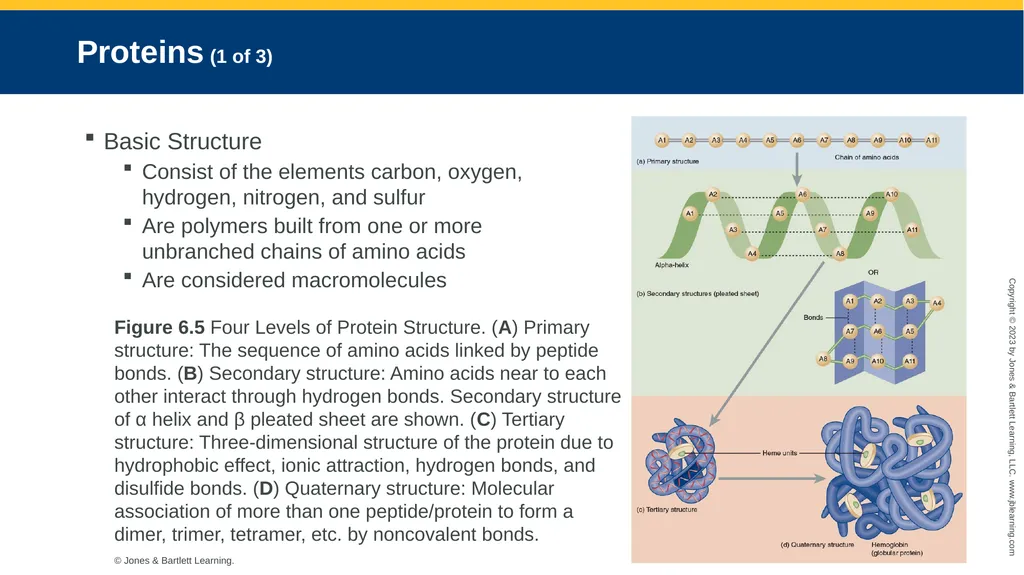
Are considered macromolecules Figure 6.5 Four Levels of Protein Structure. (A) Primary structure: The sequence of amino acids linked by peptide bonds. (B) Secondary structure: Amino acids near to each other interact through hydrogen bonds. Secondary structure of α helix and β pleated sheet are shown. (C) Tertiary structure: Three-dimensional structure of the protein due to hydrophobic effect, ionic attraction, hydrogen bonds, and disulfide bonds. (D) Quaternary structure: Molecular association of more than one peptide/protein to form a dimer, trimer, tetramer, etc. by noncovalent bonds.
© Jones & Bartlett Learning.<br>
Consist of the elements carbon, oxygen, hydrogen, nitrogen, and sulfur
Are polymers built from one or more unbranched chains of amino acids
Are considered macromolecules Figure 6.5 Four Levels of Protein Structure. (A) Primary structure: The sequence of amino acids linked by peptide bonds. (B) Secondary structure: Amino acids near to each other interact through hydrogen bonds. Secondary structure of α helix and β pleated sheet are shown. (C) Tertiary structure: Three-dimensional structure of the protein due to hydrophobic effect, ionic attraction, hydrogen bonds, and disulfide bonds. (D) Quaternary structure: Molecular association of more than one peptide/protein to form a dimer, trimer, tetramer, etc. by noncovalent bonds.
© Jones & Bartlett Learning.<br>
10
Proteins (2 of 3) General Chemical Properties
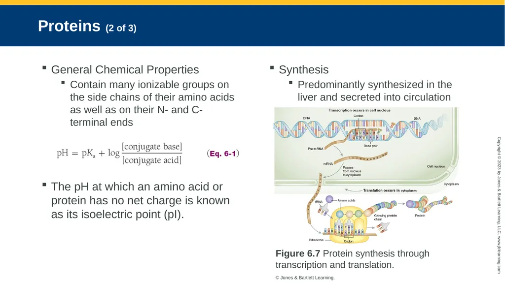
Contain many ionizable groups on the side chains of their amino acids as well as on their N- and C-terminal ends
The pH at which an amino acid or protein has no net charge is known as its isoelectric point (pI). Synthesis
Predominantly synthesized in the liver and secreted into circulation Figure 6.7 Protein synthesis through transcription and translation.
© Jones & Bartlett Learning.<br>
Contain many ionizable groups on the side chains of their amino acids as well as on their N- and C-terminal ends
The pH at which an amino acid or protein has no net charge is known as its isoelectric point (pI). Synthesis
Predominantly synthesized in the liver and secreted into circulation Figure 6.7 Protein synthesis through transcription and translation.
© Jones & Bartlett Learning.<br>
11
Proteins (3 of 3) Catabolism and Nitrogen Balance
Proteins in the body are repetitively synthesized (anabolism) and then degraded (catabolism) allowing for efficient recycling of amino acids.
When in excess, amino acids can be converted to urea for excretion by the kidneys and into glucose or ketones to be used as an alternative source of energy. Classification
Classified based on their structure, composition, and function
Simple proteins contain peptide chains composed of only amino acids and may be globular or fibrous in shape.
Complex proteins consist of a protein and a nonprotein. The non-amino part of a conjugated protein is generally referred to as the prosthetic group.<br>
Proteins in the body are repetitively synthesized (anabolism) and then degraded (catabolism) allowing for efficient recycling of amino acids.
When in excess, amino acids can be converted to urea for excretion by the kidneys and into glucose or ketones to be used as an alternative source of energy. Classification
Classified based on their structure, composition, and function
Simple proteins contain peptide chains composed of only amino acids and may be globular or fibrous in shape.
Complex proteins consist of a protein and a nonprotein. The non-amino part of a conjugated protein is generally referred to as the prosthetic group.<br>
12
Plasma Proteins (1 of 2) Prealbumin
Prealbumin, or transthyretin, is so named because it migrates before albumin in classic serum protein electrophoresis (SPE).
Is a transport protein for the thyroid hormones, thyroxine (T4) and triiodothyronine (T3)
Low concentration may also indicate poor nutritional status. Albumin
Is a negative acute-phase reactant, meaning it decreases in the presence of acute disease states, such as inflammation, infection, or injury
Albumin is responsible for nearly 80% of the colloid osmotic pressure.
Increased renal loss of albumin, termed albuminuria, commonly occurs in renal disease.<br>
Prealbumin, or transthyretin, is so named because it migrates before albumin in classic serum protein electrophoresis (SPE).
Is a transport protein for the thyroid hormones, thyroxine (T4) and triiodothyronine (T3)
Low concentration may also indicate poor nutritional status. Albumin
Is a negative acute-phase reactant, meaning it decreases in the presence of acute disease states, such as inflammation, infection, or injury
Albumin is responsible for nearly 80% of the colloid osmotic pressure.
Increased renal loss of albumin, termed albuminuria, commonly occurs in renal disease.<br>
13
Plasma Proteins (2 of 2) Globulins
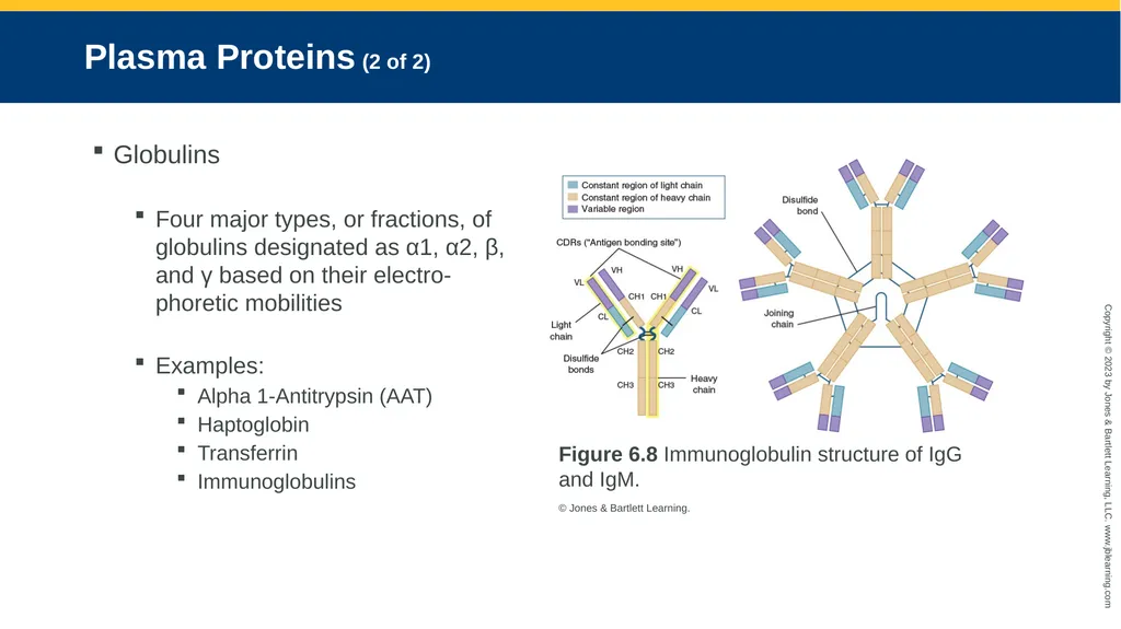
Four major types, or fractions, of globulins designated as α1, α2, β, and γ based on their electro-phoretic mobilities
Examples:
Alpha 1-Antitrypsin (AAT)
Haptoglobin
Transferrin
Immunoglobulins Figure 6.8 Immunoglobulin structure of IgG and IgM.
© Jones & Bartlett Learning.<br>
Four major types, or fractions, of globulins designated as α1, α2, β, and γ based on their electro-phoretic mobilities
Examples:
Alpha 1-Antitrypsin (AAT)
Haptoglobin
Transferrin
Immunoglobulins Figure 6.8 Immunoglobulin structure of IgG and IgM.
© Jones & Bartlett Learning.<br>
14
Other Proteins of Clinical Significance (1 of 2) Troponin
Cardiac troponin (cTn) represents a complex of regulatory proteins: troponin C (TnC), troponin I (cTnI), and troponin T (cTnT), that are specific to cardiac muscle.
cTn measurement above the decision limit, along with clinical evidence, is indicative of myocardial injury. Natriuretic Peptides
Family of structurally related hormones that include atrial natriuretic pep-tide (ANP), B-type (or brain) natriuretic peptide (BNP), C-type natriuretic peptide (CNP), and dendroaspis natriuretic peptide (DNP)
NT-proBNP and BNP are found in greatest concentration in the left ventricular myocardium.<br>
Cardiac troponin (cTn) represents a complex of regulatory proteins: troponin C (TnC), troponin I (cTnI), and troponin T (cTnT), that are specific to cardiac muscle.
cTn measurement above the decision limit, along with clinical evidence, is indicative of myocardial injury. Natriuretic Peptides
Family of structurally related hormones that include atrial natriuretic pep-tide (ANP), B-type (or brain) natriuretic peptide (BNP), C-type natriuretic peptide (CNP), and dendroaspis natriuretic peptide (DNP)
NT-proBNP and BNP are found in greatest concentration in the left ventricular myocardium.<br>
15
Other Proteins of Clinical Significance (2 of 2) Fibronectin
Proteins are found in plasma and on cell surfaces and can be synthesized by hepatocytes, endothelial cells, peritoneal macrophages, and fibroblasts.
Fetal fibronectin (fFN) is used to help predict the short-term risk of premature delivery. Cystatin C
Cystatin C may be used to evaluate glomerular function in individuals for whom creatinine measurements may be misleading.<br>
Proteins are found in plasma and on cell surfaces and can be synthesized by hepatocytes, endothelial cells, peritoneal macrophages, and fibroblasts.
Fetal fibronectin (fFN) is used to help predict the short-term risk of premature delivery. Cystatin C
Cystatin C may be used to evaluate glomerular function in individuals for whom creatinine measurements may be misleading.<br>
16
Plasma Total Protein Abnormalities Hypoproteinemia
Causes of hypoproteinemia include excessive loss, decreased synthesis, and increased protein catabolism.
Excessive loss of plasma proteins may occur as a result of renal disease.
May also be the result of decreased intake of proteins and amino acids associated with malnutrition, maldigestion, or malabsorption syndromes Hyperproteinemia
Is more likely the result of dehydration
Dehydration may result from a variety of conditions including vomiting, diarrhea, excessive sweating, diabetic acidosis, and hypoaldosteronism.
May also be the result of excessive production of proteins, primarily of the γ- globulins<br>
Causes of hypoproteinemia include excessive loss, decreased synthesis, and increased protein catabolism.
Excessive loss of plasma proteins may occur as a result of renal disease.
May also be the result of decreased intake of proteins and amino acids associated with malnutrition, maldigestion, or malabsorption syndromes Hyperproteinemia
Is more likely the result of dehydration
Dehydration may result from a variety of conditions including vomiting, diarrhea, excessive sweating, diabetic acidosis, and hypoaldosteronism.
May also be the result of excessive production of proteins, primarily of the γ- globulins<br>
17
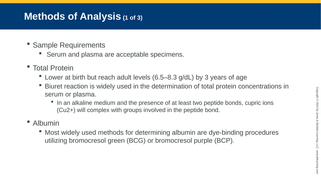
Methods of Analysis (1 of 3) Sample Requirements
Serum and plasma are acceptable specimens.
Total Protein
Lower at birth but reach adult levels (6.5–8.3 g/dL) by 3 years of age
Biuret reaction is widely used in the determination of total protein concentrations in serum or plasma.
In an alkaline medium and the presence of at least two peptide bonds, cupric ions (Cu2+) will complex with groups involved in the peptide bond.
Albumin
Most widely used methods for determining albumin are dye-binding procedures utilizing bromocresol green (BCG) or bromocresol purple (BCP).<br>
Serum and plasma are acceptable specimens.
Total Protein
Lower at birth but reach adult levels (6.5–8.3 g/dL) by 3 years of age
Biuret reaction is widely used in the determination of total protein concentrations in serum or plasma.
In an alkaline medium and the presence of at least two peptide bonds, cupric ions (Cu2+) will complex with groups involved in the peptide bond.
Albumin
Most widely used methods for determining albumin are dye-binding procedures utilizing bromocresol green (BCG) or bromocresol purple (BCP).<br>
18
Methods of Analysis (2 of 3) Electrophoresis
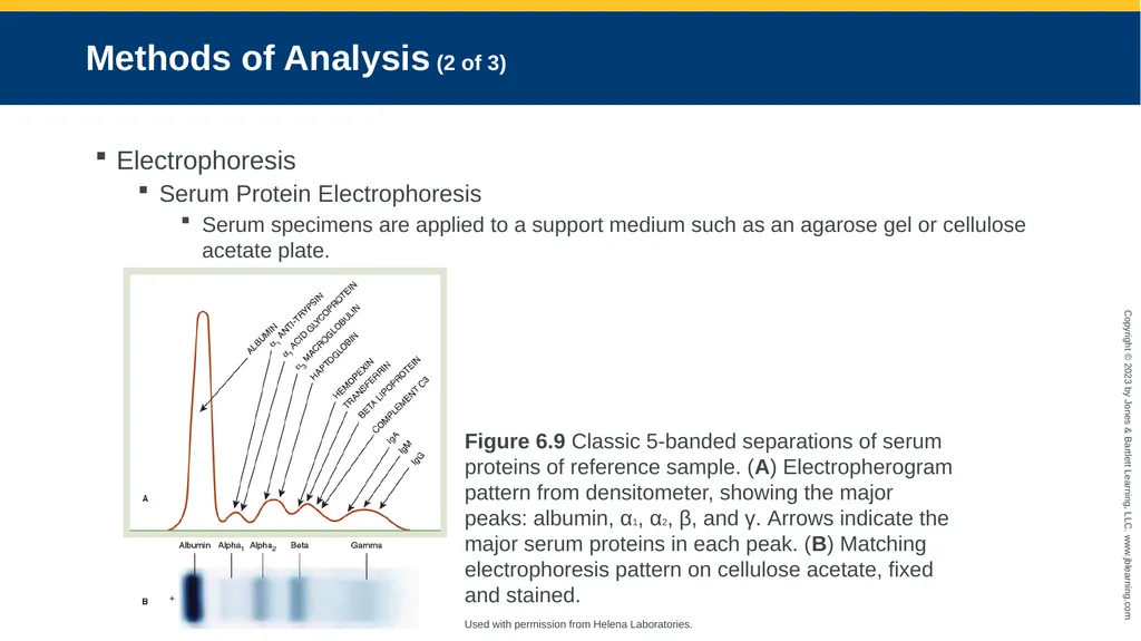
Serum Protein Electrophoresis
Serum specimens are applied to a support medium such as an agarose gel or cellulose acetate plate. Figure 6.9 Classic 5-banded separations of serum proteins of reference sample. (A) Electropherogram pattern from densitometer, showing the major peaks: albumin, α1, α2, β, and γ. Arrows indicate the major serum proteins in each peak. (B) Matching electrophoresis pattern on cellulose acetate, fixed and stained.
Used with permission from Helena Laboratories.<br>
Serum Protein Electrophoresis
Serum specimens are applied to a support medium such as an agarose gel or cellulose acetate plate. Figure 6.9 Classic 5-banded separations of serum proteins of reference sample. (A) Electropherogram pattern from densitometer, showing the major peaks: albumin, α1, α2, β, and γ. Arrows indicate the major serum proteins in each peak. (B) Matching electrophoresis pattern on cellulose acetate, fixed and stained.
Used with permission from Helena Laboratories.<br>
19
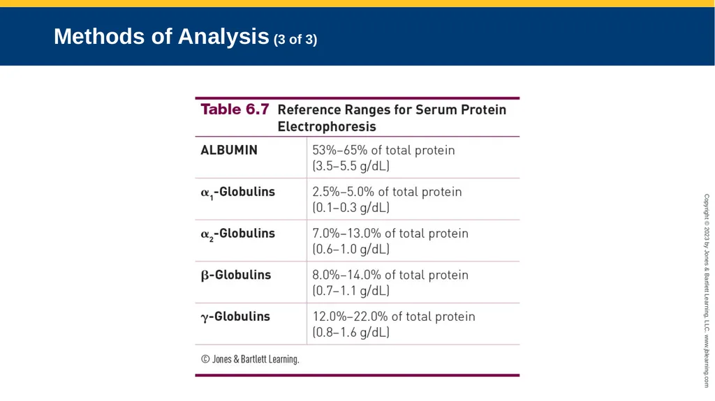
Methods of Analysis (3 of 3)<br>
20
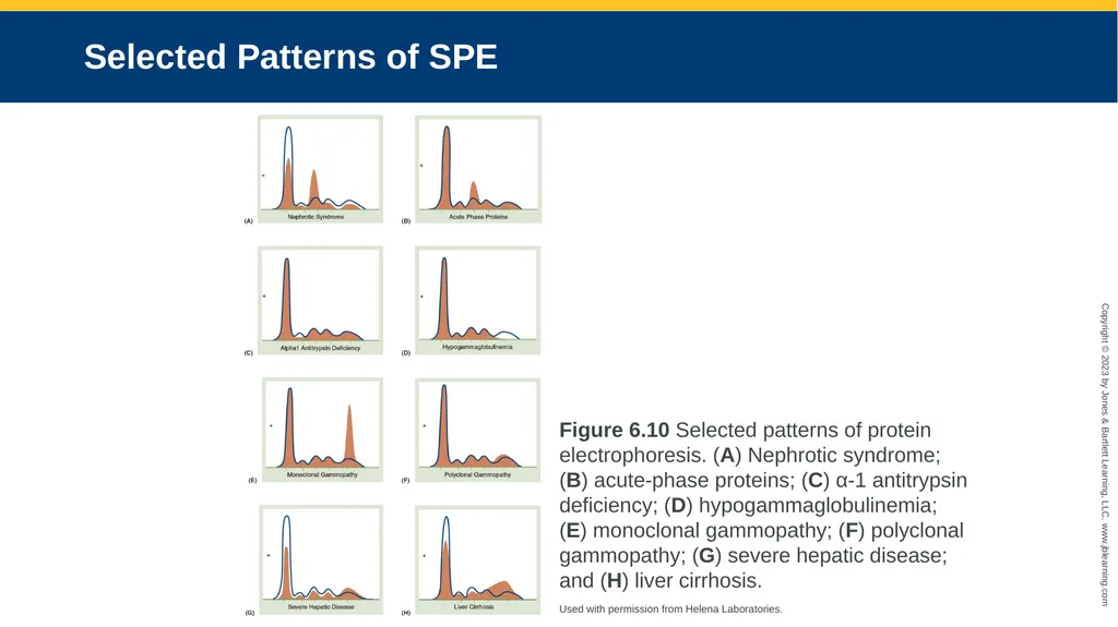
Selected Patterns of SPE Figure 6.10 Selected patterns of protein electrophoresis. (A) Nephrotic syndrome; (B) acute-phase proteins; (C) α-1 antitrypsin deficiency; (D) hypogammaglobulinemia; (E) monoclonal gammopathy; (F) polyclonal gammopathy; (G) severe hepatic disease; and (H) liver cirrhosis.
Used with permission from Helena Laboratories.<br>
Used with permission from Helena Laboratories.<br>
21
Methods of Analysis Capillary Electrophoresis
A collection of techniques in which separation takes place inside a silica capillary
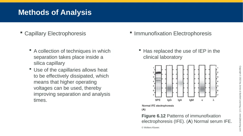
Use of the capillaries allows heat to be effectively dissipated, which means that higher operating voltages can be used, thereby improving separation and analysis times. Immunofixation Electrophoresis
Has replaced the use of IEP in the clinical laboratory Figure 6.12 Patterns of immunofixation electrophoresis (IFE). (A) Normal serum IFE.
© Wolters Kluwer.<br>
A collection of techniques in which separation takes place inside a silica capillary
Use of the capillaries allows heat to be effectively dissipated, which means that higher operating voltages can be used, thereby improving separation and analysis times. Immunofixation Electrophoresis
Has replaced the use of IEP in the clinical laboratory Figure 6.12 Patterns of immunofixation electrophoresis (IFE). (A) Normal serum IFE.
© Wolters Kluwer.<br>
22
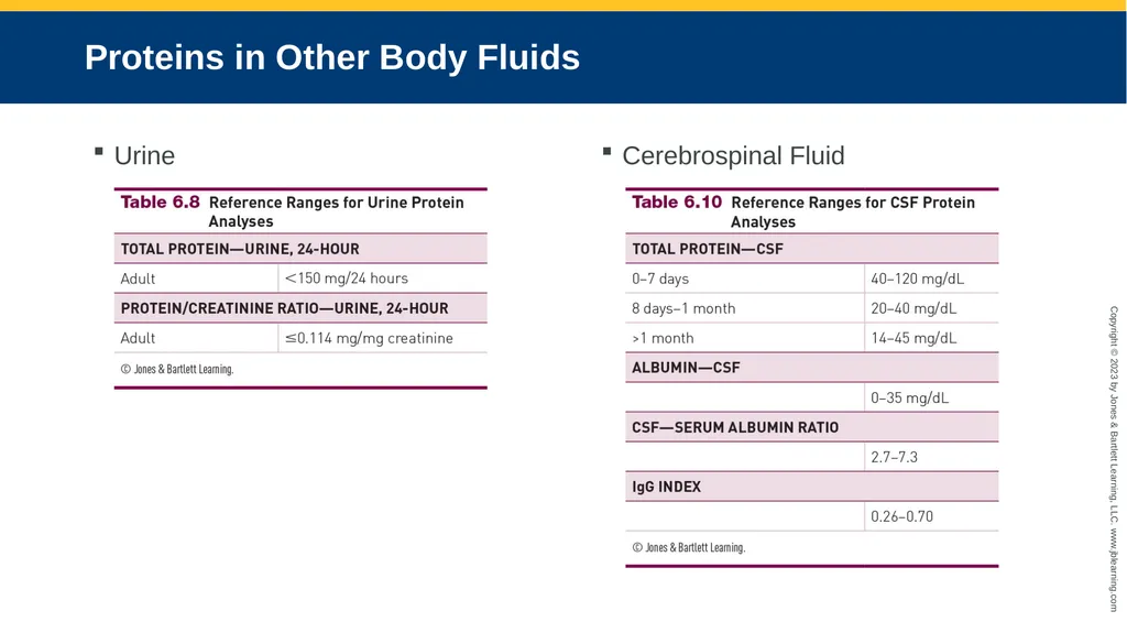
Proteins in Other Body Fluids Urine Cerebrospinal Fluid<br>
23
Hemoglobin Is classified as a transport protein. Its role is to transport oxygen from the lungs to the tissues, and transport carbon dioxide back to the lungs.
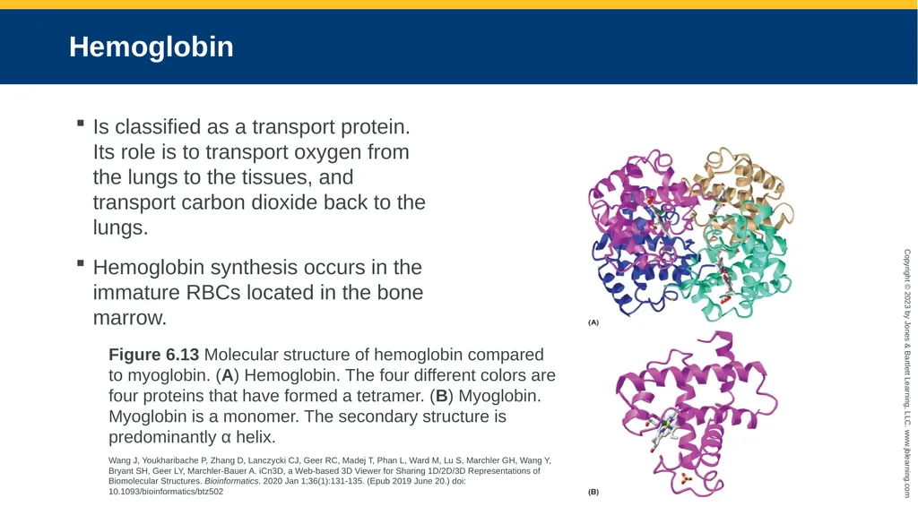
Hemoglobin synthesis occurs in the immature RBCs located in the bone marrow. Figure 6.13 Molecular structure of hemoglobin compared to myoglobin. (A) Hemoglobin. The four different colors are four proteins that have formed a tetramer. (B) Myoglobin. Myoglobin is a monomer. The secondary structure is predominantly α helix.
Wang J, Youkharibache P, Zhang D, Lanczycki CJ, Geer RC, Madej T, Phan L, Ward M, Lu S, Marchler GH, Wang Y, Bryant SH, Geer LY, Marchler-Bauer A. iCn3D, a Web-based 3D Viewer for Sharing 1D/2D/3D Representations of Biomolecular Structures. Bioinformatics. 2020 Jan 1;36(1):131-135. (Epub 2019 June 20.) doi: 10.1093/bioinformatics/btz502<br>
Hemoglobin synthesis occurs in the immature RBCs located in the bone marrow. Figure 6.13 Molecular structure of hemoglobin compared to myoglobin. (A) Hemoglobin. The four different colors are four proteins that have formed a tetramer. (B) Myoglobin. Myoglobin is a monomer. The secondary structure is predominantly α helix.
Wang J, Youkharibache P, Zhang D, Lanczycki CJ, Geer RC, Madej T, Phan L, Ward M, Lu S, Marchler GH, Wang Y, Bryant SH, Geer LY, Marchler-Bauer A. iCn3D, a Web-based 3D Viewer for Sharing 1D/2D/3D Representations of Biomolecular Structures. Bioinformatics. 2020 Jan 1;36(1):131-135. (Epub 2019 June 20.) doi: 10.1093/bioinformatics/btz502<br>
24
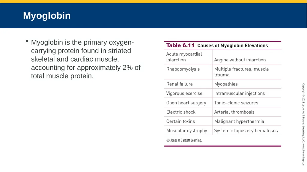
Myoglobin Myoglobin is the primary oxygen-carrying protein found in striated skeletal and cardiac muscle, accounting for approximately 2% of total muscle protein.<br>
25
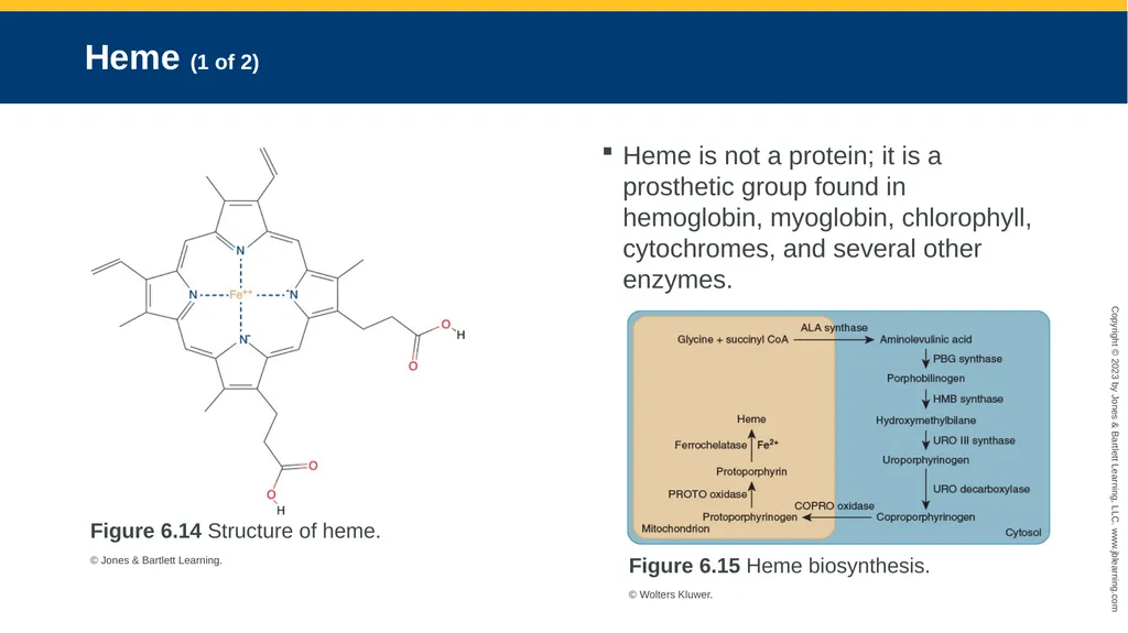
Heme (1 of 2) Figure 6.14 Structure of heme.
© Jones & Bartlett Learning. Heme is not a protein; it is a prosthetic group found in hemoglobin, myoglobin, chlorophyll, cytochromes, and several other enzymes. Figure 6.15 Heme biosynthesis.
© Wolters Kluwer.<br>
© Jones & Bartlett Learning. Heme is not a protein; it is a prosthetic group found in hemoglobin, myoglobin, chlorophyll, cytochromes, and several other enzymes. Figure 6.15 Heme biosynthesis.
© Wolters Kluwer.<br>
26
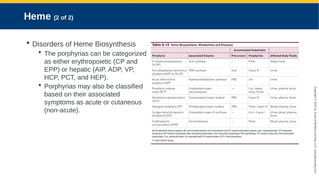
Heme (2 of 2) Disorders of Heme Biosynthesis
The porphyrias can be categorized as either erythropoietic (CP and EPP) or hepatic (AIP, ADP, VP, HCP, PCT, and HEP).
Porphyrias may also be classified based on their associated symptoms as acute or cutaneous (non-acute).<br>
The porphyrias can be categorized as either erythropoietic (CP and EPP) or hepatic (AIP, ADP, VP, HCP, PCT, and HEP).
Porphyrias may also be classified based on their associated symptoms as acute or cutaneous (non-acute).<br>
27
Conclusion<br>