
Chapter 9 Congenital and Hereditary Diseases
MT
Published · 50 slides · 0 views

1 / 1




Description
Chapter 9 Congenital and Hereditary Diseases Learning Objectives (1 of 2) Describe Common causes of congenital malformations and their incidence Abnormalities of sex chromosomes and manifestations Common genetic abnormalities and
Related Topics
Share
Embed code
Download this presentation From Below
"Chapter 9 Congenital and Hereditary Diseases" is the property of its rightful owner. Permission is granted to download and print the materials on this website for personal, non-commercial use only, and to display it on your personal computer provided you do not modify the materials and that you retain all copyright notices contained in the materials. By downloading content from our website, you accept the terms of this agreement.
Presentation Transcript
01
Chapter 9 Congenital and Hereditary Diseases<br>
02
Learning Objectives (1 of 2) Describe
Common causes of congenital malformations and their incidence
Abnormalities of sex chromosomes and manifestations
Common genetic abnormalities and transmission
Compare phenylketonuria versus hemophilia in terms of transmission and clinical manifestations
Describe congenital malformations resulting from uterine injury<br>
Common causes of congenital malformations and their incidence
Abnormalities of sex chromosomes and manifestations
Common genetic abnormalities and transmission
Compare phenylketonuria versus hemophilia in terms of transmission and clinical manifestations
Describe congenital malformations resulting from uterine injury<br>
03
Learning Objectives (2 of 2) Explain
Amniocentesis
Multifactorial inheritance
Example of multifactorial defect and relevant factors
List causes and clinical manifestations of Down syndrome
Discuss reasons for identifying 14/21 chromosome translocation carrier
Explain methods for diagnosing congenital abnormalities.<br>
Amniocentesis
Multifactorial inheritance
Example of multifactorial defect and relevant factors
List causes and clinical manifestations of Down syndrome
Discuss reasons for identifying 14/21 chromosome translocation carrier
Explain methods for diagnosing congenital abnormalities.<br>
04
Hereditary and Congenital Malformations Congenital disease: abnormality present at birth, even though it may not be detected until some time after birth
Hereditary or genetic disease: resulting from a chromosome abnormality or a defective gene<br>
Hereditary or genetic disease: resulting from a chromosome abnormality or a defective gene<br>
05
Genetics (1 of 5) Genetics: study of heredity; transmission of physical, biochemical, and physiologic traits from biological parents to their children
Disorders can be transmitted by gene mutations that can result in disability or death
Genetic information is carried in genes strung together on strands of DNA to form chromosomes
Except reproductive cells, every normal human cell has 46 chromosomes<br>
Disorders can be transmitted by gene mutations that can result in disability or death
Genetic information is carried in genes strung together on strands of DNA to form chromosomes
Except reproductive cells, every normal human cell has 46 chromosomes<br>
06
Genetics (2 of 5) Chromosomes: composed of double coils of DNA
Genes: segments of DNA chains
Genome: sum total of all genes contained in a cell’s chromosomes; the same in all cells
In human beings, normal chromosome component:
22 pairs of autosomes
1 pair of sex chromosomes (XX in females and XY in males)
Karyotype: a representation of a person’s set of chromosomes<br>
Genes: segments of DNA chains
Genome: sum total of all genes contained in a cell’s chromosomes; the same in all cells
In human beings, normal chromosome component:
22 pairs of autosomes
1 pair of sex chromosomes (XX in females and XY in males)
Karyotype: a representation of a person’s set of chromosomes<br>
07
Genetics (3 of 5) In somatic cells: chromosomes exist in pairs, one member of each pair is derived from male parent and other from female parent
With 22 pairs called autosomes
Except for the sex chromosomes, members of the pair are similar in size, shape, and appearance (homologous chromosomes)
Mitosis: cell division of somatic cells
Each of two new cells or daughter cells receives the same chromosomes as the parent cell<br>
With 22 pairs called autosomes
Except for the sex chromosomes, members of the pair are similar in size, shape, and appearance (homologous chromosomes)
Mitosis: cell division of somatic cells
Each of two new cells or daughter cells receives the same chromosomes as the parent cell<br>
08
Genetics (4 of 5) Not all genes are expressed in all cells and not all genes active all the time
Meiosis: cell division that occurs during development of egg and sperm
Number of chromosomes is reduced
Daughter cells receive only half of chromosomes possessed by the parent cell
DNA ultimately controls formation of essential substances throughout the life of every cell in the body through the genetic code (precise sequence of AT and CG pairs on the DNA molecule)<br>
Meiosis: cell division that occurs during development of egg and sperm
Number of chromosomes is reduced
Daughter cells receive only half of chromosomes possessed by the parent cell
DNA ultimately controls formation of essential substances throughout the life of every cell in the body through the genetic code (precise sequence of AT and CG pairs on the DNA molecule)<br>
09
Genetics (5 of 5) Genes control
Hereditary traits, cell reproduction, and daily functions of all cells
Cell function, through structures and chemicals made within the cell
RNA formation that controls formation of specific proteins; most are enzymes that assist in chemical reactions in cells<br>
Hereditary traits, cell reproduction, and daily functions of all cells
Cell function, through structures and chemicals made within the cell
RNA formation that controls formation of specific proteins; most are enzymes that assist in chemical reactions in cells<br>
10
Trait Predominance (1 of 2) Each parent contributes 1 set of chromosomes (or a set of genes) so that every child has two genes for every locus on the autosomal chromosomes
Some characteristics or traits of the child are determined by 1 gene that may have many variants (e.g. eye color)
Polygenic traits require interaction of ≥ 1 genes
Environmental factors may affect how genes are expressed
Alleles: variations in a particular gene
Homozygous: has identical alleles on each chromosome
Heterozygous: alleles are different<br>
Some characteristics or traits of the child are determined by 1 gene that may have many variants (e.g. eye color)
Polygenic traits require interaction of ≥ 1 genes
Environmental factors may affect how genes are expressed
Alleles: variations in a particular gene
Homozygous: has identical alleles on each chromosome
Heterozygous: alleles are different<br>
11
Trait Predominance (2 of 2) Children will express a dominant allele when one or both chromosomes in a pair carry it
A recessive allele is expressed only if both chromosomes carry the recessive alleles
For example, a child may receive a gene for brown eyes from one parent and a gene for blue eyes from the other parent
Gene for brown eyes is dominant
Gene for blue eyes is recessive
The dominant gene is more likely to be expressed and child is more likely to have brown eyes<br>
A recessive allele is expressed only if both chromosomes carry the recessive alleles
For example, a child may receive a gene for brown eyes from one parent and a gene for blue eyes from the other parent
Gene for brown eyes is dominant
Gene for blue eyes is recessive
The dominant gene is more likely to be expressed and child is more likely to have brown eyes<br>
12
Autosomal Inheritance For unknown reasons, on autosomal chromosomes, one allele may be more influential than the other in determining a specific trait
The more powerful or dominant gene is more likely to be expressed than the recessive gene<br>
The more powerful or dominant gene is more likely to be expressed than the recessive gene<br>
13
Sex-Linked Inheritance X and Y chromosomes are not literally a pair, as X is much larger than the Y chromosome
One X chromosome is inactivated (Lyon Hypothesis)
In males: one copy of most genes is on the X chromosome
Inheritance of these genes is called X-linked
A man will transmit one copy of each X-linked gene to his daughters and none to his sons
A woman will transmit one copy to each daughter or son<br>
One X chromosome is inactivated (Lyon Hypothesis)
In males: one copy of most genes is on the X chromosome
Inheritance of these genes is called X-linked
A man will transmit one copy of each X-linked gene to his daughters and none to his sons
A woman will transmit one copy to each daughter or son<br>
14
Multifactorial Inheritance Environmental factors can affect the expression of some genes
Example: child’s height will be within the range of height of both parents, but environmental factors such as nutritional patterns and health care also influence development
The better nourished, healthier children of two short parents may be taller than either<br>
Example: child’s height will be within the range of height of both parents, but environmental factors such as nutritional patterns and health care also influence development
The better nourished, healthier children of two short parents may be taller than either<br>
15
Genetically Determined Diseases Result from abnormalities of individual genes on the chromosomes
Chromosomes appear normal.
Some defects arise spontaneously.
Others may be caused by environmental teratogens (agents or influences that cause physical defects in the developing embryo)
Mutation: permanent change in genetic material that may occur spontaneously or after exposure of a cell to radiation, certain chemicals, or viruses<br>
Chromosomes appear normal.
Some defects arise spontaneously.
Others may be caused by environmental teratogens (agents or influences that cause physical defects in the developing embryo)
Mutation: permanent change in genetic material that may occur spontaneously or after exposure of a cell to radiation, certain chemicals, or viruses<br>
16
Factors in Congenital Malformations Genetic or hereditary disorders or diseases caused by abnormalities in an individual’s genetic material (genome)
Congenital disease or malformation: any abnormality present at birth
Four factors in congenital malformations
1. Chromosomal abnormalities
2. Abnormalities of individual genes
3. Intrauterine injury to embryo or fetus
4. Environmental factors<br>
Congenital disease or malformation: any abnormality present at birth
Four factors in congenital malformations
1. Chromosomal abnormalities
2. Abnormalities of individual genes
3. Intrauterine injury to embryo or fetus
4. Environmental factors<br>
17
Causes of Congenital Malformations 2-3% of all newborn infants have congenital defects
Additional 2-3% defects: NOT recognized at birth; developmental defects demonstrated later as infants grow older
25% to 50% spontaneously aborted embryos, fetuses, and stillborn infants have major malformations<br>
Additional 2-3% defects: NOT recognized at birth; developmental defects demonstrated later as infants grow older
25% to 50% spontaneously aborted embryos, fetuses, and stillborn infants have major malformations<br>
18
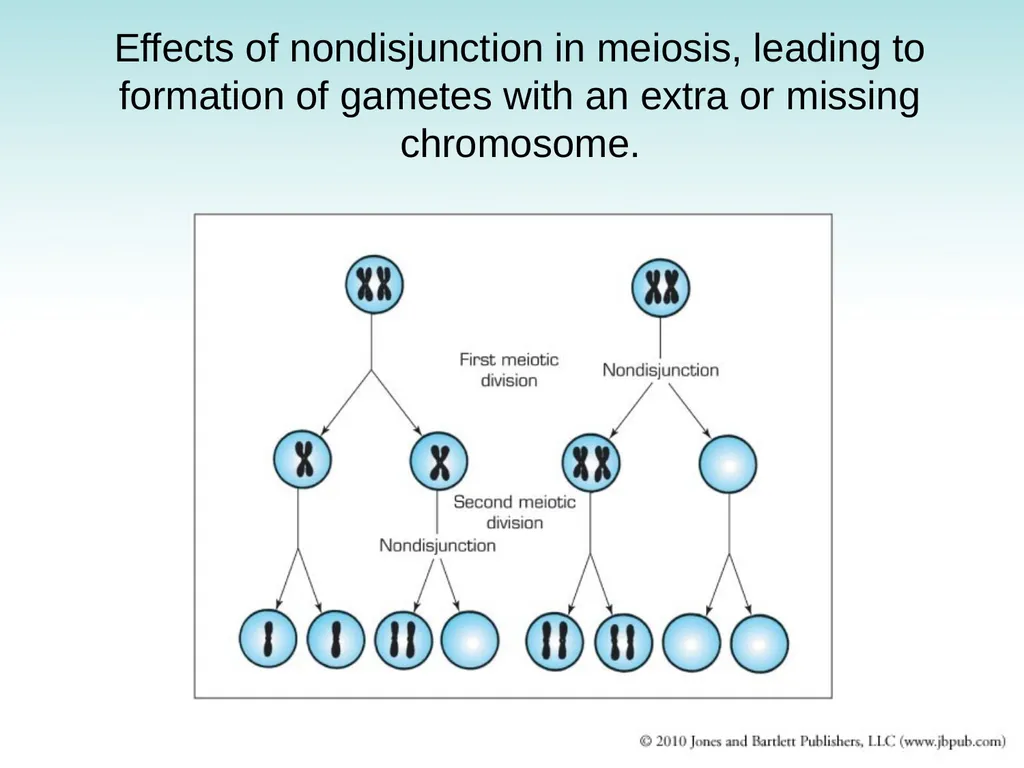
Chromosomal Abnormalities Nondisjunction: failure of homologous chromosomes in germ cells to separate in first or second meiotic division
May involve either sex chromosomes or autosomes
Causes abnormalities in distribution of chromosomes between germ cells
One of two germ cells has an extra chromosome while the other lacks a chromosome<br>
May involve either sex chromosomes or autosomes
Causes abnormalities in distribution of chromosomes between germ cells
One of two germ cells has an extra chromosome while the other lacks a chromosome<br>
19
Chromosomal Abnormalities Monosomy: absence of a chromosome in a cell
Trisomy: presence of an extra chromosome in a cell
Deletions: chromosome breaks during meiosis and broken piece is lost
Translocations: misplaced chromosome or part of it attaches to another chromosome<br>
Trisomy: presence of an extra chromosome in a cell
Deletions: chromosome breaks during meiosis and broken piece is lost
Translocations: misplaced chromosome or part of it attaches to another chromosome<br>
20
Sex Chromosome Abnormalities
(1 of 3) Variations in normal number of sex chromosomes are often associated with some reduction of intelligence
Y chromosome: directs masculine sexual differentiation, associated with male body configuration regardless of number of X chromosomes present
Extra Y: no significant effect as it mainly carries genes concerned with male sexual differentiation
Absent Y: body configuration is female
Extra X in female: has little effect (one X chromosome is inactivated)
Extra X in male: has adverse effects on male development<br>
Y chromosome: directs masculine sexual differentiation, associated with male body configuration regardless of number of X chromosomes present
Extra Y: no significant effect as it mainly carries genes concerned with male sexual differentiation
Absent Y: body configuration is female
Extra X in female: has little effect (one X chromosome is inactivated)
Extra X in male: has adverse effects on male development<br>
21
Sex Chromosome Abnormalities
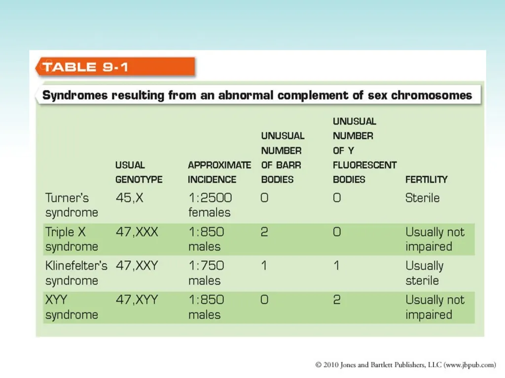
(2 of 3) Two most common ones in the female
1. Turner’s Syndrome: absence of one X chromosome
2. Triple X Syndrome: extra X chromosome
Two most common ones in the male
1. Klinefelter’s Syndrome: extra X chromosome
2. XYY Syndrome: extra Y chromosome<br>
1. Turner’s Syndrome: absence of one X chromosome
2. Triple X Syndrome: extra X chromosome
Two most common ones in the male
1. Klinefelter’s Syndrome: extra X chromosome
2. XYY Syndrome: extra Y chromosome<br>
22
Sex Chromosome Abnormalities
(3 of 3) Fragile X Syndrome (x-linked mental deficiency)
Not related to either excess or deficiency of sex chromosomes
Associated with a characteristic abnormality of the X chromosome
Second to Down syndrome as a major cause of mental deficiency<br>
Not related to either excess or deficiency of sex chromosomes
Associated with a characteristic abnormality of the X chromosome
Second to Down syndrome as a major cause of mental deficiency<br>
23
Autosomal Abnormalities (1 of 3) Absence of an autosome results in the loss of several genes that development is generally not possible and the embryo is aborted.
Deletion of a small part of an autosome may be compatible with development but usually results in multiple severe congenital abnormalities.
Down syndrome: most common chromosomal abnormality, an autosomal trisomy<br>
Deletion of a small part of an autosome may be compatible with development but usually results in multiple severe congenital abnormalities.
Down syndrome: most common chromosomal abnormality, an autosomal trisomy<br>
24
Autosomal Abnormalities (2 of 3) With trisomy of small chromosome 21
Many fetuses are aborted early in pregnancy (70%)
Those who live have Down syndrome
Nondisjunction during oogenesis occurs in 95% of cases
Increased frequency with advancing maternal age: 1 in 50 if mother is > 40 years old
Extra chromosome 21 acquired as part of the translocation chromosome<br>
Many fetuses are aborted early in pregnancy (70%)
Those who live have Down syndrome
Nondisjunction during oogenesis occurs in 95% of cases
Increased frequency with advancing maternal age: 1 in 50 if mother is > 40 years old
Extra chromosome 21 acquired as part of the translocation chromosome<br>
25
Autosomal Abnormalities (3 of 3) Nondisjunction occurring in zygote
Most common chromosomal abnormality: 1:600 births
Manifestations: mental deficiency, cardiac malformation, major defects in other organ systems
Trisomy of chromosome 13: Cleft lip and palate; abnormal development of skull, brain, and eyes; congenital heart defect; polydactyly
Trisomy of chromosome 18:
Both 13 and 18 trisomies are usually fatal in the neonatal period or in early infancy<br>
Most common chromosomal abnormality: 1:600 births
Manifestations: mental deficiency, cardiac malformation, major defects in other organ systems
Trisomy of chromosome 13: Cleft lip and palate; abnormal development of skull, brain, and eyes; congenital heart defect; polydactyly
Trisomy of chromosome 18:
Both 13 and 18 trisomies are usually fatal in the neonatal period or in early infancy<br>
26
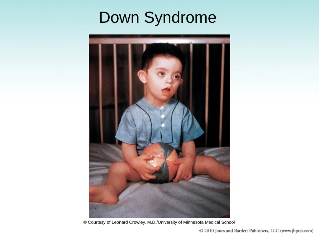
Down Syndrome © Courtesy of Leonard Crowley, M.D./University of Minnesota Medical School<br>
27
Effects of nondisjunction in meiosis, leading to formation of gametes with an extra or missing chromosome.<br>
28
Translocation Down syndrome
(1 of 3) Occurs in small number of persons with Down syndrome
Extra chromosome: chromosome 21 fused with chromosome 14 or another chromosome
Total number of chromosomes not increased but genetic material is equivalent to 47 chromosomes
Causes
Normal chromosomes in cells of both parents
Translocation occurred accidentally during gametogenesis in the germ cells of one of the parents
14/21 carrier in one of the parents
Carrier parent has only 45 chromosomes because one is the fusion of chromosome 21 with 14<br>
Extra chromosome: chromosome 21 fused with chromosome 14 or another chromosome
Total number of chromosomes not increased but genetic material is equivalent to 47 chromosomes
Causes
Normal chromosomes in cells of both parents
Translocation occurred accidentally during gametogenesis in the germ cells of one of the parents
14/21 carrier in one of the parents
Carrier parent has only 45 chromosomes because one is the fusion of chromosome 21 with 14<br>
29
Translocation Down syndrome
(2 of 3) 14/21 carrier in one of the parents
Carrier parent is capable of transmitting abnormal chromosome to his or her children resulting in translocation Down syndrome<br>
Carrier parent is capable of transmitting abnormal chromosome to his or her children resulting in translocation Down syndrome<br>
30
Translocation Down syndrome
(3 of 3) Possible outcomes of pregnancy involving a female carrier
Translocation chromosome is nor always transmitted
Normal
Carrier
Nonviable
Down Syndrome<br>
Translocation chromosome is nor always transmitted
Normal
Carrier
Nonviable
Down Syndrome<br>
31
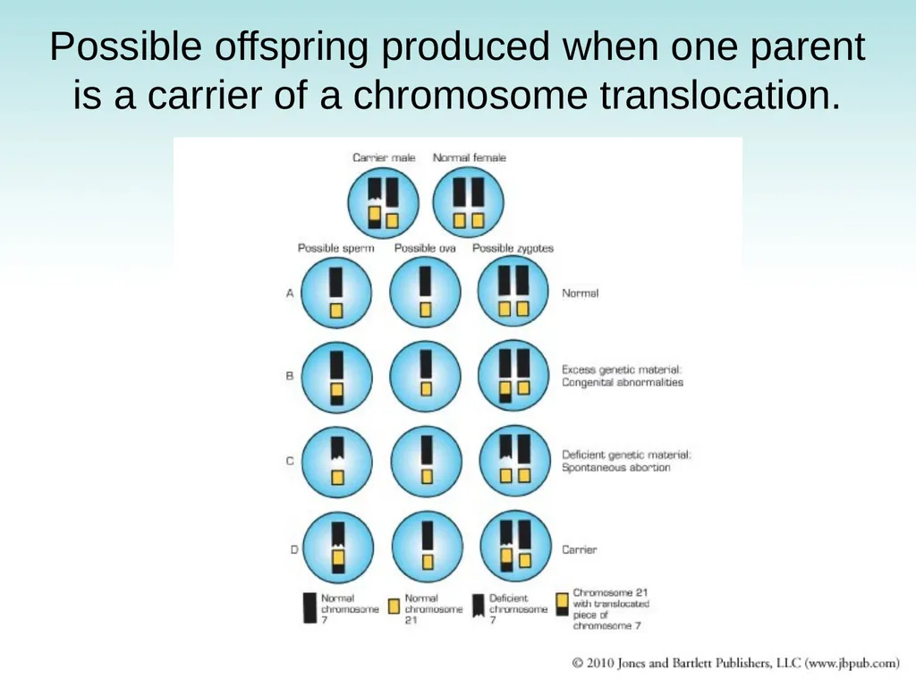
Possible offspring produced when one parent is a carrier of a chromosome translocation.<br>
32
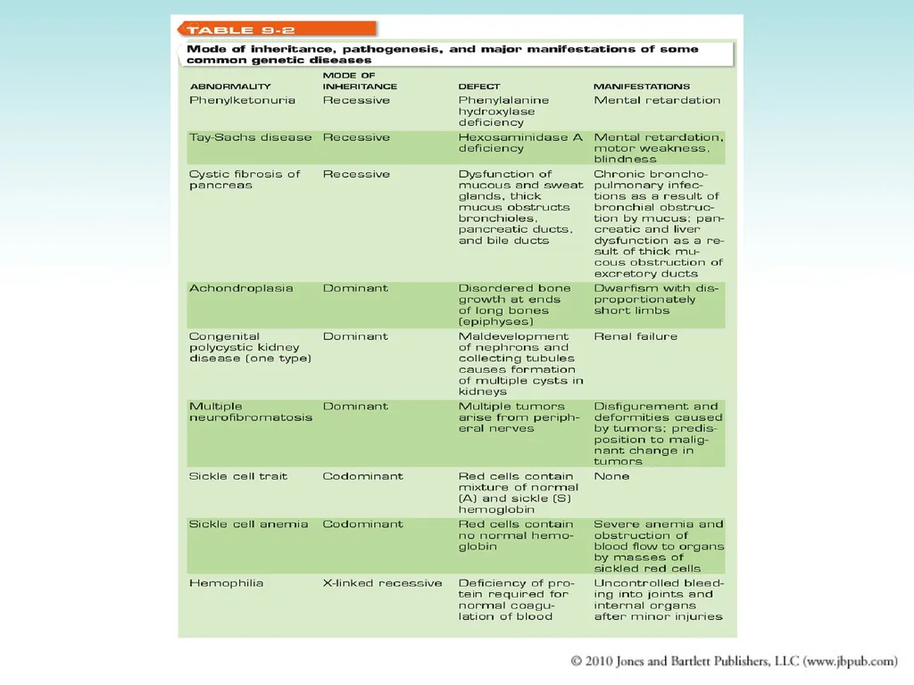
Transmission of Genetically Determined Diseases Autosomal dominant inheritance
Autosomal recessive inheritance
Codominant inheritance
X-linked inheritance
Most hereditary diseases are transmitted on autosomes
Few are carried on sex chromosomes<br>
Autosomal recessive inheritance
Codominant inheritance
X-linked inheritance
Most hereditary diseases are transmitted on autosomes
Few are carried on sex chromosomes<br>
33
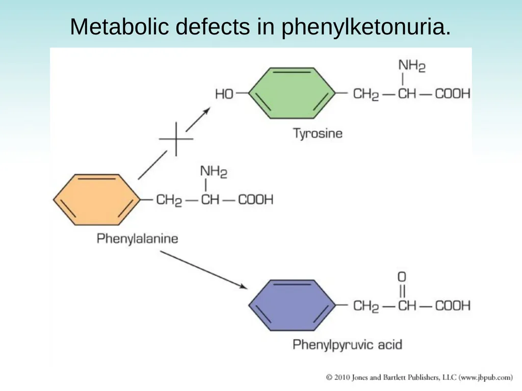
Metabolic defects in phenylketonuria.<br>
34
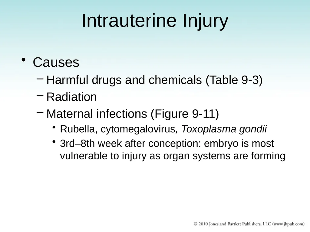
Intrauterine Injury Causes
Harmful drugs and chemicals (Table 9-3)
Radiation
Maternal infections (Figure 9-11)
Rubella, cytomegalovirus, Toxoplasma gondii
3rd–8th week after conception: embryo is most vulnerable to injury as organ systems are forming<br>
Harmful drugs and chemicals (Table 9-3)
Radiation
Maternal infections (Figure 9-11)
Rubella, cytomegalovirus, Toxoplasma gondii
3rd–8th week after conception: embryo is most vulnerable to injury as organ systems are forming<br>
35
Categories of Drugs Harmful to Fetus<br>
36
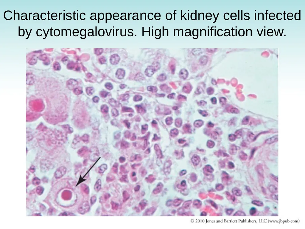
Characteristic appearance of kidney cells infected by cytomegalovirus. High magnification view.<br>
37
Cytomegalovirus infection in newborn<br>
38
Multifactorial Inheritance Combined effect of multiple genes interacting with environmental agents
Congenital abnormalities
Cleft lip, cleft palate, cardiac malformations, clubfoot, dislocation of hip, anencephaly, spina bifida<br>
Congenital abnormalities
Cleft lip, cleft palate, cardiac malformations, clubfoot, dislocation of hip, anencephaly, spina bifida<br>
39
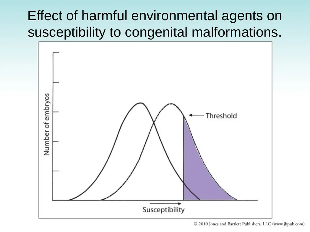
Effect of harmful environmental agents on susceptibility to congenital malformations.<br>
40
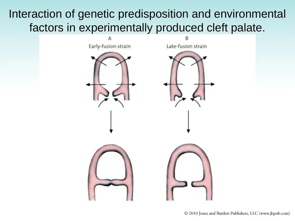
Interaction of genetic predisposition and environmental factors in experimentally produced cleft palate.<br>
41
Prenatal Diagnosis of Congenital Abnormalities (1 of 2) Examination of fetal cells: determination of biochemical abnormalities in fetal cells
Chromosomal abnormalities
Biochemical abnormalities
Analysis of DNA
Examination of amnionic fluid: products secreted into fluid by fetus that may indicate fetal abnormality<br>
Chromosomal abnormalities
Biochemical abnormalities
Analysis of DNA
Examination of amnionic fluid: products secreted into fluid by fetus that may indicate fetal abnormality<br>
42
Prenatal Diagnosis of Congenital Abnormalities (2 of 2) Ultrasound examination: detection of major structural abnormalities
Major structural abnormalities of nervous system (anencephaly; spina bifida)
Hydrocephalus
Obstruction of urinary tract
Failure of kidneys to develop
Failure of limbs to form normally
Fetal DNA analysis: determination of biochemical abnormalities by analysis of DNA of fetal cells
Amniocentesis
Chorionic villus sampling<br>
Major structural abnormalities of nervous system (anencephaly; spina bifida)
Hydrocephalus
Obstruction of urinary tract
Failure of kidneys to develop
Failure of limbs to form normally
Fetal DNA analysis: determination of biochemical abnormalities by analysis of DNA of fetal cells
Amniocentesis
Chorionic villus sampling<br>
43
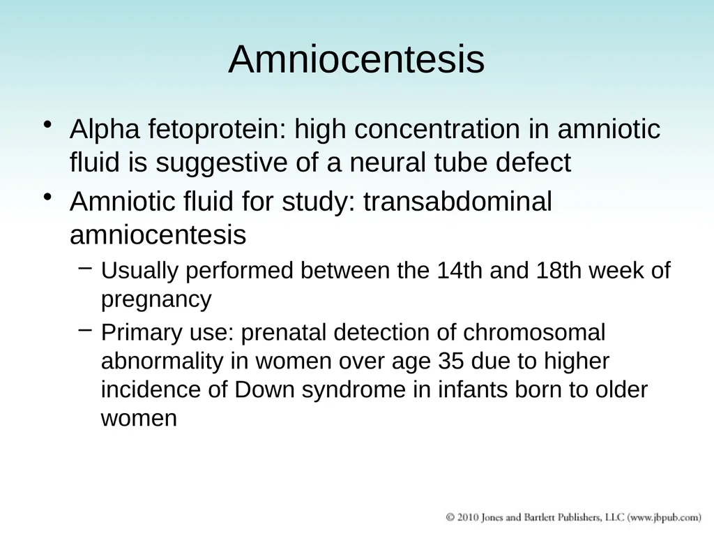
Amniocentesis Alpha fetoprotein: high concentration in amniotic fluid is suggestive of a neural tube defect
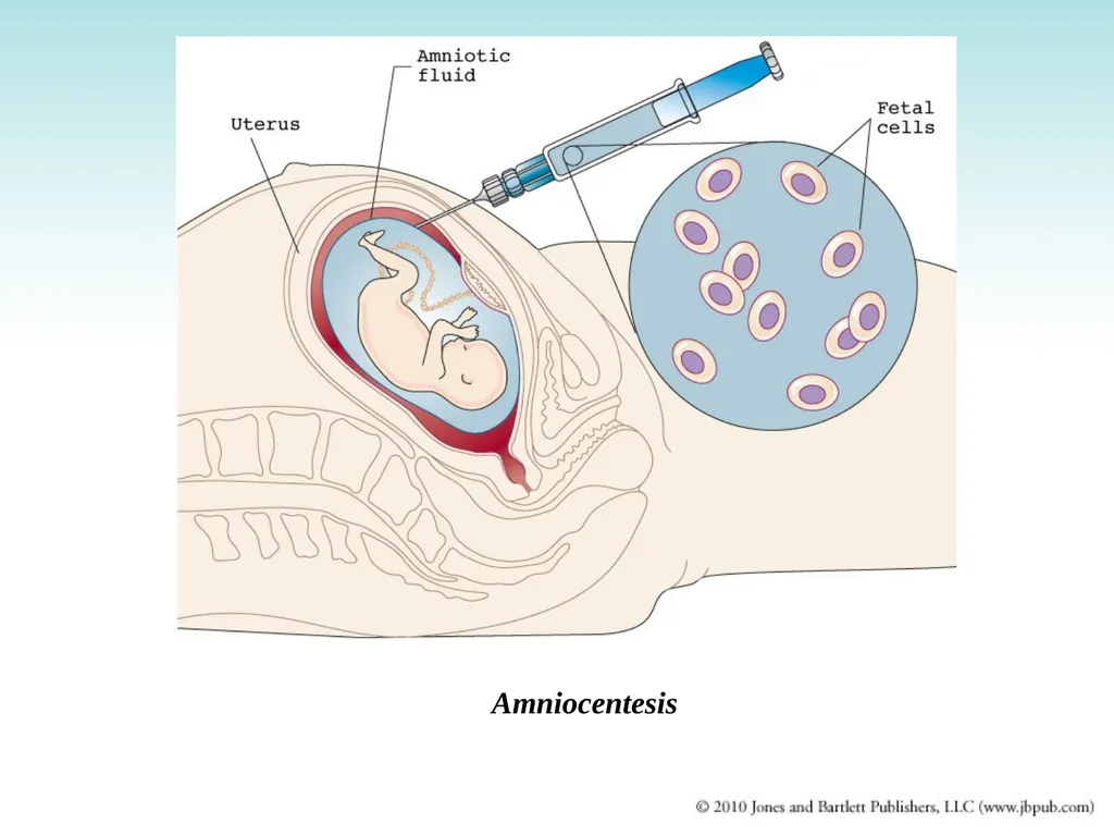
Amniotic fluid for study: transabdominal amniocentesis
Usually performed between the 14th and 18th week of pregnancy
Primary use: prenatal detection of chromosomal abnormality in women over age 35 due to higher incidence of Down syndrome in infants born to older women<br>
Amniotic fluid for study: transabdominal amniocentesis
Usually performed between the 14th and 18th week of pregnancy
Primary use: prenatal detection of chromosomal abnormality in women over age 35 due to higher incidence of Down syndrome in infants born to older women<br>
44
Amniocentesis<br>
45
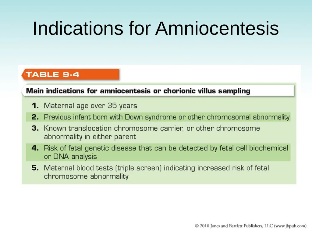
Indications for Amniocentesis<br>
46
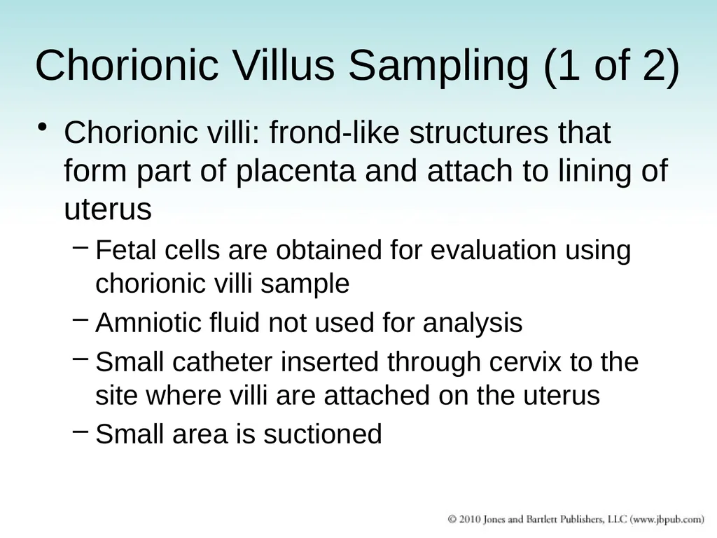
Chorionic Villus Sampling (1 of 2) Chorionic villi: frond-like structures that form part of placenta and attach to lining of uterus
Fetal cells are obtained for evaluation using chorionic villi sample
Amniotic fluid not used for analysis
Small catheter inserted through cervix to the site where villi are attached on the uterus
Small area is suctioned<br>
Fetal cells are obtained for evaluation using chorionic villi sample
Amniotic fluid not used for analysis
Small catheter inserted through cervix to the site where villi are attached on the uterus
Small area is suctioned<br>
47
Chorionic Villus Sampling (2 of 2) Advantages
Can be performed earlier than amniocentesis (8th to 10th week)
Carries less risk from abortion if parent decides to terminate pregnancy in case of congenital abnormality
Disadvantages
More difficult technically than amniocentesis
Complications: spontaneous abortion, limb deformities in fetus<br>
Can be performed earlier than amniocentesis (8th to 10th week)
Carries less risk from abortion if parent decides to terminate pregnancy in case of congenital abnormality
Disadvantages
More difficult technically than amniocentesis
Complications: spontaneous abortion, limb deformities in fetus<br>
48
Discussion What are the consequences of chromosome nondisjunction?
What is the karyotype of an individual with Down syndrome? Why does this happen? What are the possible outcomes of a pregnancy involving a 14/21 translocation chromosome?
What are the causes of congenital abnormalities and their approximate incidences?
What are the indications for amniocentesis?<br>
What is the karyotype of an individual with Down syndrome? Why does this happen? What are the possible outcomes of a pregnancy involving a 14/21 translocation chromosome?
What are the causes of congenital abnormalities and their approximate incidences?
What are the indications for amniocentesis?<br>