04
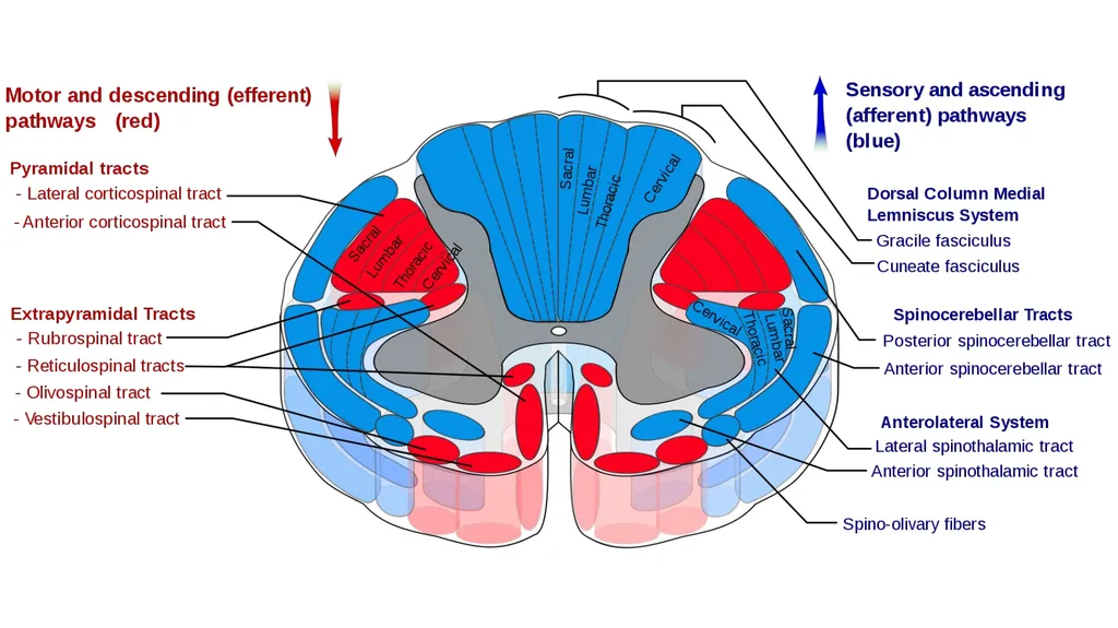
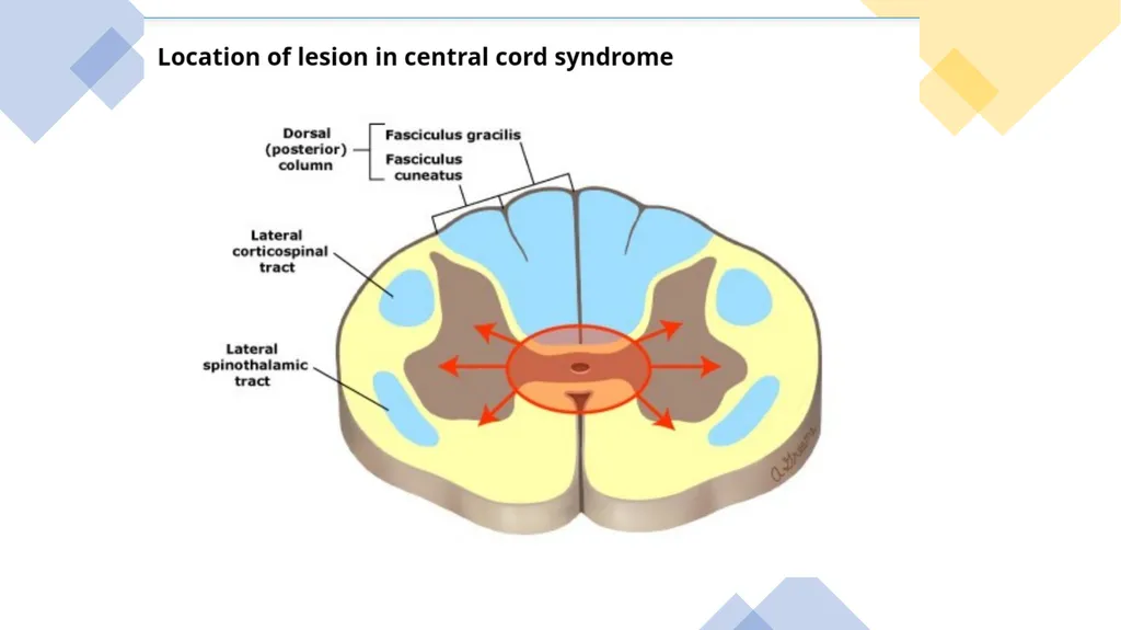
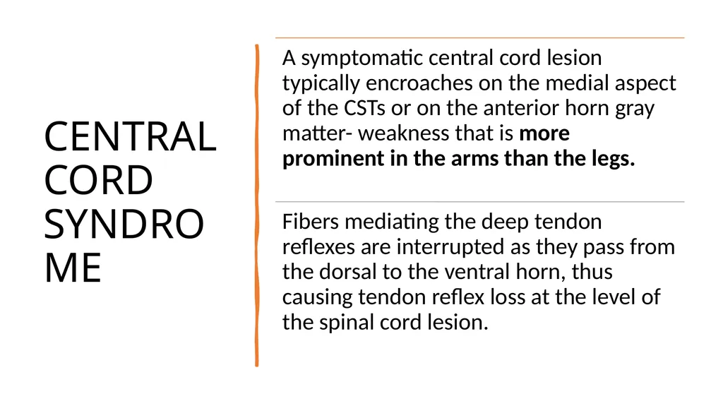
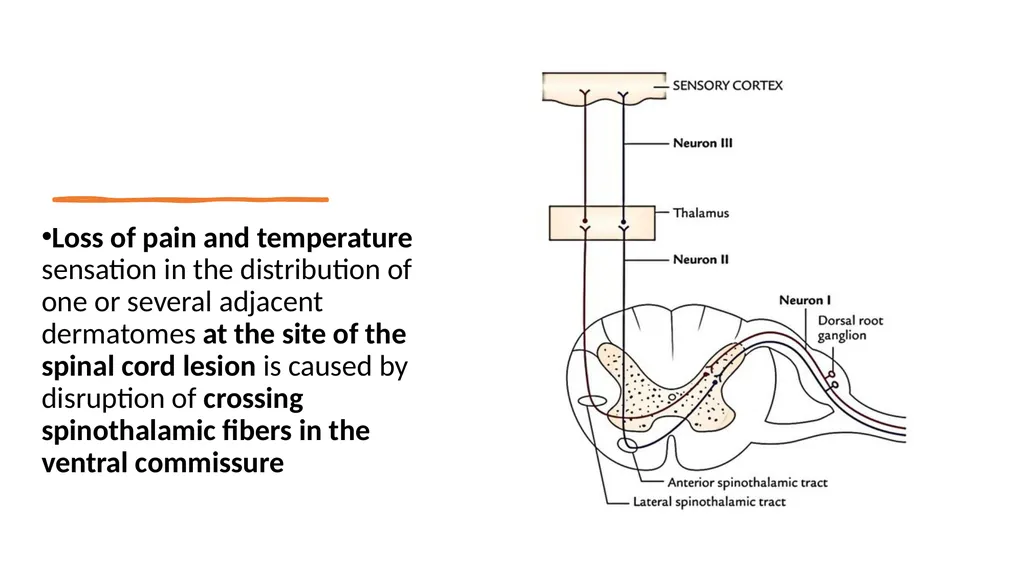
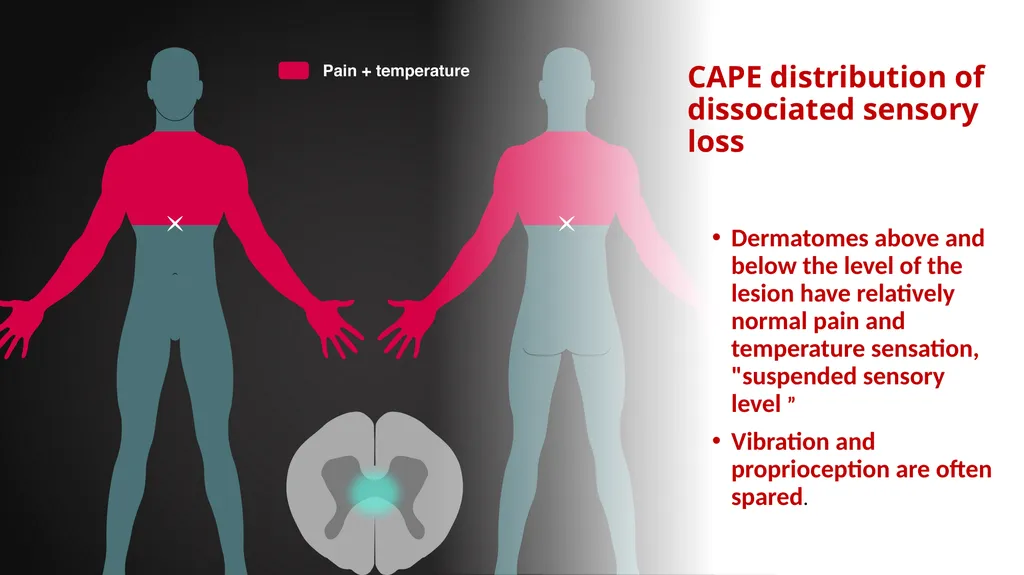
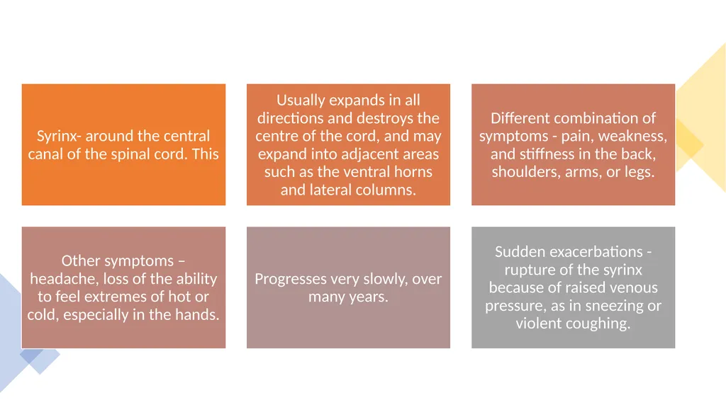
Loss of pain and temperature sensation in the distribution of one or several adjacent dermatomes at the site of the spinal cord lesion is caused by disruption of crossing spinothalamic fibers in the ventral commissure<br>
05
CAPE distribution of dissociated sensory loss Dermatomes above and below the level of the lesion have relatively normal pain and temperature sensation, "suspended sensory level ”
Vibration and proprioception are often spared.<br>
06
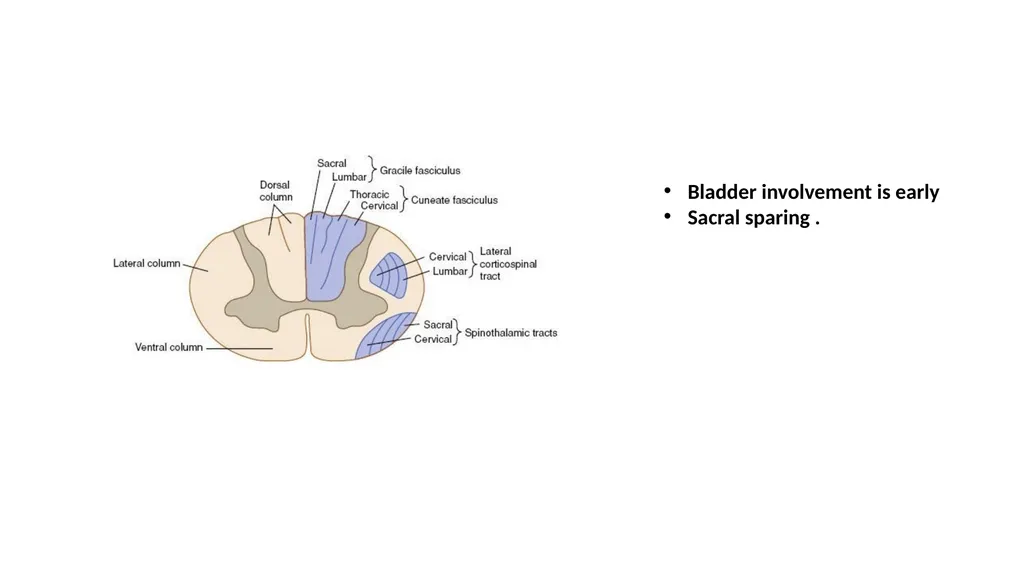
Bladder involvement is early
Sacral sparing .<br>
07
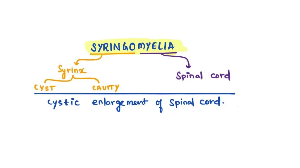
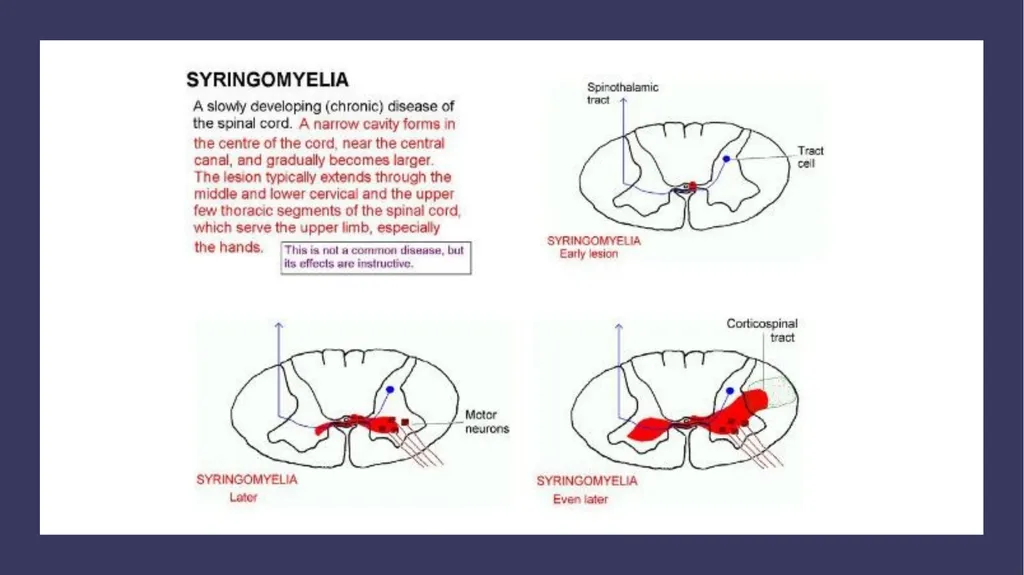
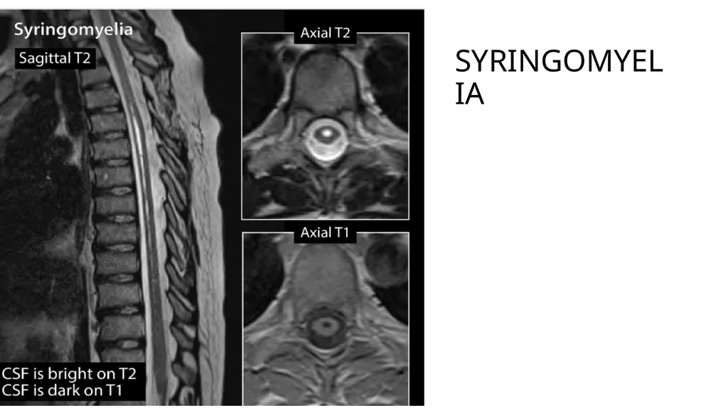
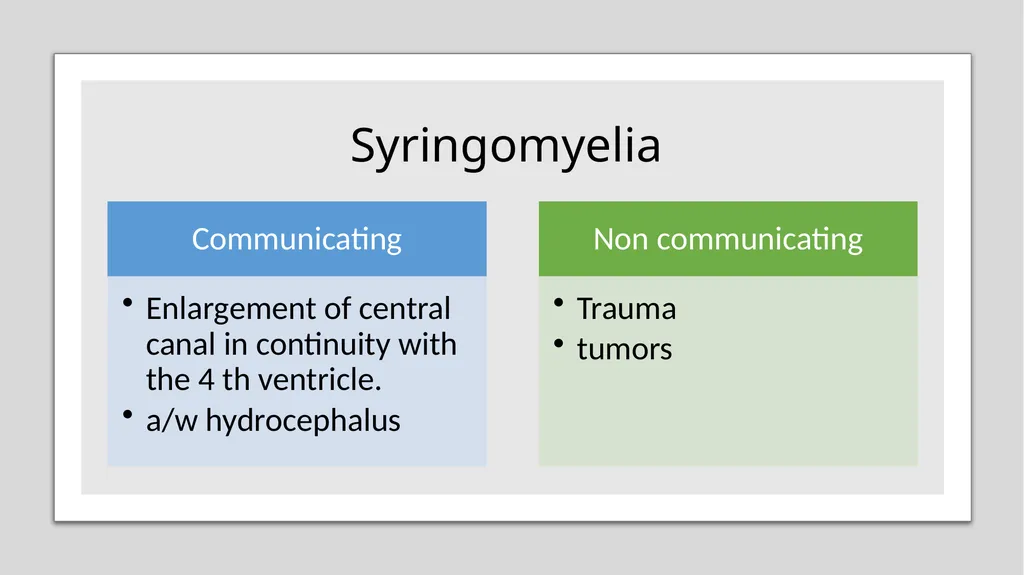
Syringomyelia Fluid-filled, cavity within the spinal cord lined by glial cells.
Most lesions are between C2 and T9.
Descend down or extend upward into the brainstem (syringobulbia).
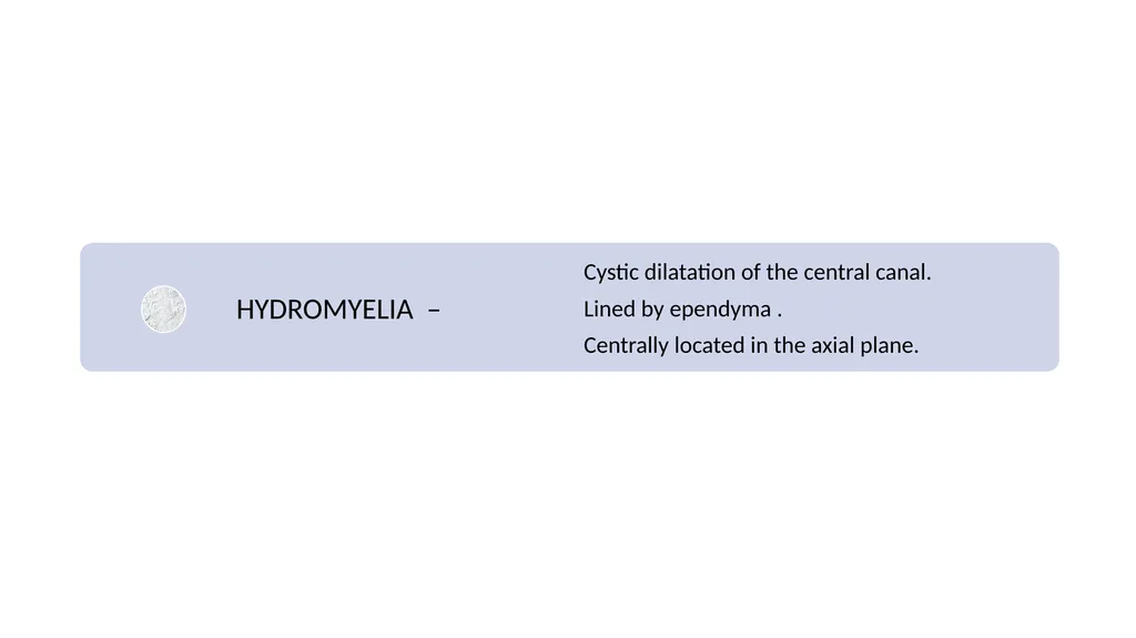
A syrinx can represent a focal dilation of the central canal, or it may lie separately, within the spinal cord parenchyma<br>
08
Sensory features
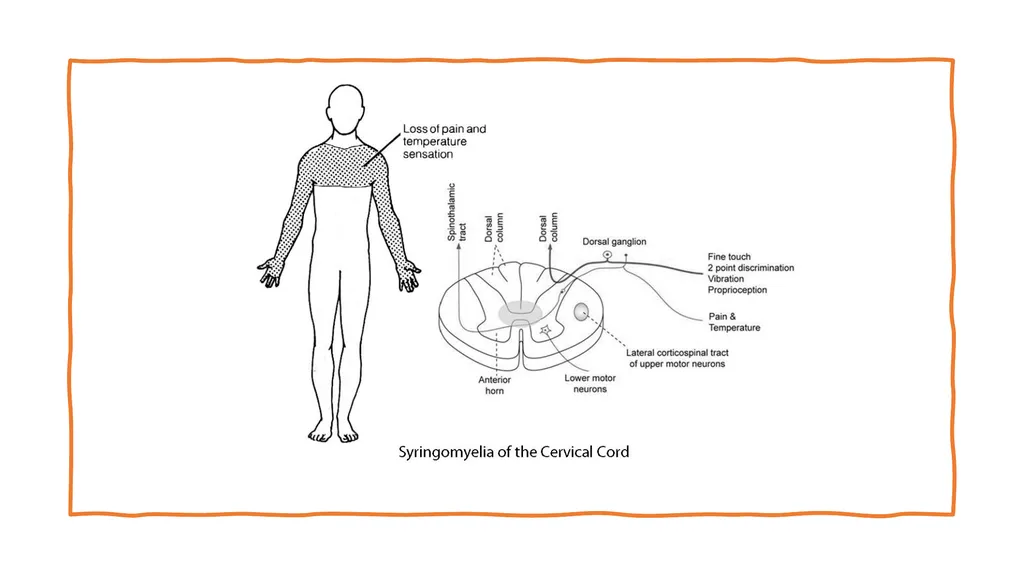
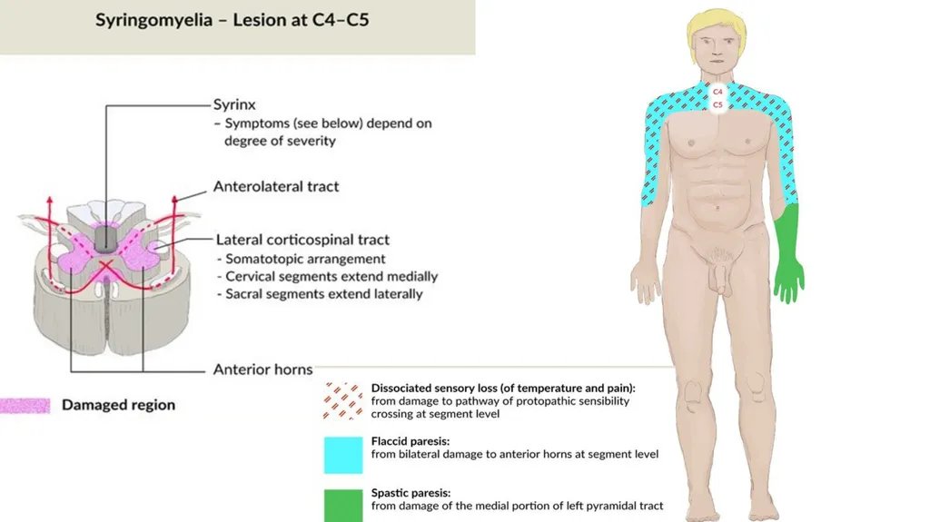
Pain and temperature sensation are lost - spinothalamic tract damage.
One side may be affected more than the other.
Classically, the sensation loss is experienced in a shawl-like distribution over the arms, shoulders and upper body.
Spares the dorsal column/medial lemniscus of the spinal cord, leaving pressure, vibration, touch and proprioception intact in the upper extremities.
Light touch, vibration and position senses in the feet are affected as the syrinx enlarges into the dorsal columns.<br>
09
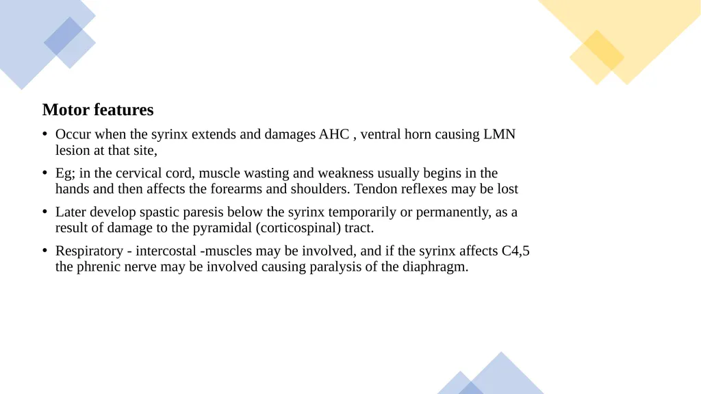
Motor features
Occur when the syrinx extends and damages AHC , ventral horn causing LMN lesion at that site,
Eg; in the cervical cord, muscle wasting and weakness usually begins in the hands and then affects the forearms and shoulders. Tendon reflexes may be lost
Later develop spastic paresis below the syrinx temporarily or permanently, as a result of damage to the pyramidal (corticospinal) tract.
Respiratory - intercostal -muscles may be involved, and if the syrinx affects C4,5 the phrenic nerve may be involved causing paralysis of the diaphragm.<br>
10
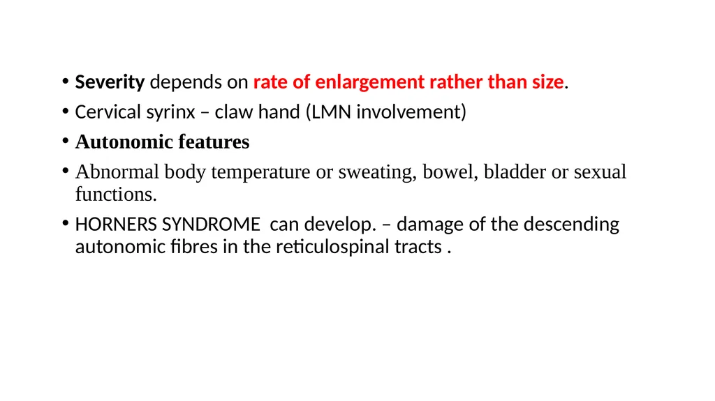
Severity depends on rate of enlargement rather than size.
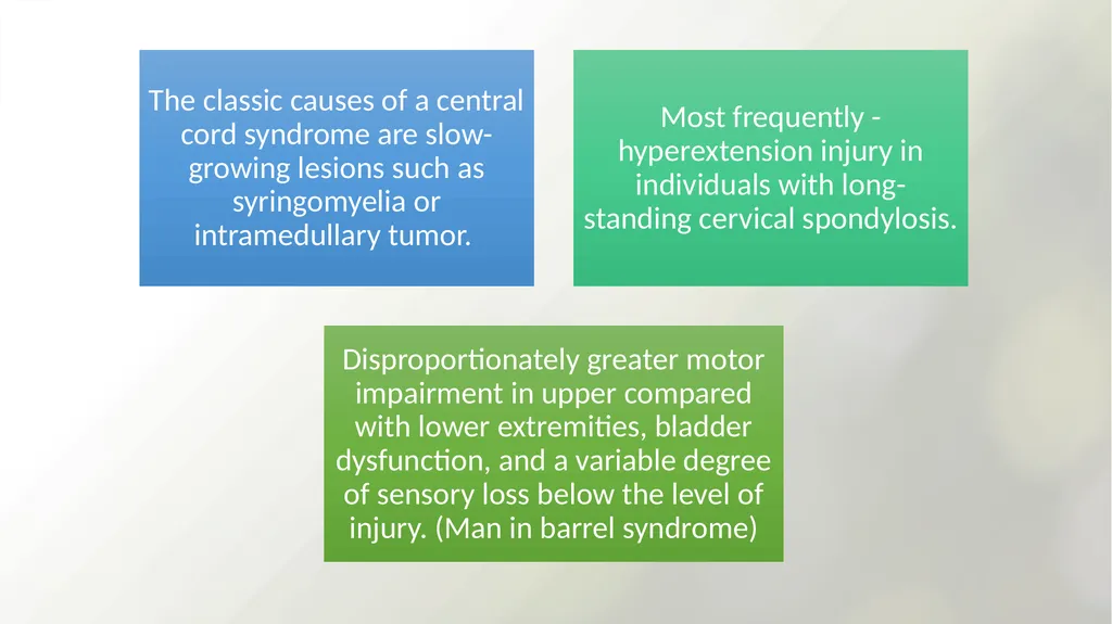
Cervical syrinx – claw hand (LMN involvement)
Autonomic features
Abnormal body temperature or sweating, bowel, bladder or sexual functions.
HORNERS SYNDROME can develop. – damage of the descending autonomic fibres in the reticulospinal tracts .<br>
13
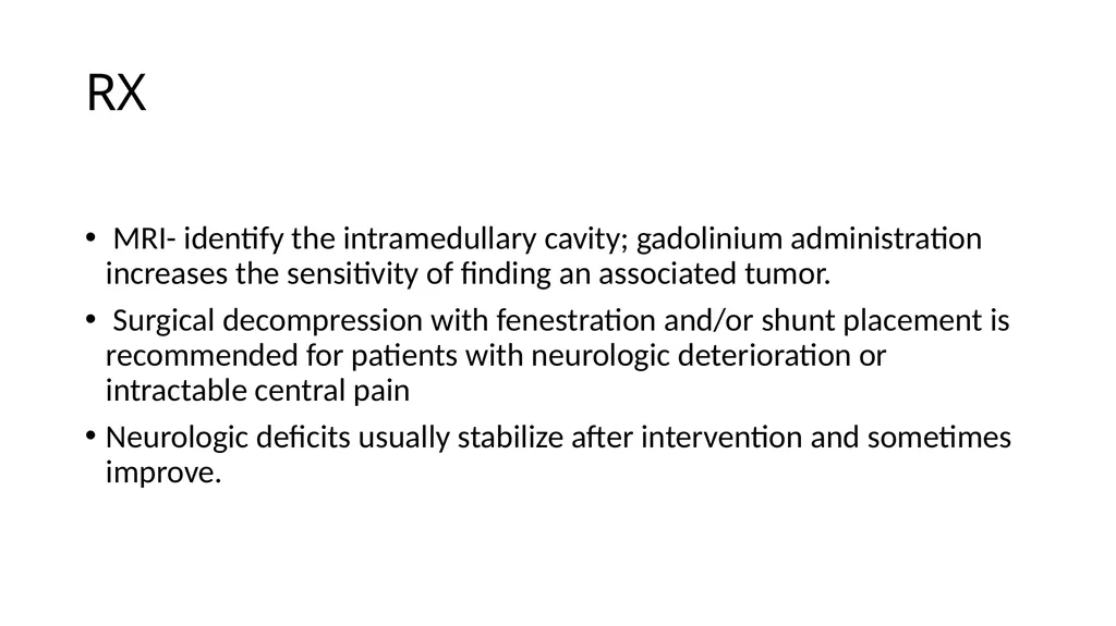
RX MRI- identify the intramedullary cavity; gadolinium administration increases the sensitivity of finding an associated tumor.
Surgical decompression with fenestration and/or shunt placement is recommended for patients with neurologic deterioration or intractable central pain
Neurologic deficits usually stabilize after intervention and sometimes improve.<br>
14
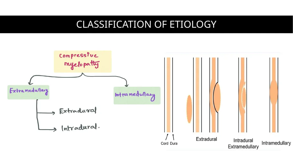
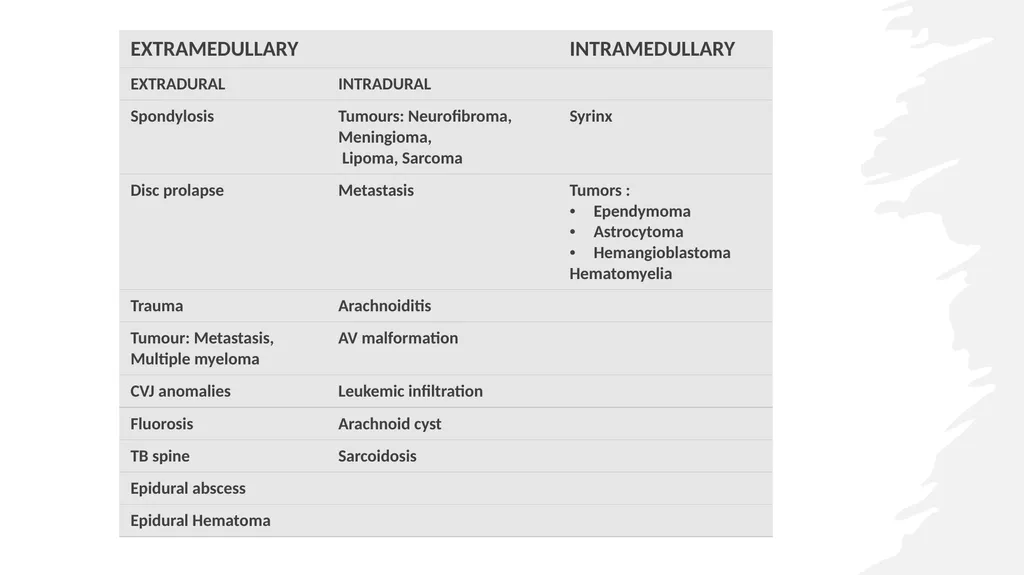
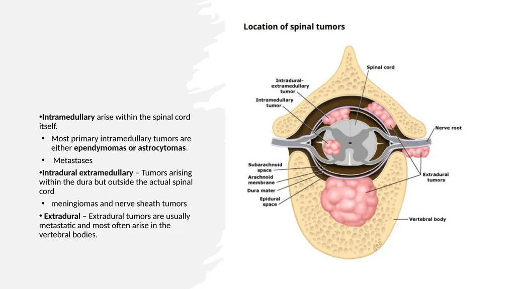
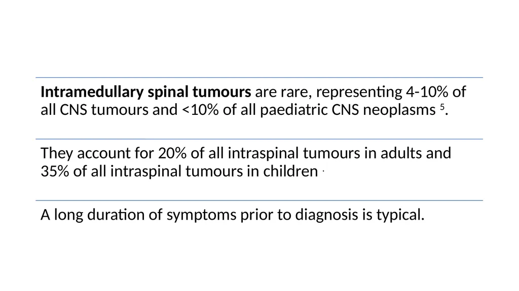
Intramedullary arise within the spinal cord itself.
Most primary intramedullary tumors are either ependymomas or astrocytomas.
Metastases
Intradural extramedullary – Tumors arising within the dura but outside the actual spinal cord
meningiomas and nerve sheath tumors
Extradural – Extradural tumors are usually metastatic and most often arise in the vertebral bodies.<br>
15
Intraaxial in location and can be either primary or metastatic.
Primary spinal cord tumors account for 2 to 4 % of all primary central nervous system (CNS) tumors, one-third of which are located in the intramedullary compartment.<br>
16
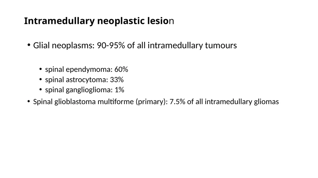
Intramedullary neoplastic lesion Glial neoplasms: 90-95% of all intramedullary tumours
spinal ependymoma: 60%
spinal astrocytoma: 33%
spinal ganglioglioma: 1%
Spinal glioblastoma multiforme (primary): 7.5% of all intramedullary gliomas<br>
17
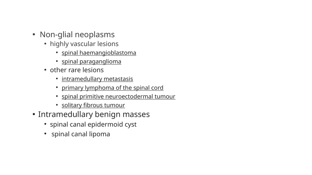
Non-glial neoplasms
highly vascular lesions
spinal haemangioblastoma
spinal paraganglioma
other rare lesions
intramedullary metastasis
primary lymphoma of the spinal cord
spinal primitive neuroectodermal tumour
solitary fibrous tumour
Intramedullary benign masses
spinal canal epidermoid cyst
spinal canal lipoma<br>
18
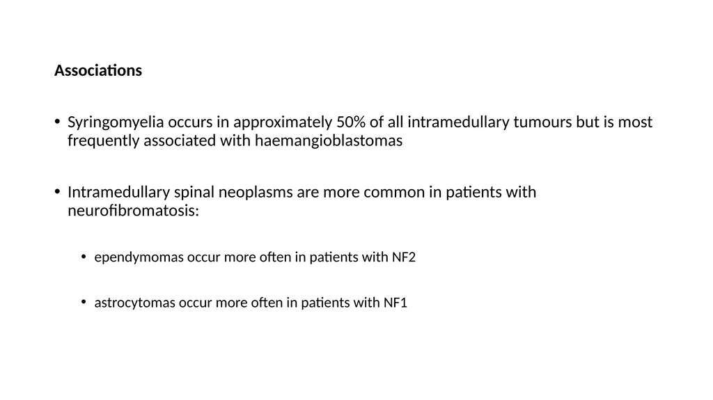
Associations
Syringomyelia occurs in approximately 50% of all intramedullary tumours but is most frequently associated with haemangioblastomas
Intramedullary spinal neoplasms are more common in patients with neurofibromatosis:
ependymomas occur more often in patients with NF2
astrocytomas occur more often in patients with NF1<br>
20
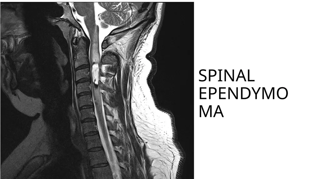
One third of primary ependymomas arise in the spinal cord. The other two thirds arise from the brain parenchyma in the ependymal cells lining the ventricles.
May be located anywhere along the spinal cord.
Approximately 50% occur in the lumbosacral spinal cord or filum terminale
50 % occur anywhere in the cervical or thoracic spinal cord.<br>
22
Approximately one-half of spinal cord astrocytomas are pilocytic and one-half are infiltrative astrocytomas
Pilocytic astrocytomas are well circumscribed and low grade with nonaggressive clinical behavior. On MRI these tumors enhance intensely with gadolinium
Diffuse fibrillary astrocytomas of the spinal cord usually appear as nonencapsulated lesions that enhance minimally or heterogeneously on MRI. Approximately one-third are histologically high grade (anaplastic astrocytoma or glioblastoma)<br>
23
The most important factor associated with a better prognosis was low tumor grade (WHO grade I). Other factors associated with a better prognosis included tumor location other than the cervical region, limited extent of tumor involvement along the spinal cord, and longer (>180 days) duration of symptoms.
Postoperative RT did not affect outcomes in patients with pilocytic astrocytomas, although it did appear to improve the outcome for infiltrative astrocytomas.
Chemotherapy ineffective.<br>
25
VHL - cerebellar hemangioblastomas, retinal angiomatosis, renal cell carcinomas, pheochromocytoma, or spinal cord hemangioblastomas.
In the spine, the most common locations are the intramedullary cervical and thoracic spinal cord, although they can occur in any space
Approximately two thirds of intramedullary hemangioblastomas are associated with syringomyelia.
Hemangiomas are highly vascular tumors thus engorged vessels may be seen, particularly along the dorsal surface of the cord.
Spinal angiography often shows large feeding vessels and draining veins, as well as a prominent vascular blush<br>
26
Metastases —
rare, although the increased use of MRI has resulted in more frequent recognition of such lesions
Approximately one-half of cases are associated with lung cancer.
Breast cancer, renal cell carcinoma, lymphoma, and melanoma<br>
27
Differential diagnosis Various lesions may mimic intramedullary tumours.
vascular lesions
cavernous malformation (cavernoma)
dural arteriovenous fistula (DAVF)-(type I AVM)
spinal cord infarction
inflammatory lesions
demyelination (e.g. multiple sclerosis)
transverse myelitis
spinal cord abscess
spinal cord tuberculoma
spinal cord contusion (acute)<br>
28
Radiographic features Plain radiograph
Widening of the interpedicular distance may be seen in less than 10% of cases
CT
Generally not useful because bony changes are relatively rare.
Myelography
The spinal cord can be enlarged
Gradual subarachnoid space effacement<br>
29
MRI
Three general characteristics of intramedullary neoplasms are recognised on MRI:
They cause either focal or diffuse spinal cord expansion
They produce high signal intensity on proton density and T2 weighted images
Vast majority show at least some contrast enhancement, in contrast to intracranial neoplasms, even low-grade intramedullary tumours enhance to some degree; however, the absence of enhancement does not exclude an intramedullary neoplasm in the presence of cord expansion<br>