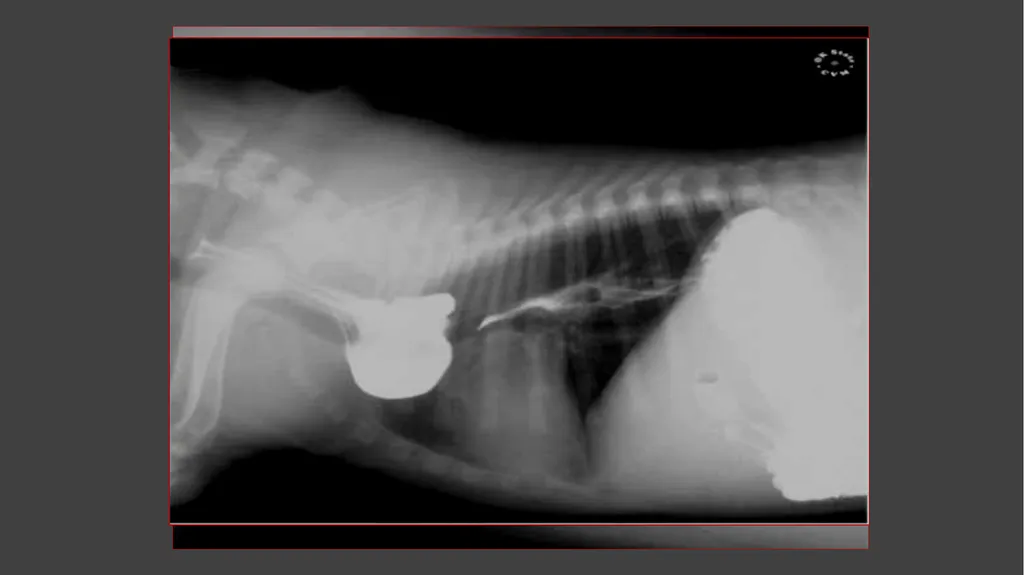
Contrast Radiography Dr. Gulshan Kumar MVSc, PhD When the radio-density of the tissue itself or its surrounding structures is changed to obtain a radiograph with improved visualization and demarcation, it is called contrast radiography.
"Contrast Radiography Dr. Gulshan Kumar MVSc, PhD" is the property of its rightful owner. Permission is granted to
download and print the materials on this website for personal, non-commercial use only, and to display it
on your personal computer provided you do not modify the materials and that you retain all copyright
notices contained in the materials. By downloading content from our website, you accept the terms of this
agreement.
Presentation Transcript
01
Contrast Radiography Dr. Gulshan Kumar
MVSc, PhD<br>
02
When the radio-density of the tissue itself or its surrounding structures is changed to obtain a radiograph with improved visualization and demarcation, it is called contrast radiography.
The substance used for the purpose is called contrast medium.
A contrast agent is a chemical substance of very high or very low atomic number or weight, which either increases or decreases the density of the organ under examination.<br>
03
Positive contrast media - Materials which increases radio-density of the tissue under question in relation to surrounding tissue.
Negative contrast media- Materials which relatively decrease the radio-density.
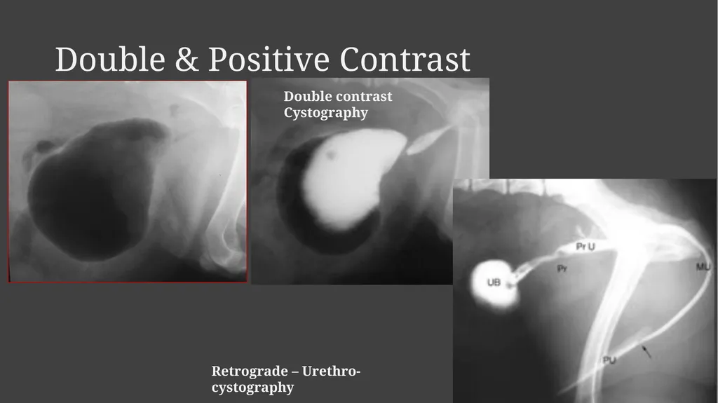
When both contrast media are used together it is termed as double contrast radiography.<br>
04
Advantages Structures or organs can be evaluated more effectively for their size, shape and position.
Valuable information can be gained regarding serosal and mucosal surfaces of hollow organs or their contents which otherwise are not apparent on plain radiographs.
In some instances some idea of the function of the organ can be formed.<br>
05
Positive contrast media The atomic number of body part is generally positively correlated with the radio-density of a compound.
The highest atomic number in the body is that of the bone i.e. 138.
The atomic number of air filled lungs is 4.
For compounds to be used as positive contrast agents, the atomic number of the element has to be above 50 e.g. for barium it is 56 and that of iodine 53<br>
06
Ideal qualities of positive contrast medium It should have desired atomic number (Z) number.
It should be inert or its metabolic byproducts should not be toxic.
Uranium (Z = 92) could be the best positive contrast medium but it is radioactive
Similarly lead (Z= 82) compounds produce excellent contrast but are toxic to living beings.<br>
07
Ideal qualities of positive contrast medium Emulsion of lead oxide and soap in water is a cheap contrast agent used for angiography in experimental animals to be euthanised.
In general, high osmolar contrast media are more toxic than low osmolar contrast media.
It should be retained in the area of interest only for a desired period.
Agents used for outlining excretory organs must specifically be excreted through that route in sufficient concentration so as to produce radio-density of diagnostic value, e.g. barium sulphate for alimentary canal, meglumine iodipamide for biliary system<br>
08
Adverse reactions Chemo-toxic or local reactions and
Systemic or hypersensitivity reactions
Adverse reactions can be minimized by pre administration of corticosteroids.
Non ionic low osmolarity contrast media (iohexol, iopamidol) cause fewer side effects<br>
09
Broad classification of positive contrast media Barium sulfate preparations<br>
10
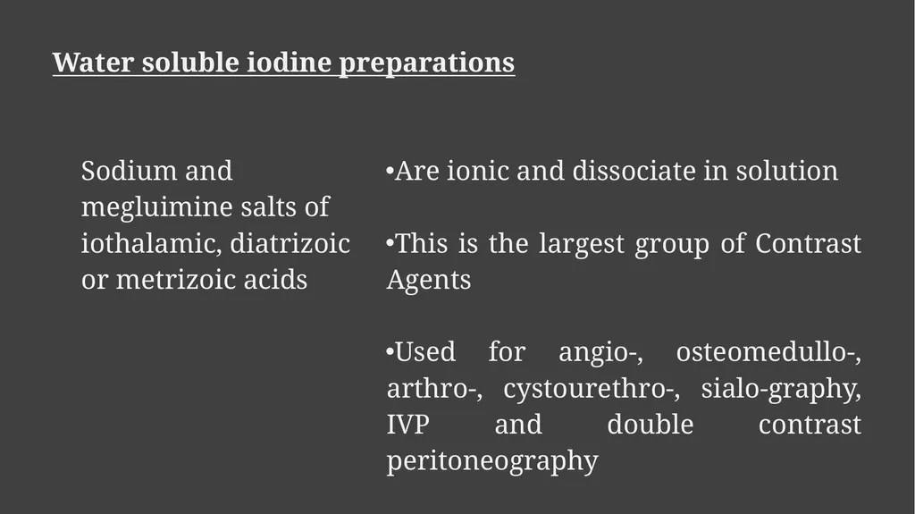
Water soluble iodine preparations<br>
11
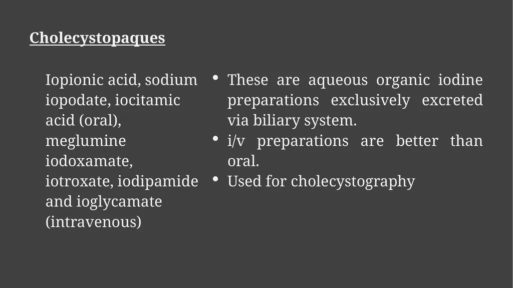
Cholecystopaques<br>
12
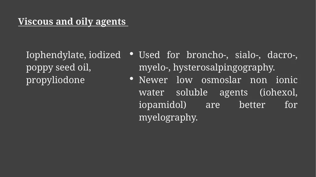
Viscous and oily agents<br>
13
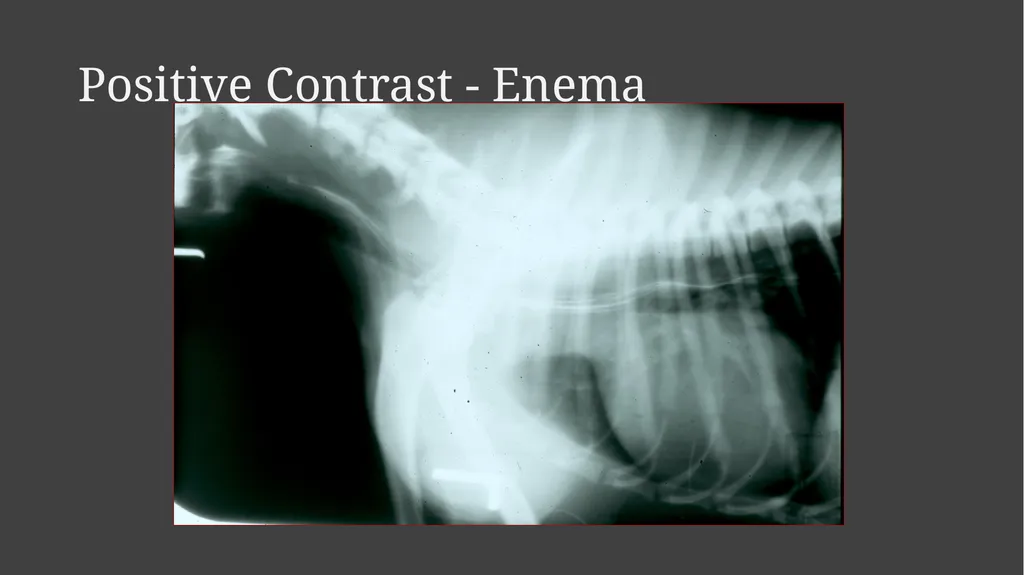
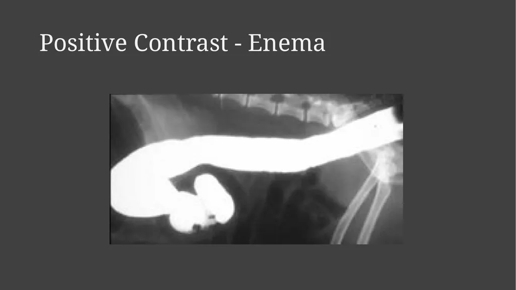
Positive Contrast - Enema<br>
14
Positive Contrast - Enema<br>
15
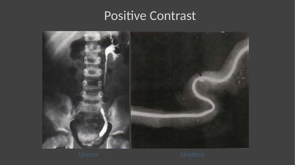
Ureter Urethra Positive Contrast<br>
16
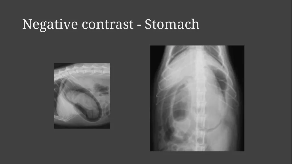
Negative contrast media Room air, carbon dioxide and oxygen are most commonly used negative contrast media.
Nitrogen can also be used.
An ideal negative contrast media should be inert, quickly dissolved in the body fluids and quickly eliminated from the body.
Room air is cheapest, is safe but less readily absorbed.
After a gas has been infused into the tissues or a body cavity, it is eliminated.
………….<br>
17
Negative contrast media Since solubility of carbon dioxide in water is very high, it is eliminated quickly.
Gases provide poor contrast and are best used in double contrast studies.
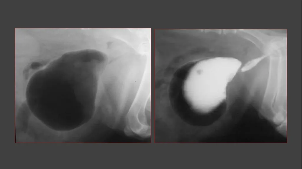
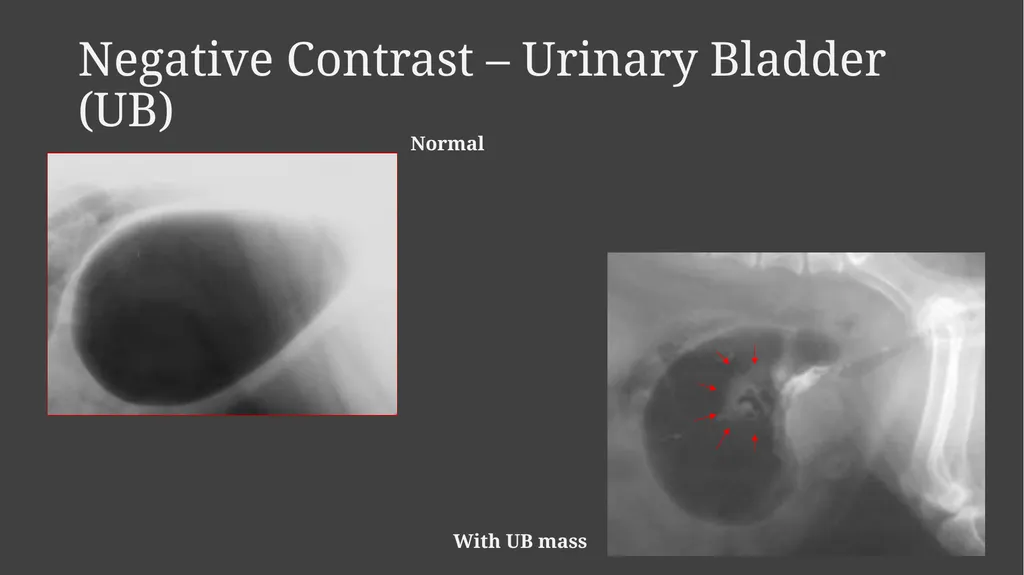
Indications include arthrography, fasciagraphy, pneumo-peritoneography, pneumocystography etc.<br>
18
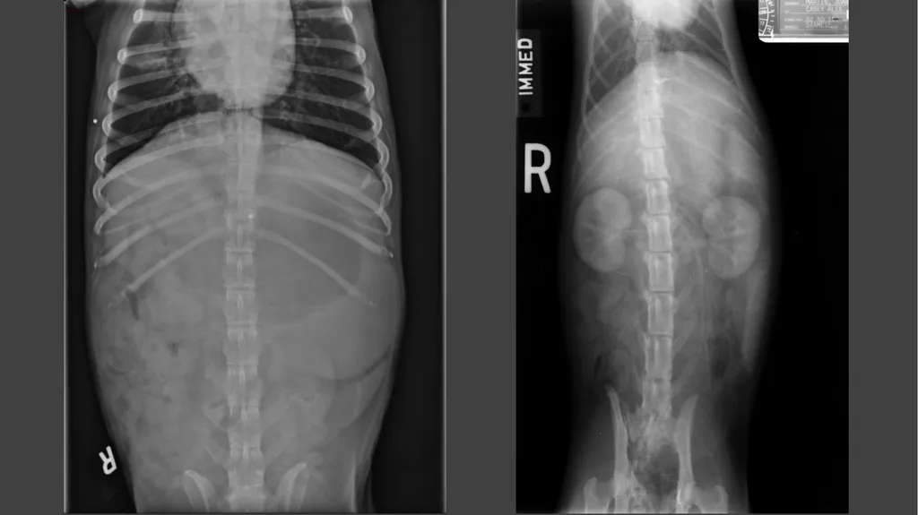
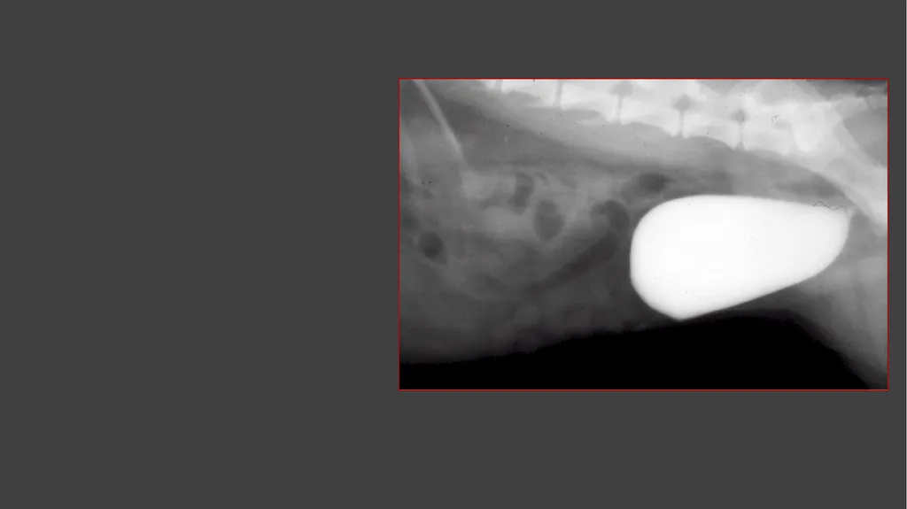
Negative contrast - Stomach<br>
19
Negative Contrast – Urinary Bladder (UB) Normal With UB mass<br>