04
Clinical Features: History Symptoms
Dyspnoea
Cough with/without sputum
Tight chest
Wheeze
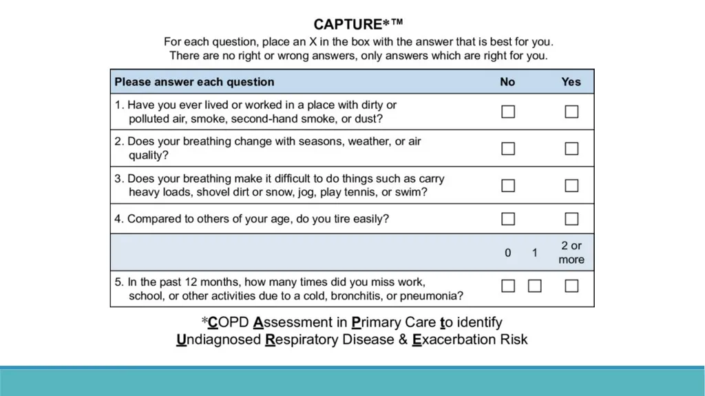
Inhalation exposure history
SMOKING
Exposure to occupational fumes
Air pollution<br>
05
Clinical Features: Examination Low saturation
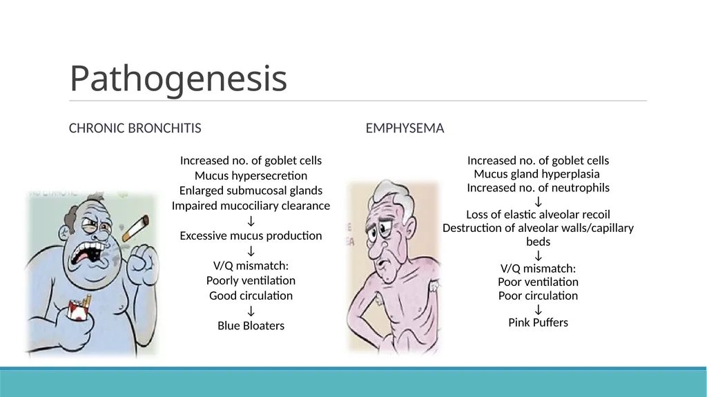
Cyanosis (chronic bronchitis)
Pursed-lip breathing (emphysema)
Nicotine staining of fingers
Signs of respiratory distress
Signs of hyperinflation
Low-pitched wheezes
Early- inspiratory crackles
Signs of right heart failure<br>
06
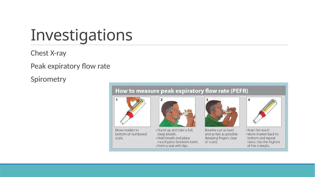
Investigations Chest X-ray
Peak expiratory flow rate
Spirometry<br>
07
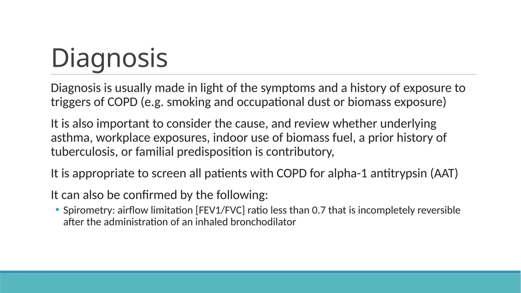
Diagnosis Diagnosis is usually made in light of the symptoms and a history of exposure to triggers of COPD (e.g. smoking and occupational dust or biomass exposure)
It is also important to consider the cause, and review whether underlying asthma, workplace exposures, indoor use of biomass fuel, a prior history of tuberculosis, or familial predisposition is contributory,
It is appropriate to screen all patients with COPD for alpha-1 antitrypsin (AAT)
It can also be confirmed by the following:
Spirometry: airflow limitation [FEV1/FVC] ratio less than 0.7 that is incompletely reversible after the administration of an inhaled bronchodilator<br>
08
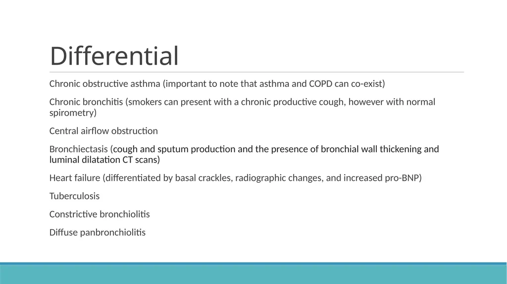
Differential Chronic obstructive asthma (important to note that asthma and COPD can co-exist)
Chronic bronchitis (smokers can present with a chronic productive cough, however with normal spirometry)
Central airflow obstruction
Bronchiectasis (cough and sputum production and the presence of bronchial wall thickening and luminal dilatation CT scans)
Heart failure (differentiated by basal crackles, radiographic changes, and increased pro-BNP)
Tuberculosis
Constrictive bronchiolitis
Diffuse panbronchiolitis<br>
09
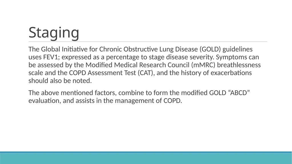
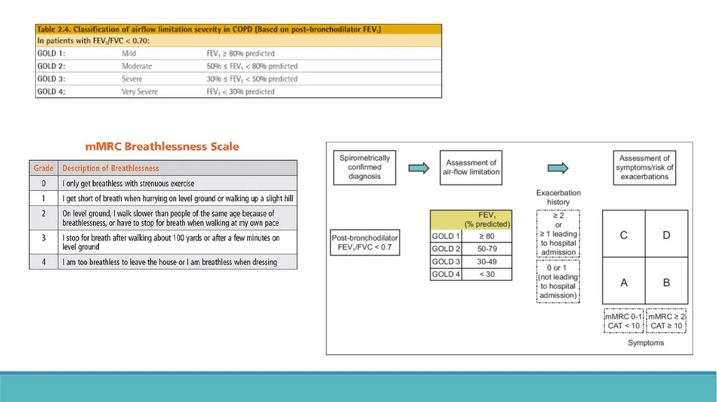
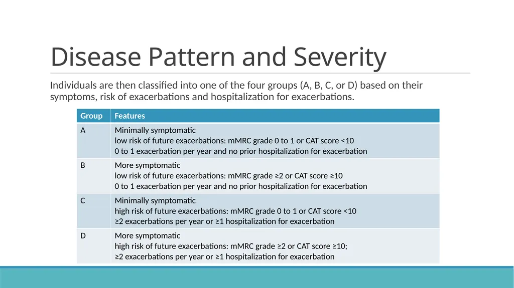
Staging The Global Initiative for Chronic Obstructive Lung Disease (GOLD) guidelines uses FEV1; expressed as a percentage to stage disease severity. Symptoms can be assessed by the Modified Medical Research Council (mMRC) breathlessness scale and the COPD Assessment Test (CAT), and the history of exacerbations should also be noted.
The above mentioned factors, combine to form the modified GOLD “ABCD” evaluation, and assists in the management of COPD.<br>
10
Disease Pattern and Severity Individuals are then classified into one of the four groups (A, B, C, or D) based on their symptoms, risk of exacerbations and hospitalization for exacerbations.<br>
11
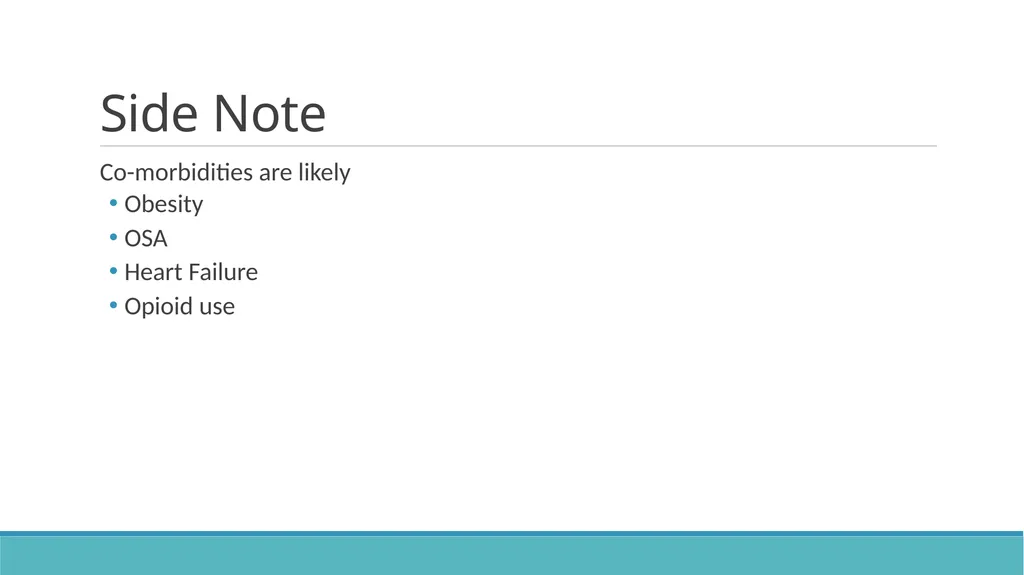
Side Note Co-morbidities are likely
Obesity
OSA
Heart Failure
Opioid use<br>
12
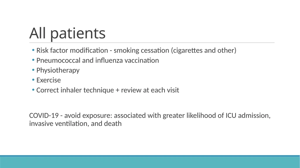
All patients Risk factor modification - smoking cessation (cigarettes and other)
Pneumococcal and influenza vaccination
Physiotherapy
Exercise
Correct inhaler technique + review at each visit
COVID-19 - avoid exposure: associated with greater likelihood of ICU admission, invasive ventilation, and death<br>
13
Stable COPD Aim of therapy
Improve symptoms
Decrease exacerbations
Improve patient function and quality of life<br>
14
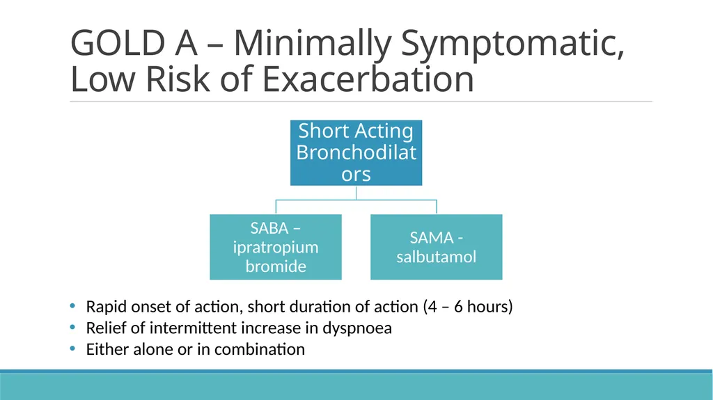
GOLD A – Minimally Symptomatic, Low Risk of Exacerbation Rapid onset of action, short duration of action (4 – 6 hours)
Relief of intermittent increase in dyspnoea
Either alone or in combination<br>
15
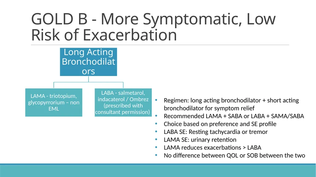
GOLD B - More Symptomatic, Low Risk of Exacerbation Regimen: long acting bronchodilator + short acting bronchodilator for symptom relief
Recommended LAMA + SABA or LABA + SAMA/SABA
Choice based on preference and SE profile
LABA SE: Resting tachycardia or tremor
LAMA SE: urinary retention
LAMA reduces exacerbations > LABA
No difference between QOL or SOB between the two<br>
16
GOLD C - Minimally Symptomatic, High Risk of Exacerbation LAMA suggested<br>
17
GOLD D - More Symptomatic, High Risk of Exacerbation LABA/LAMA combination preferred over LABA/ICS
LABA/ICS combination associated with reduced mortality and exacerbations compared with single bronchodilator therapy<br>
18
Non – pharmacological therapy Supplemental oxygen - chronic hypoxaemia
Nocturnal hypoxaemia - various options based on co-morbidities and sleep studies; CPAP as an example
Surgical - lung volume reduction or transplant<br>
19
Monitoring Three-to-six monthly visits: assess symptoms (mMRC) and oximetry, smoking status, adherence to medication
Annual spirometry - assess degree of airflow limitation
Screening for lung cancer and co-morbidities like heart failure, ILD, cardiac arrhythmias and, bronchiectasis<br>
20
COPD Exacerbation The WHO defines an exacerbation of COPD as "an acute event characterized by a worsening of the patient's respiratory symptoms that is beyond normal day-to-day variations and leads to a change in medication.”
This includes an acute change in one or more of the following symptoms:
Cough increases in frequency and severity
Sputum production increases in volume and/or changes character
Dyspnoea increases<br>
21
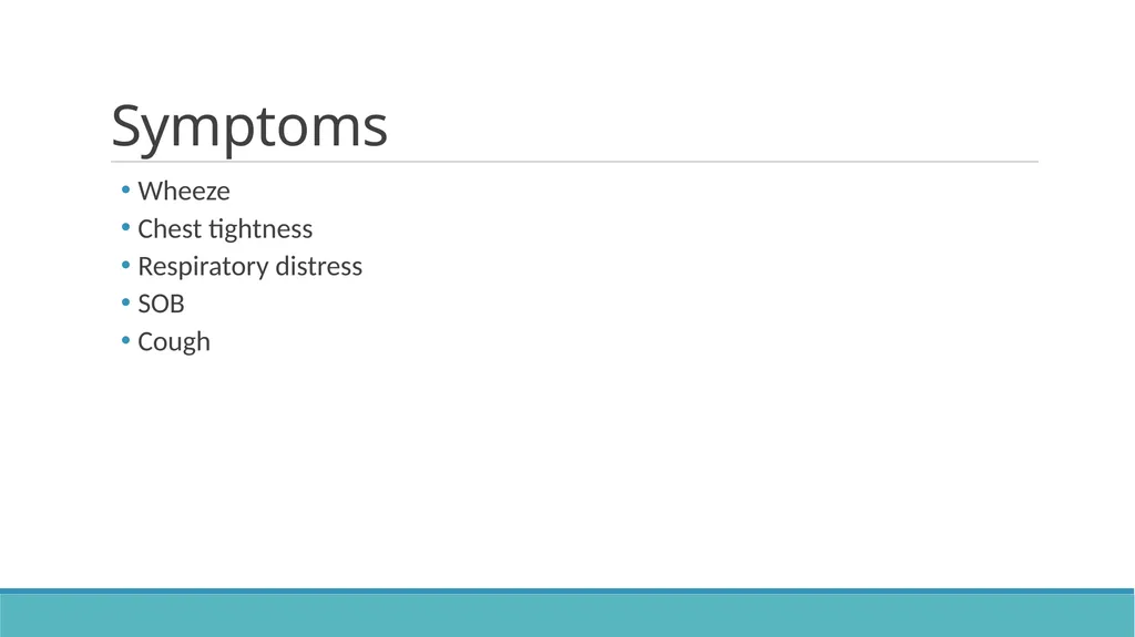
Symptoms Wheeze
Chest tightness
Respiratory distress
SOB
Cough<br>
22
Home vs Hospital Clinical severity
Respiratory failure<br>
23
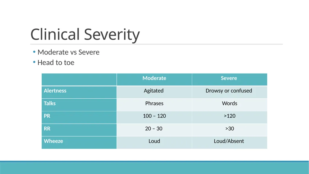
Clinical Severity Moderate vs Severe
Head to toe<br>
24
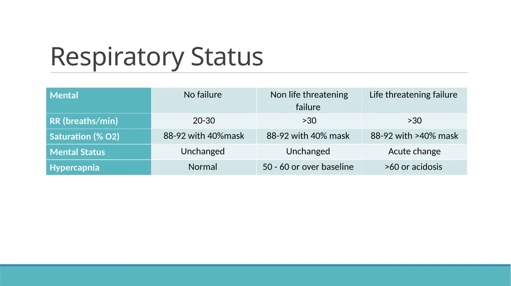
Respiratory Status<br>
25
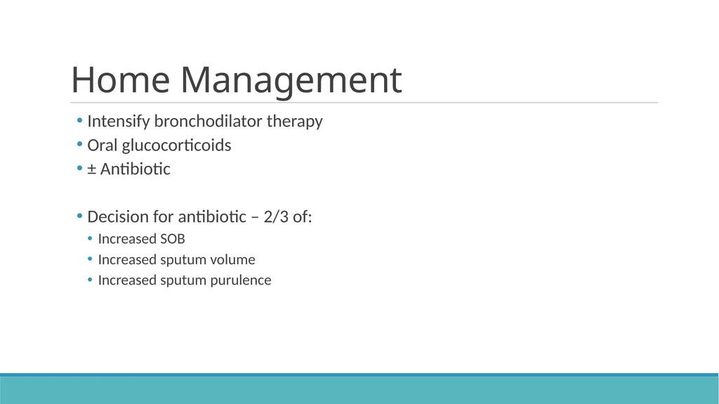
Home Management Intensify bronchodilator therapy
Oral glucocorticoids
± Antibiotic
Decision for antibiotic – 2/3 of:
Increased SOB
Increased sputum volume
Increased sputum purulence<br>
26
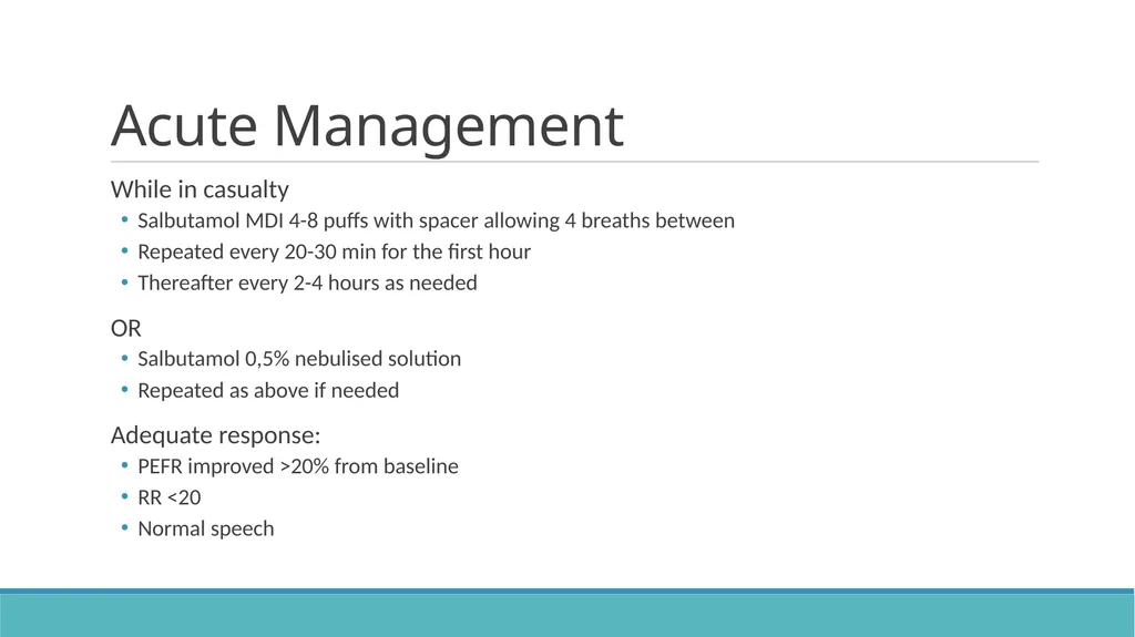
Acute Management While in casualty
Salbutamol MDI 4-8 puffs with spacer allowing 4 breaths between
Repeated every 20-30 min for the first hour
Thereafter every 2-4 hours as needed
OR
Salbutamol 0,5% nebulised solution
Repeated as above if needed
Adequate response:
PEFR improved >20% from baseline
RR <20
Normal speech<br>
27
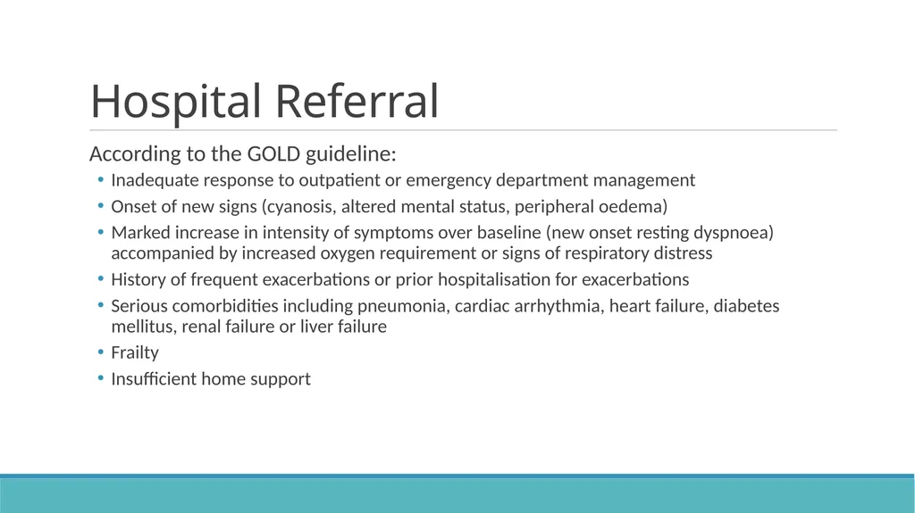
Hospital Referral According to the GOLD guideline:
Inadequate response to outpatient or emergency department management
Onset of new signs (cyanosis, altered mental status, peripheral oedema)
Marked increase in intensity of symptoms over baseline (new onset resting dyspnoea) accompanied by increased oxygen requirement or signs of respiratory distress
History of frequent exacerbations or prior hospitalisation for exacerbations
Serious comorbidities including pneumonia, cardiac arrhythmia, heart failure, diabetes mellitus, renal failure or liver failure
Frailty
Insufficient home support<br>
28
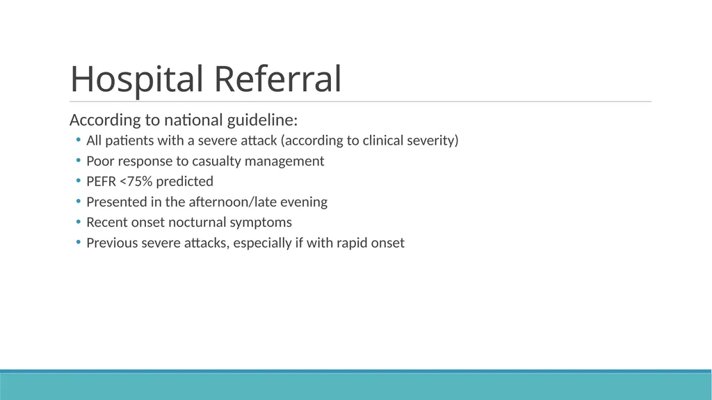
Hospital Referral According to national guideline:
All patients with a severe attack (according to clinical severity)
Poor response to casualty management
PEFR <75% predicted
Presented in the afternoon/late evening
Recent onset nocturnal symptoms
Previous severe attacks, especially if with rapid onset<br>
29
While awaiting referral Same as acute management in casualty (salbutamol etc.)
In addition:
Oxygen – enough to maintain O2 saturation of 88 – 92%
Prevents worsening hypercapnia – maintaining these saturations are associated with a lower mortality rate
Steroid therapy – oral or hydrocortisone 100mg IVI (slow infusion)
Ipratropium bromide nebulised if poor response to salbutamol<br>
30
References https://goldcopd.org/wp-content/uploads/2016/12/wms-GOLD-2017-Pocket-Guide-1.pdf
https://www.researchgate.net/publication/325345675_An_Update_on_the_Global_Initiative_for_Chronic_Obstructive_Lung_Disease_2017_Guidelines_With_a_Focus_on_Classification_and_Management_of_Stable_COPD<br>