Corneal opacity Dr. Adeeb A Assistant professor Dept. of Shalakya tantra Scarring or Opacity If the ulcer is very superficial and involves epithelium only, it heals without leaving any opacity. When the Bowmans and few superficial stromal
"Corneal opacity Dr. Adeeb A Assistant professor" is the property of its rightful owner. Permission is granted to
download and print the materials on this website for personal, non-commercial use only, and to display it
on your personal computer provided you do not modify the materials and that you retain all copyright
notices contained in the materials. By downloading content from our website, you accept the terms of this
agreement.
Presentation Transcript
01
Corneal opacity Dr. Adeeb A
Assistant professor
Dept. of Shalakya tantra<br>
02
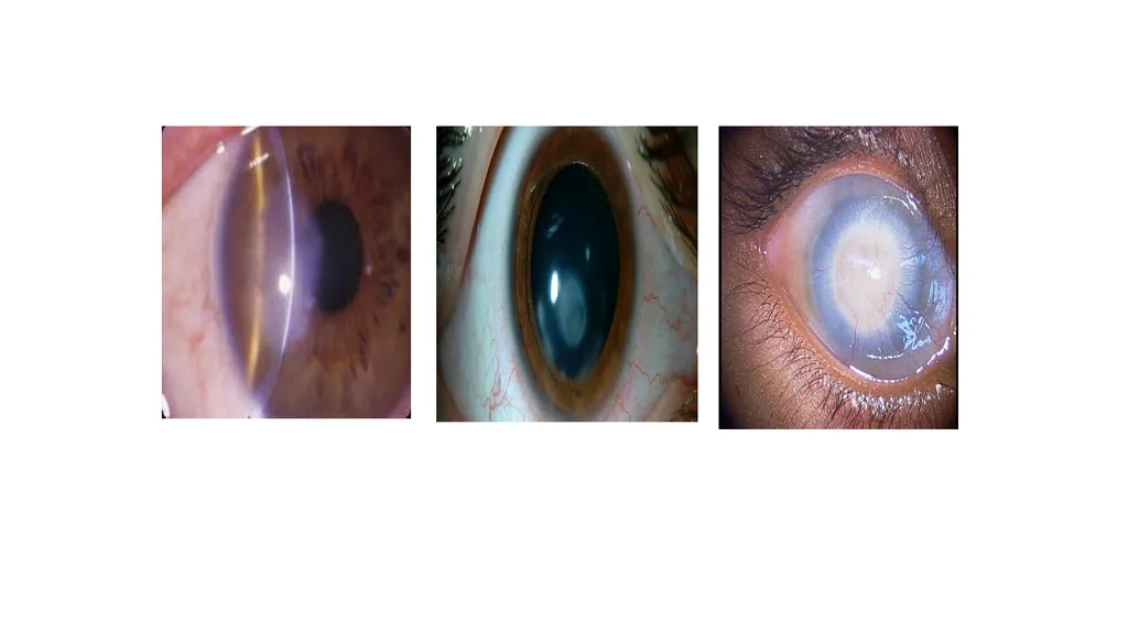
Scarring or Opacity If the ulcer is very superficial and involves epithelium only, it heals without leaving any opacity.
When the Bowman’s and few superficial stromal cells are involved (up to 1/3rd), the resultant opacity is called Nebula (fog or mist) - describes a hard-to-see corneal scar - one where slit-lamp detection is required.<br>
03
If it involves >1/3rd of Stroma, the resultant opacity is called Macula (stain or spot). It can be seen with proper illumination (Torch light)
If the Descemets and endothelium is involved, it is called Leucoma (white) is a white scar that is easily seen just by looking at the eye.<br>
04
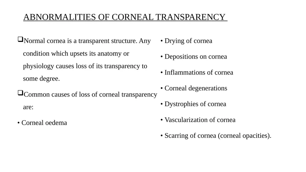
ABNORMALITIES OF CORNEAL TRANSPARENCY Normal cornea is a transparent structure. Any condition which upsets its anatomy or physiology causes loss of its transparency to some degree.
Common causes of loss of corneal transparency are:
• Corneal oedema
• Drying of cornea
• Depositions on cornea
• Inflammations of cornea
• Corneal degenerations
• Dystrophies of cornea
• Vascularization of cornea
• Scarring of cornea (corneal opacities).<br>
05
CORNEAL OPACITY<br>
06
CORNEAL OPACITY The word 'corneal opacification' literally means loss of normal transparency of cornea, which can occur in many conditions.
Corneal opacity is used particularly for the loss of transparency of cornea due to scarring.<br>
07
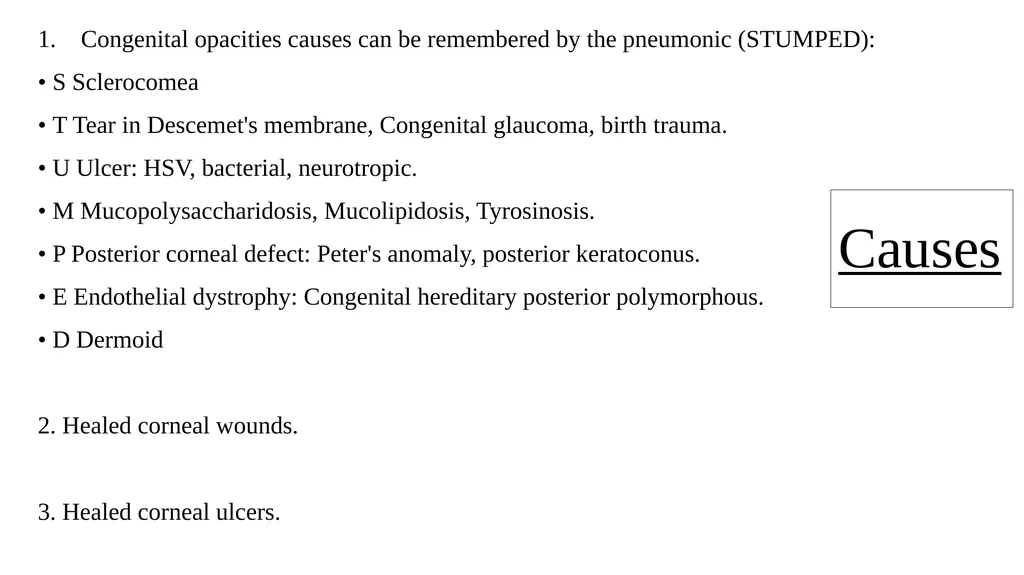
Causes Congenital opacities causes can be remembered by the pneumonic (STUMPED):
• S Sclerocomea
• T Tear in Descemet's membrane, Congenital glaucoma, birth trauma.
• U Ulcer: HSV, bacterial, neurotropic.
• M Mucopolysaccharidosis, Mucolipidosis, Tyrosinosis.
• P Posterior corneal defect: Peter's anomaly, posterior keratoconus.
• E Endothelial dystrophy: Congenital hereditary posterior polymorphous.
• D Dermoid
2. Healed corneal wounds.
3. Healed corneal ulcers.<br>
08
Clinical features Corneal opacity may produce
loss of vision (when dense opacity covers the pupillary area) or blurred vision (due to astigmatic effect).<br>
09
Types of corneal opacity Depending on the density, corneal opacity is graded as
Nebula
Macula
Leucoma.<br>
10
Nebular corneal opacity It is a faint opacity which results due to superficial scars involving Bowman's layer and superficial stroma.
A thin, diffuse nebula covering the pupillary area interferes more with vision than the localised leucoma away from pupillary area. Further, the nebula produces more discomfort to patient due to blurred image owing to irregular astigmatism than the leucoma which completely curs off the light rays.<br>
11
Macular corneal opacity. It is a semi-dense opacity produced when scarring involves about half the corneal stroma<br>
12
Leucomatous corneal opacity (leucoma simplex) It is a dense white opacity which results due co scarring of more rhan half of the stroma<br>
13
Adherent leucoma It results when healing occurs after perforation of cornea with incarceration of iris<br>
14
Corneal facet Sometimes, the corneal surface is depressed at the site of healing (due to less fibrous tissue); such a scar is called facet.<br>
15
Kerectasia ln this condition, corneal curvature is increased at the site of opacity (bulge due to weak scar).<br>
16
Anterior staphyloma. An ectasia of pseudocornea (the scar formed from organised exudates and fibrous tissue covered with epithelium) which results after total sloughing of cornea, with iris plastered behind it is called anterior staphyloma<br>
17
Treatment Optical iridectomy. It may be performed in cases with central macular or leucomatous corneal opacities, provided vision improves with pupillary dilatation.
Phototherapeutic keratectomy (PTK) performed with excimer laser is useful in superficial (nebular) corneal opacities.
Keratoplasty provides good vis11t1/ results in uncomplicated cases with corneal opacities, where optical iridecromy is not of much use.
Cosmetic-coloured contact lens gives very good cosmetic appearance in an eye with ugly scar having no potential for vision. Presently, this is considered the best option, even over and above the tattooing for cosmetic purpose.<br>
18
5. Tattooing of scar. It was performed for cosmeric purposes in the past. It is suitable only for firm scars in a quiet eye without usefu l vision. For tattooing Indian black ink, gold or platinum may be used. To perform tattooing, first of all, the epithelium covering the opacity is removed under topical anaesthesia (2% or 4% xylocaine). Then a piece of blotting paper of same size and shape, soaked in 4% gold chloride (for brown colour) or 2% platinum chloride (for dark colour) is applied over it. After 2-3 minutes, the piece of filter paper is removed and a few drops of freshly prepared 2% hydrazine hydrate solution are poured over it. Lastly, eye is irrigated vvith normal saline and patched after instilling antibiotic and atropine eye ointment. Epithelium grows over the pigmented area.<br>