
Decompressive craniectomy: Indication, technique,
CB
Published · 47 slides · 0 views
1 / 1

Description
Decompressive craniectomy: Indication, technique, present status and controversies History Decompressive craniotomy first described by Annandale in 1894 Performed as palliative procedure for inoperable brain tumor Kocher(1901) first
Related Topics
Share
Embed code
Download this presentation From Below
"Decompressive craniectomy: Indication, technique," is the property of its rightful owner. Permission is granted to download and print the materials on this website for personal, non-commercial use only, and to display it on your personal computer provided you do not modify the materials and that you retain all copyright notices contained in the materials. By downloading content from our website, you accept the terms of this agreement.
Presentation Transcript
01
Decompressive craniectomy: Indication, technique, present status and controversies<br>
02
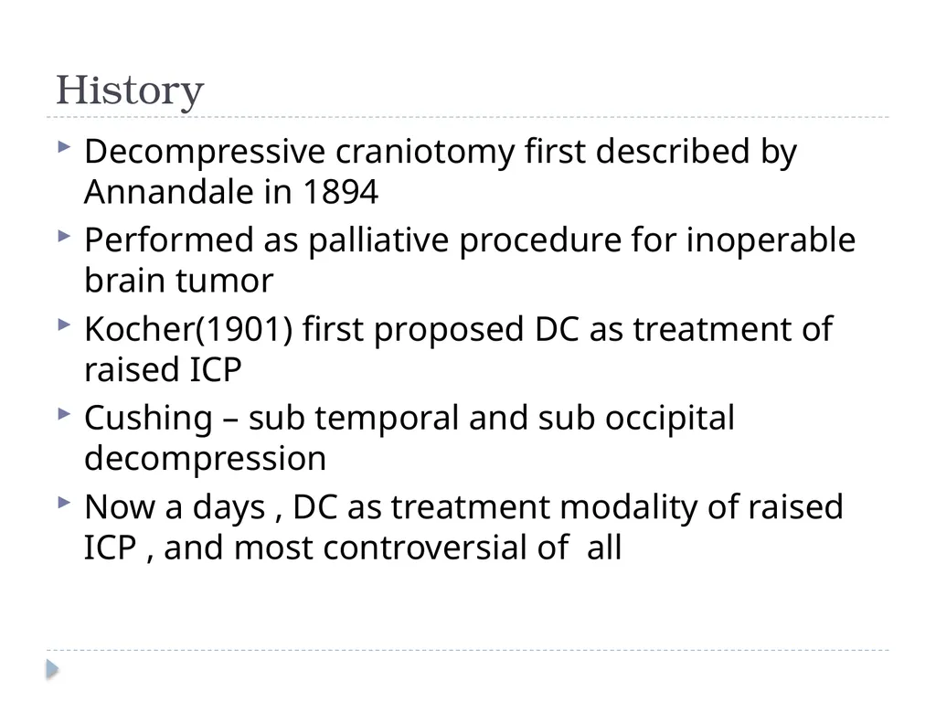
History Decompressive craniotomy first described by Annandale in 1894
Performed as palliative procedure for inoperable brain tumor
Kocher(1901) first proposed DC as treatment of raised ICP
Cushing – sub temporal and sub occipital decompression
Now a days , DC as treatment modality of raised ICP , and most controversial of all<br>
Performed as palliative procedure for inoperable brain tumor
Kocher(1901) first proposed DC as treatment of raised ICP
Cushing – sub temporal and sub occipital decompression
Now a days , DC as treatment modality of raised ICP , and most controversial of all<br>
03
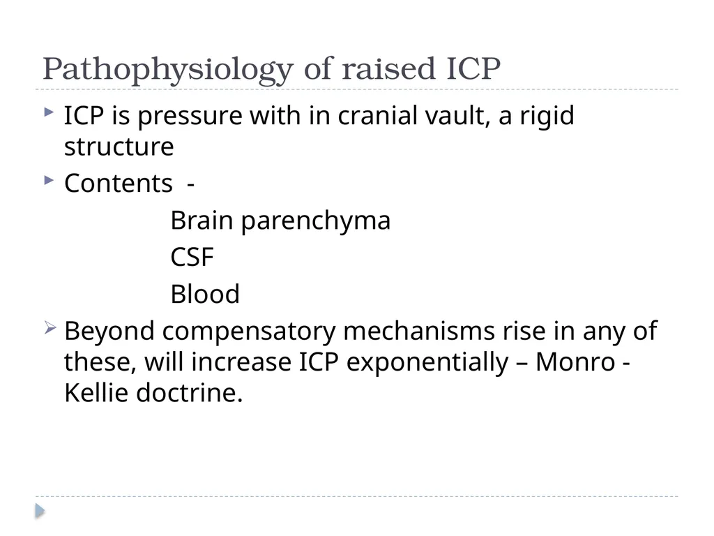
Pathophysiology of raised ICP ICP is pressure with in cranial vault, a rigid structure
Contents -
Brain parenchyma
CSF
Blood
Beyond compensatory mechanisms rise in any of these, will increase ICP exponentially – Monro - Kellie doctrine.<br>
Contents -
Brain parenchyma
CSF
Blood
Beyond compensatory mechanisms rise in any of these, will increase ICP exponentially – Monro - Kellie doctrine.<br>
04
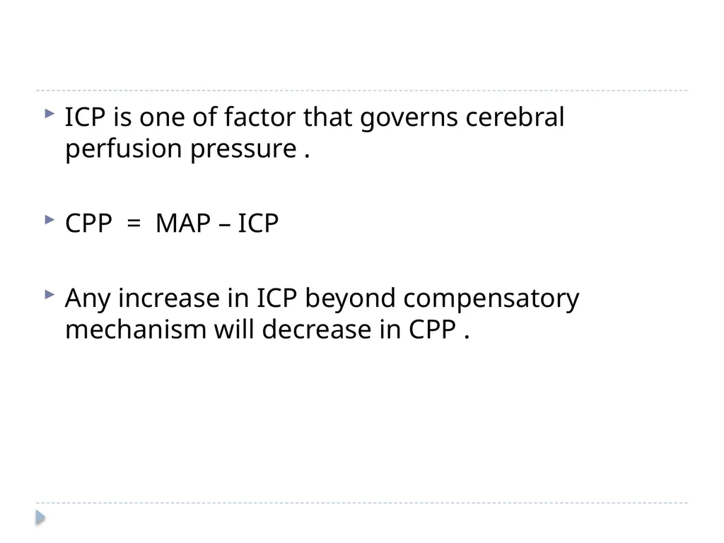
ICP is one of factor that governs cerebral perfusion pressure .
CPP = MAP – ICP
Any increase in ICP beyond compensatory mechanism will decrease in CPP .<br>
CPP = MAP – ICP
Any increase in ICP beyond compensatory mechanism will decrease in CPP .<br>
05
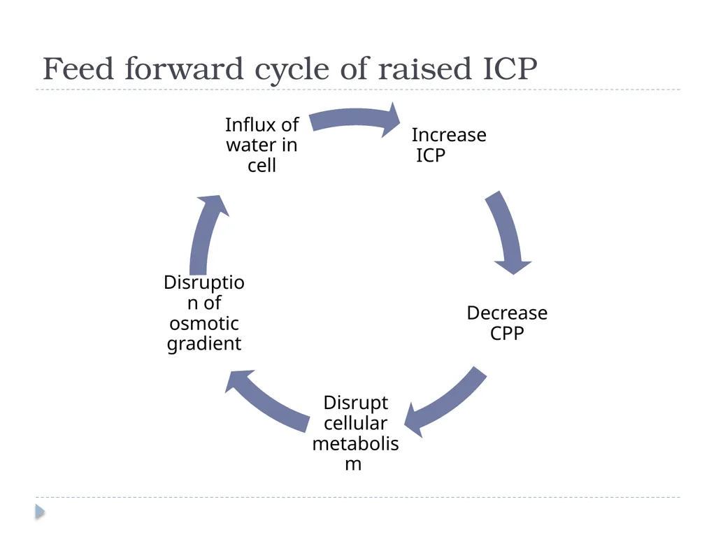
Feed forward cycle of raised ICP<br>
06
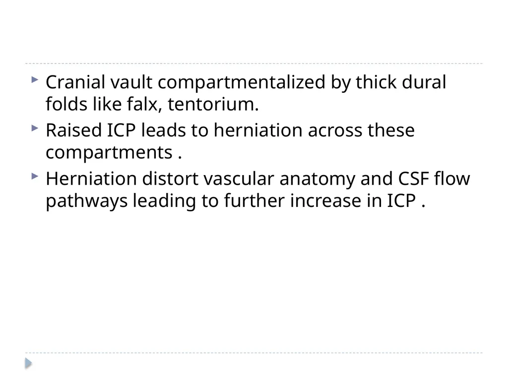
Cranial vault compartmentalized by thick dural folds like falx, tentorium.
Raised ICP leads to herniation across these compartments .
Herniation distort vascular anatomy and CSF flow pathways leading to further increase in ICP .<br>
Raised ICP leads to herniation across these compartments .
Herniation distort vascular anatomy and CSF flow pathways leading to further increase in ICP .<br>
07
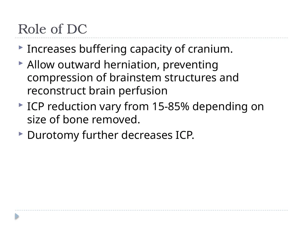
Role of DC Increases buffering capacity of cranium.
Allow outward herniation, preventing compression of brainstem structures and reconstruct brain perfusion
ICP reduction vary from 15-85% depending on size of bone removed.
Durotomy further decreases ICP.<br>
Allow outward herniation, preventing compression of brainstem structures and reconstruct brain perfusion
ICP reduction vary from 15-85% depending on size of bone removed.
Durotomy further decreases ICP.<br>
08
Indication of DC Traumatic brain injury
Malignant cerebral infarction
Others – Cerebral venous sinus thrombosis, intracerebral hematoma, metabolic encepahlopathies .<br>
Malignant cerebral infarction
Others – Cerebral venous sinus thrombosis, intracerebral hematoma, metabolic encepahlopathies .<br>
09
Role of DC in Traumatic brain injury TBI is the most common indication for decompressive craniectomy
Pathophysiology of TBI –
Primary injury
Secondary injury
Secondary injury is amenable to treatment, aim of the treatment is to prevent secondary injury.<br>
Pathophysiology of TBI –
Primary injury
Secondary injury
Secondary injury is amenable to treatment, aim of the treatment is to prevent secondary injury.<br>
10
Recommendation of brain trauma foundation General maneuvers –
head elevation & neutral position (to avoid venous congestion)
normothermia,
sedation,
volume resuscitation
First line therapy – CSF drainage, hypocapnia (PaCo2 30-35mmHg), mannitol administration
Second tier – high dose barbiturates, intense hyperventilation (PaCo2- <30mmHg), increase in mean arterial pressure, mild to moderate hypothermia, decomrpressive craniectomy<br>
head elevation & neutral position (to avoid venous congestion)
normothermia,
sedation,
volume resuscitation
First line therapy – CSF drainage, hypocapnia (PaCo2 30-35mmHg), mannitol administration
Second tier – high dose barbiturates, intense hyperventilation (PaCo2- <30mmHg), increase in mean arterial pressure, mild to moderate hypothermia, decomrpressive craniectomy<br>
11
Types of DC Primary or prophylactic DC
Therapeutic decompression or secondary decompressive craniectomy – continuous ICP monitoring is conducted and ICP is refractory to medical treatment.<br>
Therapeutic decompression or secondary decompressive craniectomy – continuous ICP monitoring is conducted and ICP is refractory to medical treatment.<br>
12
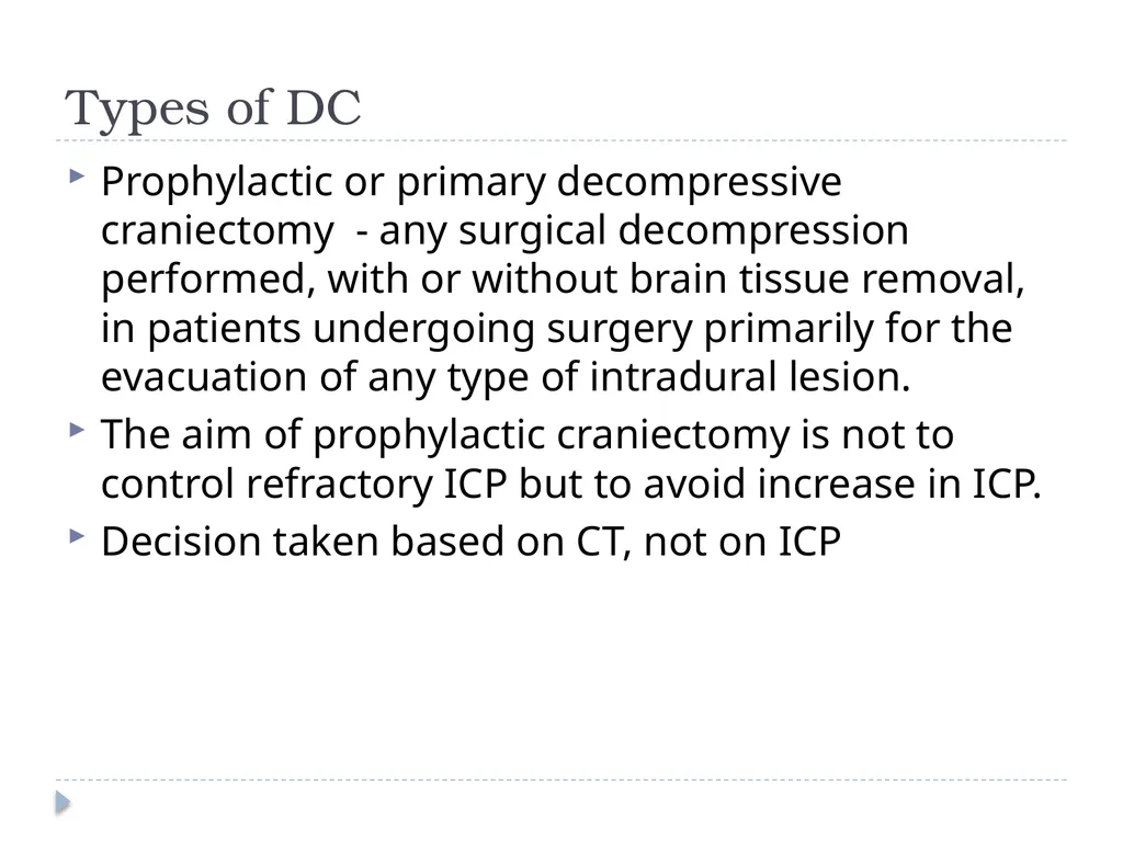
Types of DC Prophylactic or primary decompressive craniectomy - any surgical decompression performed, with or without brain tissue removal, in patients undergoing surgery primarily for the evacuation of any type of intradural lesion.
The aim of prophylactic craniectomy is not to control refractory ICP but to avoid increase in ICP.
Decision taken based on CT, not on ICP<br>
The aim of prophylactic craniectomy is not to control refractory ICP but to avoid increase in ICP.
Decision taken based on CT, not on ICP<br>
13
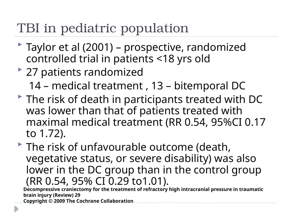
TBI in pediatric population Taylor et al (2001) – prospective, randomized controlled trial in patients <18 yrs old
27 patients randomized
14 – medical treatment , 13 – bitemporal DC
The risk of death in participants treated with DC was lower than that of patients treated with maximal medical treatment (RR 0.54, 95%CI 0.17 to 1.72).
The risk of unfavourable outcome (death, vegetative status, or severe disability) was also lower in the DC group than in the control group (RR 0.54, 95% CI 0.29 to1.01). Decompressive craniectomy for the treatment of refractory high intracranial pressure in traumatic brain injury (Review) 29
Copyright © 2009 The Cochrane Collaboration<br>
27 patients randomized
14 – medical treatment , 13 – bitemporal DC
The risk of death in participants treated with DC was lower than that of patients treated with maximal medical treatment (RR 0.54, 95%CI 0.17 to 1.72).
The risk of unfavourable outcome (death, vegetative status, or severe disability) was also lower in the DC group than in the control group (RR 0.54, 95% CI 0.29 to1.01). Decompressive craniectomy for the treatment of refractory high intracranial pressure in traumatic brain injury (Review) 29
Copyright © 2009 The Cochrane Collaboration<br>
14
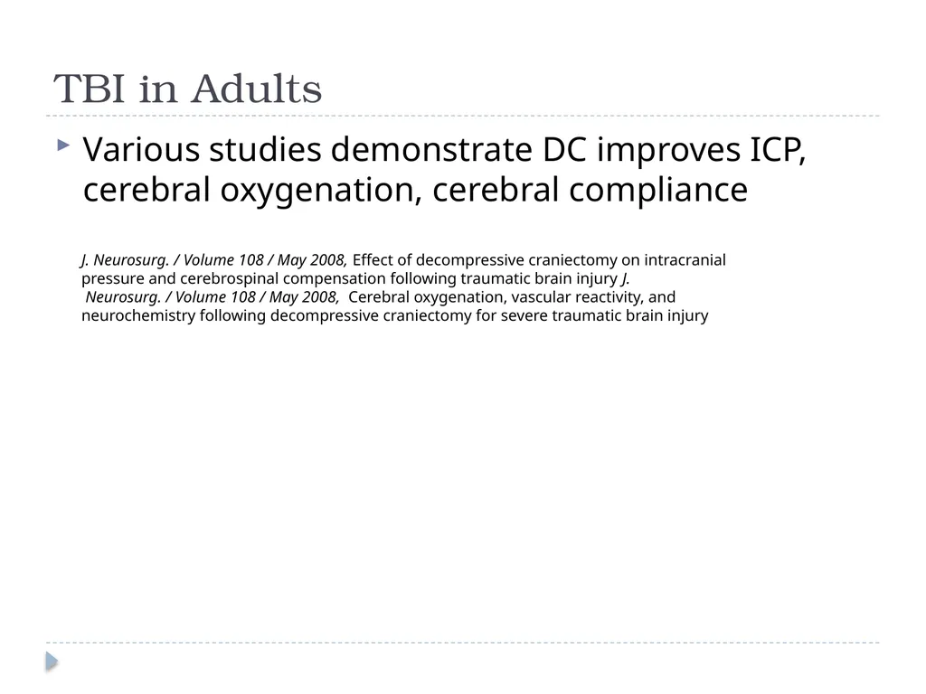
TBI in Adults Various studies demonstrate DC improves ICP, cerebral oxygenation, cerebral compliance J. Neurosurg. / Volume 108 / May 2008, Effect of decompressive craniectomy on intracranial
pressure and cerebrospinal compensation following traumatic brain injury J.
Neurosurg. / Volume 108 / May 2008, Cerebral oxygenation, vascular reactivity, and
neurochemistry following decompressive craniectomy for severe traumatic brain injury<br>
pressure and cerebrospinal compensation following traumatic brain injury J.
Neurosurg. / Volume 108 / May 2008, Cerebral oxygenation, vascular reactivity, and
neurochemistry following decompressive craniectomy for severe traumatic brain injury<br>
15
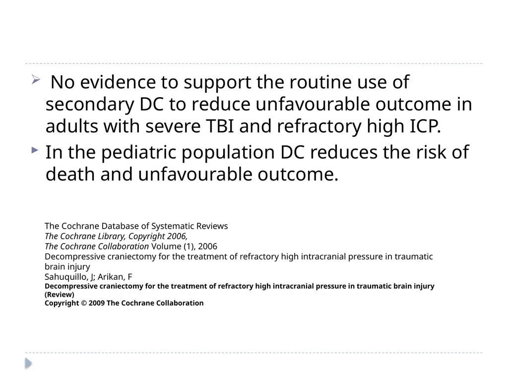
No evidence to support the routine use of secondary DC to reduce unfavourable outcome in adults with severe TBI and refractory high ICP.
In the pediatric population DC reduces the risk of death and unfavourable outcome. The Cochrane Database of Systematic Reviews The Cochrane Library, Copyright 2006, The Cochrane Collaboration Volume (1), 2006 Decompressive craniectomy for the treatment of refractory high intracranial pressure in traumatic brain injury Sahuquillo, J; Arikan, F
Decompressive craniectomy for the treatment of refractory high intracranial pressure in traumatic brain injury (Review)
Copyright © 2009 The Cochrane Collaboration<br>
In the pediatric population DC reduces the risk of death and unfavourable outcome. The Cochrane Database of Systematic Reviews The Cochrane Library, Copyright 2006, The Cochrane Collaboration Volume (1), 2006 Decompressive craniectomy for the treatment of refractory high intracranial pressure in traumatic brain injury Sahuquillo, J; Arikan, F
Decompressive craniectomy for the treatment of refractory high intracranial pressure in traumatic brain injury (Review)
Copyright © 2009 The Cochrane Collaboration<br>
16
DECRA trial (DEcompressive CRAniectomy in patient with severe traumatic brain injury )
RESCUEicp (Randomized Evaluation of Surgery with Craniectomy for Uncontrollable Elevation of ICP)<br>
RESCUEicp (Randomized Evaluation of Surgery with Craniectomy for Uncontrollable Elevation of ICP)<br>
17
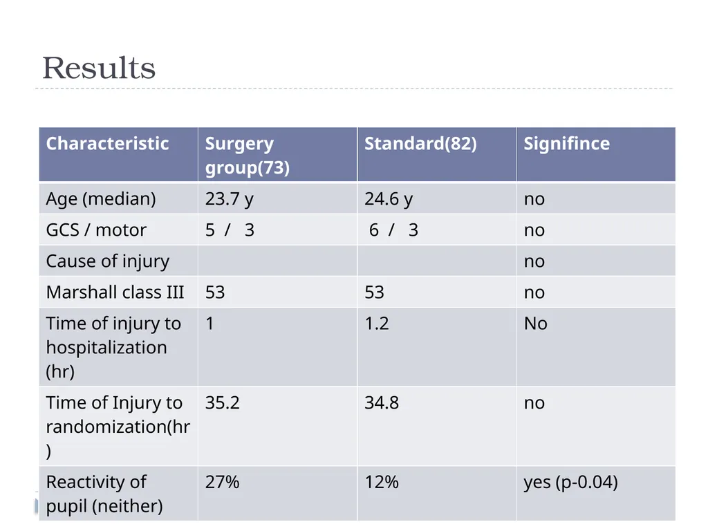
DECRA trail Multicenter, randomized, controlled Decompressive Craniectomy (DECRA) trial to test the efficacy of bifrontotemporoparietal decompressive craniectomy in adults under the age of 60 years with traumatic brain injury in whom first-tier intensive care and neurosurgical therapies had not maintained intracranial pressure below accepted targets. Decompressive Craniectomy in Diffuse Traumatic Brain Injury . N Engl J Med. 2011 Apr 21;364(16):1493-502.<br>
18
Study Design Dec 2002- April 2010 , 15 tertiary care centres in Australia, New Zealand, Saudi Arabia
Inclusion criteria
Age 15-59 yrs
Non penetrating traumatic brain injury
GCS 3-8
Marshall class III (moderate diffuse injury on CT)
Exclusion – dilated non reactive pupil, mass lesion, spinal cord injury or cardiac arrest at scene<br>
Inclusion criteria
Age 15-59 yrs
Non penetrating traumatic brain injury
GCS 3-8
Marshall class III (moderate diffuse injury on CT)
Exclusion – dilated non reactive pupil, mass lesion, spinal cord injury or cardiac arrest at scene<br>
19
Treatment protocol Standard treatment Vs surgery + standard treatment
With in 72 hrs
ICP measurement in all patient (12 before and 36 hrs after randomization)
Surgery – bifrontotemporoparetal craniectomy with bilateral Dural opening (modified Polin technique) . Sagittal sinus and falx not divided.
Results compared based on extended Glasgow outcome scale at 6 months.<br>
With in 72 hrs
ICP measurement in all patient (12 before and 36 hrs after randomization)
Surgery – bifrontotemporoparetal craniectomy with bilateral Dural opening (modified Polin technique) . Sagittal sinus and falx not divided.
Results compared based on extended Glasgow outcome scale at 6 months.<br>
20
Results<br>
21
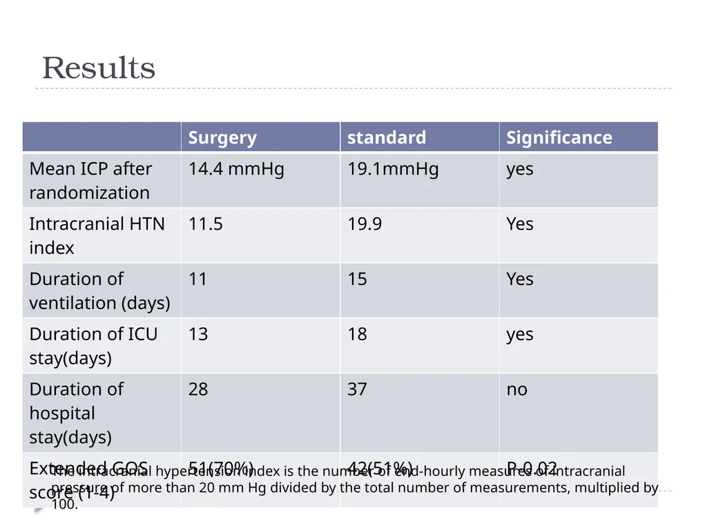
Results The intracranial hypertension index is the number of end-hourly measures of intracranial pressure of more than 20 mm Hg divided by the total number of measurements, multiplied by 100.<br>
22
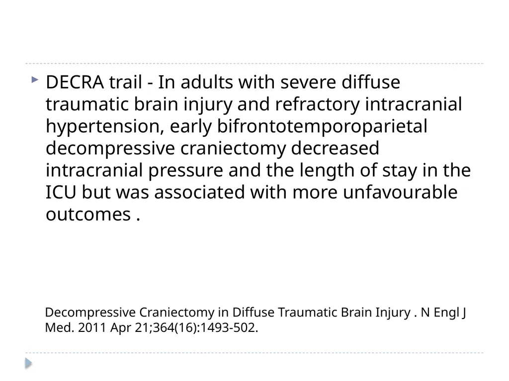
DECRA trail - In adults with severe diffuse traumatic brain injury and refractory intracranial hypertension, early bifrontotemporoparietal decompressive craniectomy decreased intracranial pressure and the length of stay in the ICU but was associated with more unfavourable outcomes . Decompressive Craniectomy in Diffuse Traumatic Brain Injury . N Engl J Med. 2011 Apr 21;364(16):1493-502.<br>
23
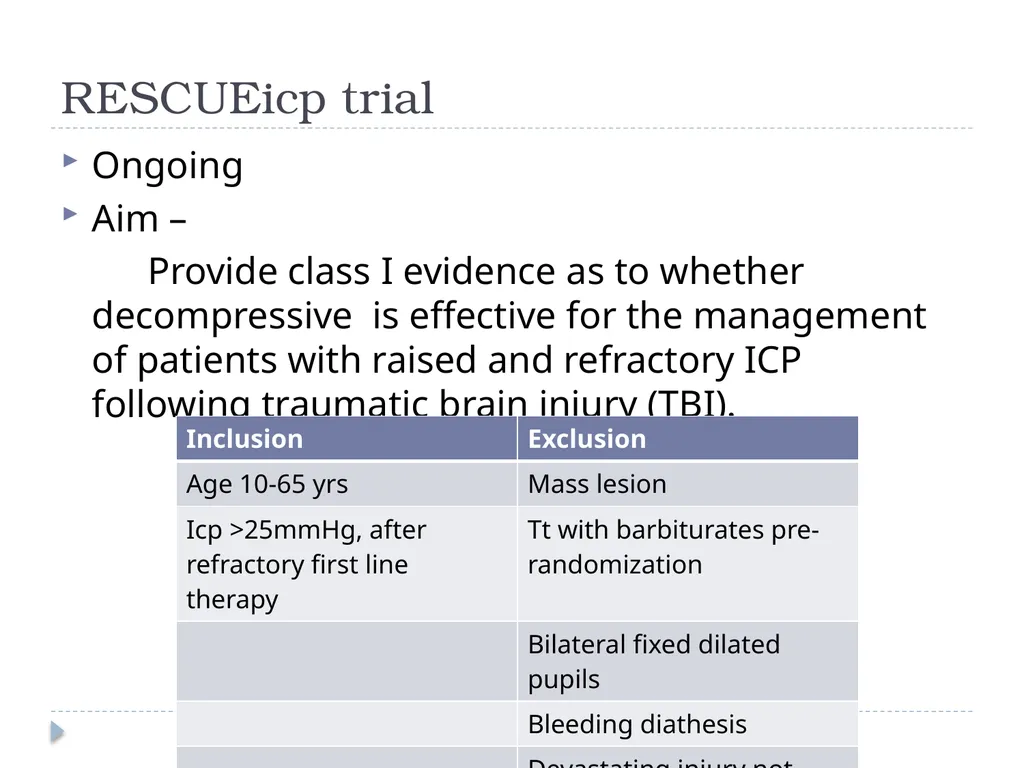
RESCUEicp trial Ongoing
Aim –
Provide class I evidence as to whether decompressive is effective for the management of patients with raised and refractory ICP following traumatic brain injury (TBI).<br>
Aim –
Provide class I evidence as to whether decompressive is effective for the management of patients with raised and refractory ICP following traumatic brain injury (TBI).<br>
24
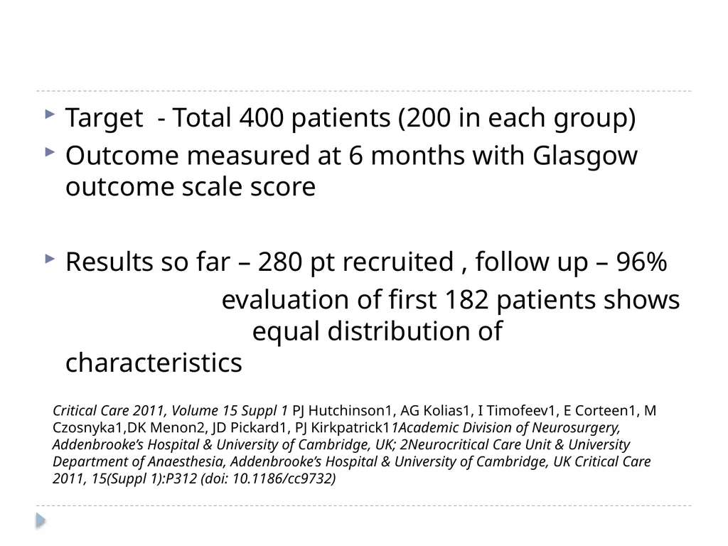
Target - Total 400 patients (200 in each group)
Outcome measured at 6 months with Glasgow outcome scale score
Results so far – 280 pt recruited , follow up – 96%
evaluation of first 182 patients shows equal distribution of characteristics Critical Care 2011, Volume 15 Suppl 1 PJ Hutchinson1, AG Kolias1, I Timofeev1, E Corteen1, M Czosnyka1,DK Menon2, JD Pickard1, PJ Kirkpatrick11Academic Division of Neurosurgery, Addenbrooke’s Hospital & University of Cambridge, UK; 2Neurocritical Care Unit & University Department of Anaesthesia, Addenbrooke’s Hospital & University of Cambridge, UK Critical Care 2011, 15(Suppl 1):P312 (doi: 10.1186/cc9732)<br>
Outcome measured at 6 months with Glasgow outcome scale score
Results so far – 280 pt recruited , follow up – 96%
evaluation of first 182 patients shows equal distribution of characteristics Critical Care 2011, Volume 15 Suppl 1 PJ Hutchinson1, AG Kolias1, I Timofeev1, E Corteen1, M Czosnyka1,DK Menon2, JD Pickard1, PJ Kirkpatrick11Academic Division of Neurosurgery, Addenbrooke’s Hospital & University of Cambridge, UK; 2Neurocritical Care Unit & University Department of Anaesthesia, Addenbrooke’s Hospital & University of Cambridge, UK Critical Care 2011, 15(Suppl 1):P312 (doi: 10.1186/cc9732)<br>
25
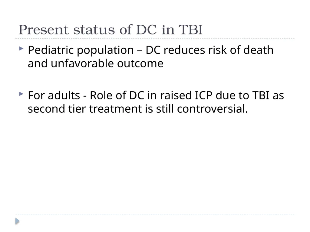
Present status of DC in TBI Pediatric population – DC reduces risk of death and unfavorable outcome
For adults - Role of DC in raised ICP due to TBI as second tier treatment is still controversial.<br>
For adults - Role of DC in raised ICP due to TBI as second tier treatment is still controversial.<br>
26
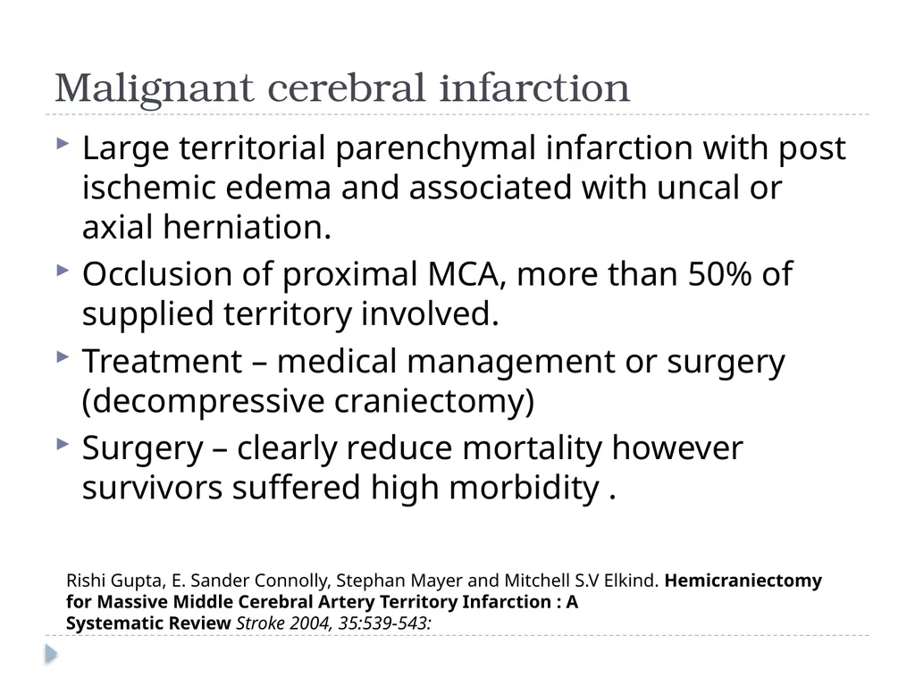
Malignant cerebral infarction Large territorial parenchymal infarction with post ischemic edema and associated with uncal or axial herniation.
Occlusion of proximal MCA, more than 50% of supplied territory involved.
Treatment – medical management or surgery (decompressive craniectomy)
Surgery – clearly reduce mortality however survivors suffered high morbidity . Rishi Gupta, E. Sander Connolly, Stephan Mayer and Mitchell S.V Elkind. Hemicraniectomy for Massive Middle Cerebral Artery Territory Infarction : A
Systematic Review Stroke 2004, 35:539-543:<br>
Occlusion of proximal MCA, more than 50% of supplied territory involved.
Treatment – medical management or surgery (decompressive craniectomy)
Surgery – clearly reduce mortality however survivors suffered high morbidity . Rishi Gupta, E. Sander Connolly, Stephan Mayer and Mitchell S.V Elkind. Hemicraniectomy for Massive Middle Cerebral Artery Territory Infarction : A
Systematic Review Stroke 2004, 35:539-543:<br>
27
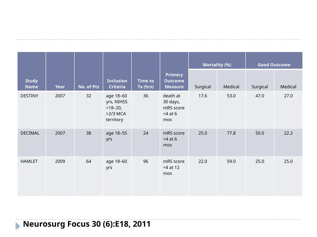
3 RCTs conducted and results published.
DECIMAL (DEcompressive Craniectomy In MALignant middle cerebral artery infarction)
DESTINY (DEcompressive Surgery for Treatment of INfarction of malignant middle cerebral arterY)
HAMLET ( Hemicraniectomy After Middle cerebral artery infarction with Life –threatening Edema Trial )<br>
DECIMAL (DEcompressive Craniectomy In MALignant middle cerebral artery infarction)
DESTINY (DEcompressive Surgery for Treatment of INfarction of malignant middle cerebral arterY)
HAMLET ( Hemicraniectomy After Middle cerebral artery infarction with Life –threatening Edema Trial )<br>
28
Neurosurg Focus 30 (6):E18, 2011<br>
29
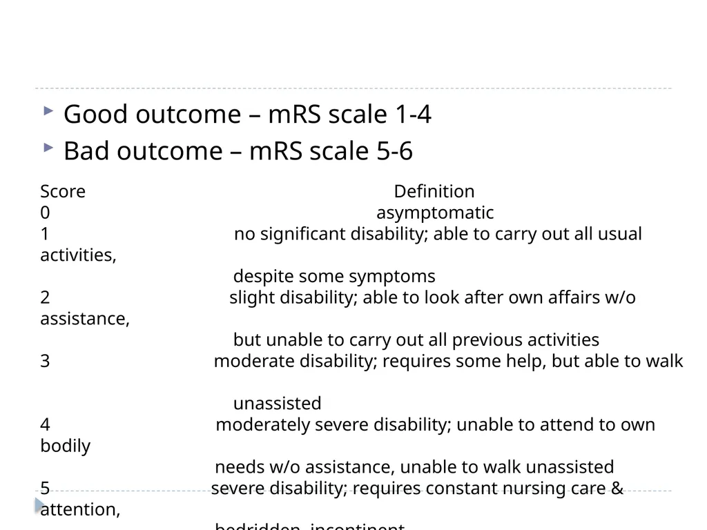
Good outcome – mRS scale 1-4
Bad outcome – mRS scale 5-6 Score Definition
0 asymptomatic
1 no significant disability; able to carry out all usual activities,
despite some symptoms
2 slight disability; able to look after own affairs w/o assistance,
but unable to carry out all previous activities
moderate disability; requires some help, but able to walk
unassisted
4 moderately severe disability; unable to attend to own bodily
needs w/o assistance, unable to walk unassisted
5 severe disability; requires constant nursing care & attention,
bedridden, incontinent
6 dead<br>
Bad outcome – mRS scale 5-6 Score Definition
0 asymptomatic
1 no significant disability; able to carry out all usual activities,
despite some symptoms
2 slight disability; able to look after own affairs w/o assistance,
but unable to carry out all previous activities
moderate disability; requires some help, but able to walk
unassisted
4 moderately severe disability; unable to attend to own bodily
needs w/o assistance, unable to walk unassisted
5 severe disability; requires constant nursing care & attention,
bedridden, incontinent
6 dead<br>
30
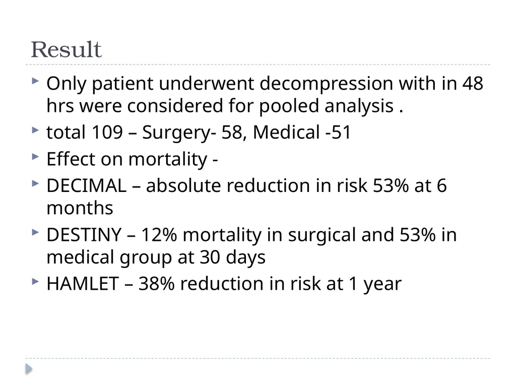
Result Only patient underwent decompression with in 48 hrs were considered for pooled analysis .
total 109 – Surgery- 58, Medical -51
Effect on mortality -
DECIMAL – absolute reduction in risk 53% at 6 months
DESTINY – 12% mortality in surgical and 53% in medical group at 30 days
HAMLET – 38% reduction in risk at 1 year<br>
total 109 – Surgery- 58, Medical -51
Effect on mortality -
DECIMAL – absolute reduction in risk 53% at 6 months
DESTINY – 12% mortality in surgical and 53% in medical group at 30 days
HAMLET – 38% reduction in risk at 1 year<br>
31
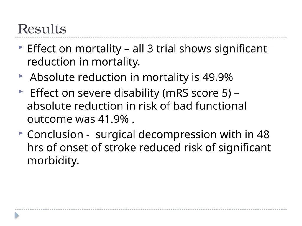
Results Effect on mortality – all 3 trial shows significant reduction in mortality.
Absolute reduction in mortality is 49.9%
Effect on severe disability (mRS score 5) – absolute reduction in risk of bad functional outcome was 41.9% .
Conclusion - surgical decompression with in 48 hrs of onset of stroke reduced risk of significant morbidity.<br>
Absolute reduction in mortality is 49.9%
Effect on severe disability (mRS score 5) – absolute reduction in risk of bad functional outcome was 41.9% .
Conclusion - surgical decompression with in 48 hrs of onset of stroke reduced risk of significant morbidity.<br>
32
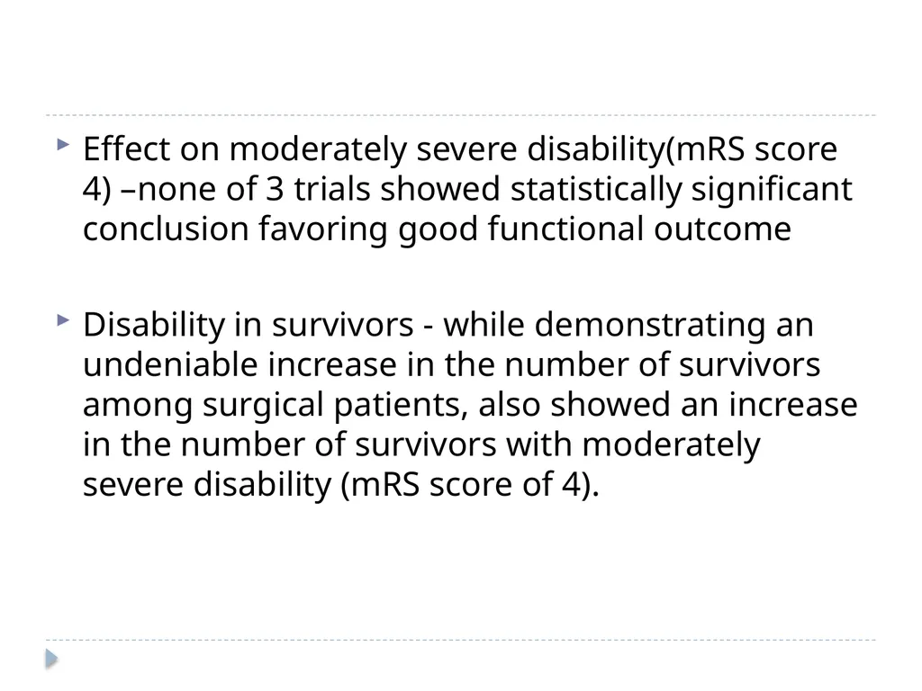
Effect on moderately severe disability(mRS score 4) –none of 3 trials showed statistically significant conclusion favoring good functional outcome
Disability in survivors - while demonstrating an undeniable increase in the number of survivors among surgical patients, also showed an increase in the number of survivors with moderately severe disability (mRS score of 4).<br>
Disability in survivors - while demonstrating an undeniable increase in the number of survivors among surgical patients, also showed an increase in the number of survivors with moderately severe disability (mRS score of 4).<br>
33
Laterality of DC – DECIMAL trail shows no difference in mRS score of survivors with or without aphasia at 1 yr.
Size of craniectomy – 12 cms diameter , FTP region reaching up to floor of middle fossa, avoiding midline. Wagner S, Schnippering H, Aschoff A, Koziol JA, Schwab S,Steiner T: Suboptimum hemicraniectomy as a cause of additional cerebral lesions in patients with malignant infarction of the middle cerebral artery. J Neurosurg 94:693–696, 200<br>
Size of craniectomy – 12 cms diameter , FTP region reaching up to floor of middle fossa, avoiding midline. Wagner S, Schnippering H, Aschoff A, Koziol JA, Schwab S,Steiner T: Suboptimum hemicraniectomy as a cause of additional cerebral lesions in patients with malignant infarction of the middle cerebral artery. J Neurosurg 94:693–696, 200<br>
34
Timing of surgery – optimal time window ?
HeADDFIRST (Hemicraniectomy And Durotomy on Deterioration From Infarction Related Swelling Trial)
window period – 96 hrs
HeMMI trial (Hemicraniectomy for Malignant Middle cerebral artery Infarcts) time period of 72 hrs.
Age of patient - <60 yrs.
DESTINY II – ongoing to study effect of Surgery in older
pt >60 yrs<br>
HeADDFIRST (Hemicraniectomy And Durotomy on Deterioration From Infarction Related Swelling Trial)
window period – 96 hrs
HeMMI trial (Hemicraniectomy for Malignant Middle cerebral artery Infarcts) time period of 72 hrs.
Age of patient - <60 yrs.
DESTINY II – ongoing to study effect of Surgery in older
pt >60 yrs<br>
35
Conclusion (present status) Pooled analysis of all 3 trials provides Class I evidence for the performance of early decompressive craniectomy in the setting of large unilateral infarcts (volume > 145 cc) within 48 hours of the ischemic event.
Further studies are needed to better define quality of life issues at long-term follow-up as well as age limit issues. Neurosurg Focus 30 (6):E18, 2011 Decompressive hemicraniectomy after malignant middle cerebral artery infarction: rationale and controversies Omar M. Arnaout, M.D., Salah G. Aoun, M.D., H. Hunt Batjer, M.D.,
and Bernard R. Bendo k, M.D. Department of Neurological Surgery, Feinberg School of Medicine and McGaw Medical Centre, North-western University, Chicago, Illinois<br>
Further studies are needed to better define quality of life issues at long-term follow-up as well as age limit issues. Neurosurg Focus 30 (6):E18, 2011 Decompressive hemicraniectomy after malignant middle cerebral artery infarction: rationale and controversies Omar M. Arnaout, M.D., Salah G. Aoun, M.D., H. Hunt Batjer, M.D.,
and Bernard R. Bendo k, M.D. Department of Neurological Surgery, Feinberg School of Medicine and McGaw Medical Centre, North-western University, Chicago, Illinois<br>
36
Technique of decompressive craniectomy Fronto-temporo-parietal approach
Bifrontal decompressive approach
Temporal approach<br>
Bifrontal decompressive approach
Temporal approach<br>
37
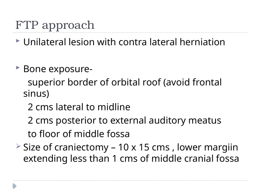
FTP approach Unilateral lesion with contra lateral herniation
Bone exposure-
superior border of orbital roof (avoid frontal sinus)
2 cms lateral to midline
2 cms posterior to external auditory meatus
to floor of middle fossa
Size of craniectomy – 10 x 15 cms , lower margiin extending less than 1 cms of middle cranial fossa<br>
Bone exposure-
superior border of orbital roof (avoid frontal sinus)
2 cms lateral to midline
2 cms posterior to external auditory meatus
to floor of middle fossa
Size of craniectomy – 10 x 15 cms , lower margiin extending less than 1 cms of middle cranial fossa<br>
38
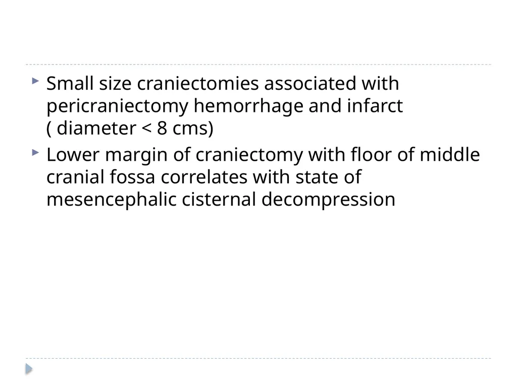
Small size craniectomies associated with pericraniectomy hemorrhage and infarct ( diameter < 8 cms)
Lower margin of craniectomy with floor of middle cranial fossa correlates with state of mesencephalic cisternal decompression<br>
Lower margin of craniectomy with floor of middle cranial fossa correlates with state of mesencephalic cisternal decompression<br>
39
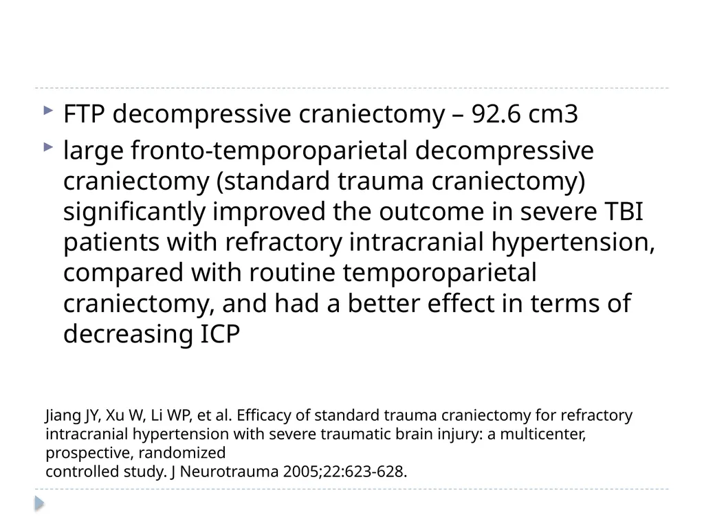
FTP decompressive craniectomy – 92.6 cm3
large fronto-temporoparietal decompressive craniectomy (standard trauma craniectomy) significantly improved the outcome in severe TBI patients with refractory intracranial hypertension, compared with routine temporoparietal craniectomy, and had a better effect in terms of decreasing ICP Jiang JY, Xu W, Li WP, et al. Efficacy of standard trauma craniectomy for refractory intracranial hypertension with severe traumatic brain injury: a multicenter, prospective, randomized
controlled study. J Neurotrauma 2005;22:623-628.<br>
large fronto-temporoparietal decompressive craniectomy (standard trauma craniectomy) significantly improved the outcome in severe TBI patients with refractory intracranial hypertension, compared with routine temporoparietal craniectomy, and had a better effect in terms of decreasing ICP Jiang JY, Xu W, Li WP, et al. Efficacy of standard trauma craniectomy for refractory intracranial hypertension with severe traumatic brain injury: a multicenter, prospective, randomized
controlled study. J Neurotrauma 2005;22:623-628.<br>
40
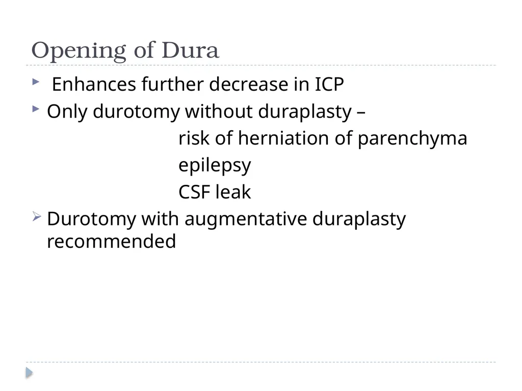
Opening of Dura Enhances further decrease in ICP
Only durotomy without duraplasty –
risk of herniation of parenchyma
epilepsy
CSF leak
Durotomy with augmentative duraplasty recommended<br>
Only durotomy without duraplasty –
risk of herniation of parenchyma
epilepsy
CSF leak
Durotomy with augmentative duraplasty recommended<br>
41
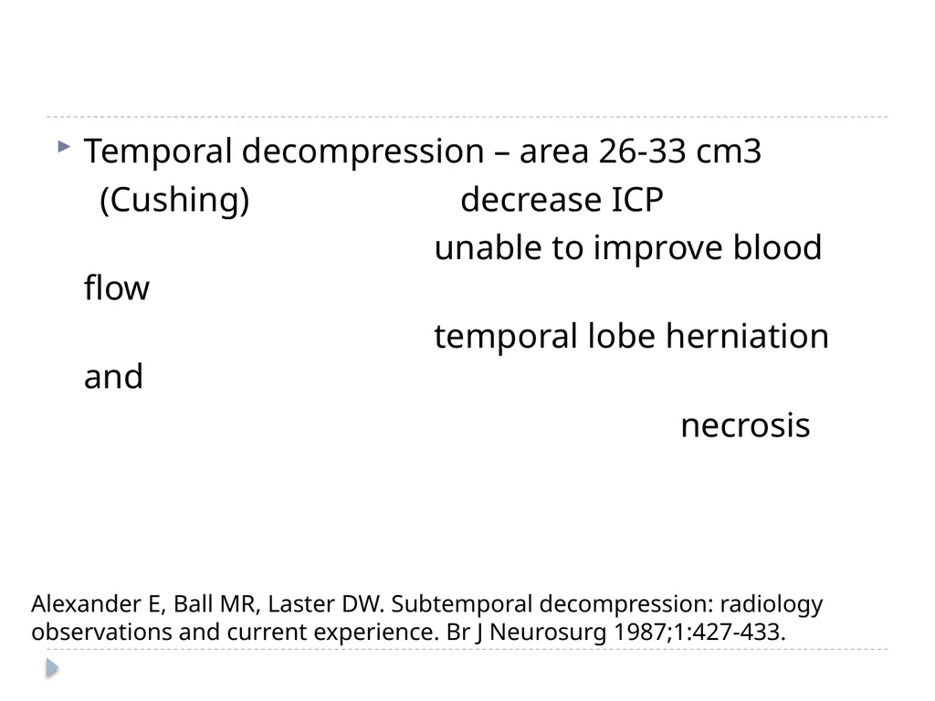
Temporal decompression – area 26-33 cm3
(Cushing) decrease ICP
unable to improve blood flow
temporal lobe herniation and
necrosis Alexander E, Ball MR, Laster DW. Subtemporal decompression: radiology observations and current experience. Br J Neurosurg 1987;1:427-433.<br>
(Cushing) decrease ICP
unable to improve blood flow
temporal lobe herniation and
necrosis Alexander E, Ball MR, Laster DW. Subtemporal decompression: radiology observations and current experience. Br J Neurosurg 1987;1:427-433.<br>
42
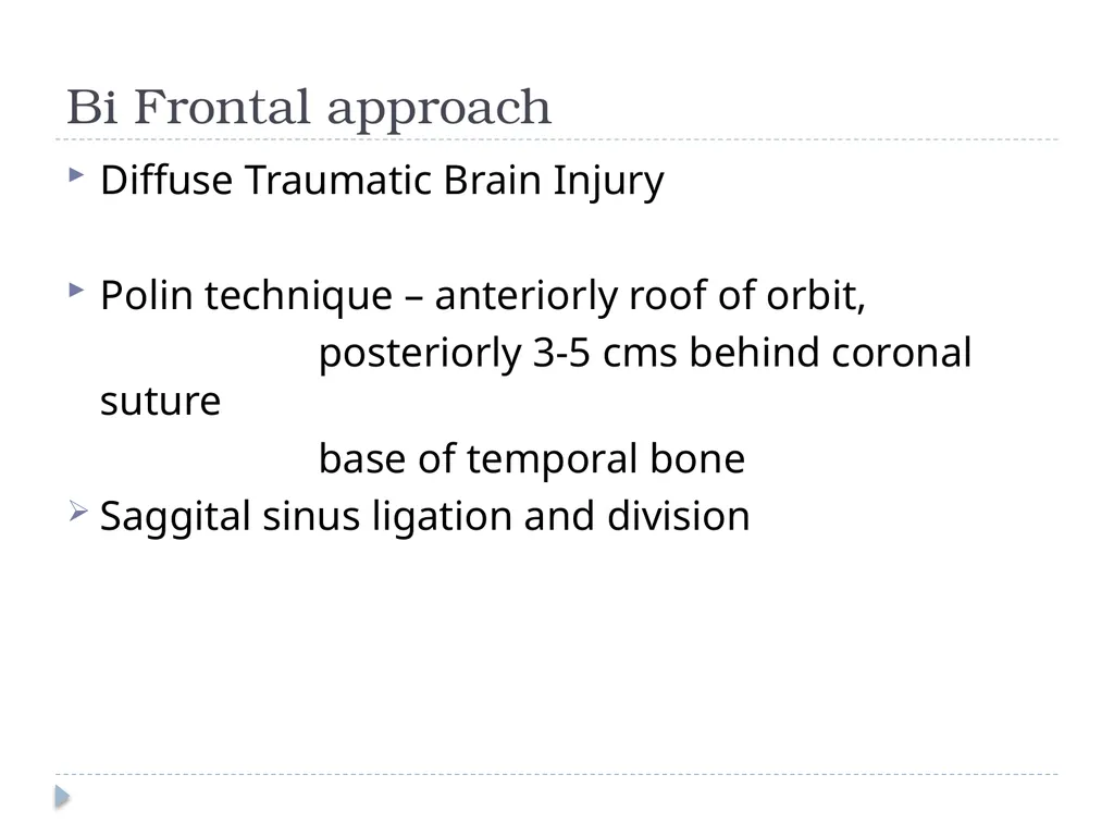
Bi Frontal approach Diffuse Traumatic Brain Injury
Polin technique – anteriorly roof of orbit,
posteriorly 3-5 cms behind coronal suture
base of temporal bone
Saggital sinus ligation and division<br>
Polin technique – anteriorly roof of orbit,
posteriorly 3-5 cms behind coronal suture
base of temporal bone
Saggital sinus ligation and division<br>
43
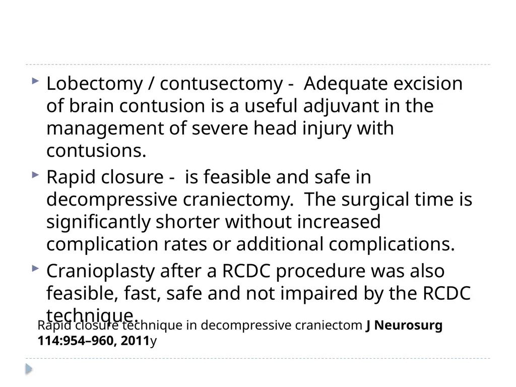
Lobectomy / contusectomy - Adequate excision of brain contusion is a useful adjuvant in the management of severe head injury with contusions.
Rapid closure - is feasible and safe in decompressive craniectomy. The surgical time is significantly shorter without increased complication rates or additional complications.
Cranioplasty after a RCDC procedure was also feasible, fast, safe and not impaired by the RCDC technique. Rapid closure technique in decompressive craniectom J Neurosurg 114:954–960, 2011y<br>
Rapid closure - is feasible and safe in decompressive craniectomy. The surgical time is significantly shorter without increased complication rates or additional complications.
Cranioplasty after a RCDC procedure was also feasible, fast, safe and not impaired by the RCDC technique. Rapid closure technique in decompressive craniectom J Neurosurg 114:954–960, 2011y<br>
44
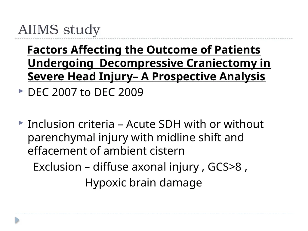
AIIMS study Factors Affecting the Outcome of Patients Undergoing Decompressive Craniectomy in Severe Head Injury– A Prospective Analysis
DEC 2007 to DEC 2009
Inclusion criteria – Acute SDH with or without parenchymal injury with midline shift and effacement of ambient cistern
Exclusion – diffuse axonal injury , GCS>8 ,
Hypoxic brain damage<br>
DEC 2007 to DEC 2009
Inclusion criteria – Acute SDH with or without parenchymal injury with midline shift and effacement of ambient cistern
Exclusion – diffuse axonal injury , GCS>8 ,
Hypoxic brain damage<br>
45
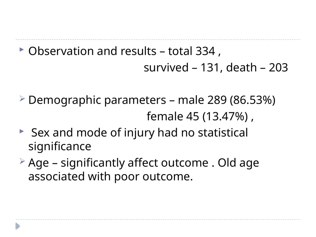
Observation and results – total 334 ,
survived – 131, death – 203
Demographic parameters – male 289 (86.53%)
female 45 (13.47%) ,
Sex and mode of injury had no statistical significance
Age – significantly affect outcome . Old age associated with poor outcome.<br>
survived – 131, death – 203
Demographic parameters – male 289 (86.53%)
female 45 (13.47%) ,
Sex and mode of injury had no statistical significance
Age – significantly affect outcome . Old age associated with poor outcome.<br>
46
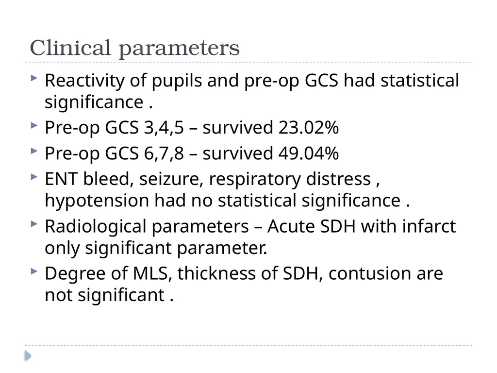
Clinical parameters Reactivity of pupils and pre-op GCS had statistical significance .
Pre-op GCS 3,4,5 – survived 23.02%
Pre-op GCS 6,7,8 – survived 49.04%
ENT bleed, seizure, respiratory distress , hypotension had no statistical significance .
Radiological parameters – Acute SDH with infarct only significant parameter.
Degree of MLS, thickness of SDH, contusion are not significant .<br>
Pre-op GCS 3,4,5 – survived 23.02%
Pre-op GCS 6,7,8 – survived 49.04%
ENT bleed, seizure, respiratory distress , hypotension had no statistical significance .
Radiological parameters – Acute SDH with infarct only significant parameter.
Degree of MLS, thickness of SDH, contusion are not significant .<br>
47
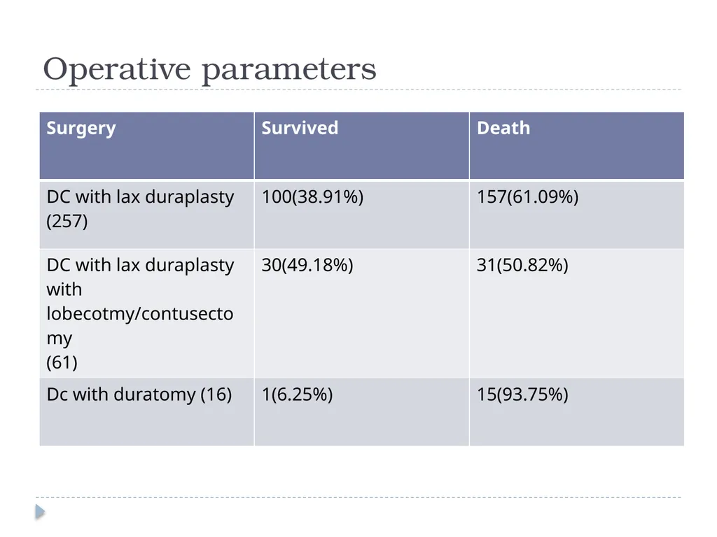
Operative parameters<br>