
Digestive system diseases Małgorzata Dybich, MA
KB
Published · 59 slides · 0 views

1 / 1





Description
Digestive system diseases Małgorzata Dybich, MA Achalasia Treats the motor function of the smooth muscles of the lower part - cardia spasm.Occurrence 1:100,000, age 30-50 years.Symptoms: :dysphagia, chest pain, food regurgitation, feeling
Related Topics
Share
Embed code
Download this presentation From Below
"Digestive system diseases Małgorzata Dybich, MA" is the property of its rightful owner. Permission is granted to download and print the materials on this website for personal, non-commercial use only, and to display it on your personal computer provided you do not modify the materials and that you retain all copyright notices contained in the materials. By downloading content from our website, you accept the terms of this agreement.
Presentation Transcript
01
Digestive system diseases Małgorzata Dybich, MA<br>
02
Achalasia Treats the motor function of the smooth muscles of the lower part - cardia spasm.Occurrence 1:100,000, age 30-50 years.Symptoms: :dysphagia, chest pain, food regurgitation, feeling of fullness. Symptoms worsen with stress and eating quickly.Complications:aspiration of the lungs, risk of cancer: cancer of the esophagus, cardia.Radiological examination of the esophagus, endoscopic examination.Conservative treatment: pureed foods, sedatives, anticholinergics, calcium channel blocking drugs.Treatmentendoscopic dilatation of the stenosis with a balloon.<br>
03
Reflux disease The disease involves regurgitation of foodfrom the stomach to the esophagus. 10% of the population.Reflux disease causes damage to the esophageal mucosa and/or symptoms resulting from reflux that are so severe that the quality of life of patients deteriorates.Symptoms:Heartburn-A burning sensation occurring after eating a meal, located in the upper abdomenor in the lower part of the chest and radiatingtowards the neck.Sour regurgitation(regurgitations)Pain behind the sternum, in the upper abdomen, cough, belching, hoarseness.<br>
04
Complications Ulceration - bleedingepithelial metaplasia in the lower esophageal mucosa - Barrett's esophagus, esophageal narrowing (dysphagia).Diagnosis:Endoscopic examination with biopsy of the mucous membrane, pH-metric determination of the lower esophagus.Treatment:-conservative - weight loss, stopping smoking, limiting coffee, sweets, mint, alcohol, mint. Avoiding eating before bed, headlessness raised.- pharmacological: drugs that block H2 receptors, drugs that accelerate gastric motility - emptyingfrom food, neutralizing drugs.<br>
05
Barrett's esophagus- regular endoscopy with biopsy of the mucosa. In the development of dysplasia - proton pump inhibitors, endoscopic ablation methods, esophageal resection.Surgery. it involves wrapping the bottom of the stomach around the esophagus for patients who do not respond to conservative treatment. Efficiency 80%.
Esophageal diverticula- recesses of the entire wall of the duct. food.caused by muscle stretches:-pharyngoesophageal (Zenker - 70%) in older men,- bifurcation diverticula - 20% asymptomatic- supradiaphragmatic - 10% associated with achalasiaSymptoms:pressure pain, dysphagia, regurgitationundigested food remains, coughing while eating, unpleasant odor from the mouth.<br>
Esophageal diverticula- recesses of the entire wall of the duct. food.caused by muscle stretches:-pharyngoesophageal (Zenker - 70%) in older men,- bifurcation diverticula - 20% asymptomatic- supradiaphragmatic - 10% associated with achalasiaSymptoms:pressure pain, dysphagia, regurgitationundigested food remains, coughing while eating, unpleasant odor from the mouth.<br>
06
Esophageal cancer Mainly affects men (5x more often)
Histopathologically: carcinoma planoepithelialen adenocarcinoma (increasingly often as a consequence of Barrett's metaplasia, which in turn is the result of reflux inflammation of the mucosa of the lower esophagus)
Risk factors: alcohol, post-inflammatory strictures, cigarette smoking)<br>
Histopathologically: carcinoma planoepithelialen adenocarcinoma (increasingly often as a consequence of Barrett's metaplasia, which in turn is the result of reflux inflammation of the mucosa of the lower esophagus)
Risk factors: alcohol, post-inflammatory strictures, cigarette smoking)<br>
07
Symptoms -Long periodasymptomatic
-In advanced stages: dysphagia, rapid weight loss leading to emaciation, odynophagia, chest pain radiating to the back, hiccups, hoarseness
-Coughing and choking may indicate the presence of a fistula into the respiratory tract
-Supraclavicular lymph node enlargementand cervical, tracheoesophageal fistulasin advanced forms.But. surgeon. 5-year survival - applies to 5% of patients., Combined chemotherapy, radiotherapy, palliative - laser removal of strictures, endoscopic enlargement.<br>
-In advanced stages: dysphagia, rapid weight loss leading to emaciation, odynophagia, chest pain radiating to the back, hiccups, hoarseness
-Coughing and choking may indicate the presence of a fistula into the respiratory tract
-Supraclavicular lymph node enlargementand cervical, tracheoesophageal fistulasin advanced forms.But. surgeon. 5-year survival - applies to 5% of patients., Combined chemotherapy, radiotherapy, palliative - laser removal of strictures, endoscopic enlargement.<br>
08
Stomach ulcer disease
and duodenum Helicobacter pylori infection
(80% of gastric ulcers and 90% of duodenal ulcers)
Ulcers that develop without the involvement of H. pylori infection:
(8% of duodenal ulcers and 30% of stomach ulcers)- alcohol, radiotherapy, stress
–nonsteroidal anti-inflammatory drugs (NSAIDs)
–ulcers in Zollinger-Ellison syndrome
–ulcers in Crohn's disease
-Acute damage (or erosion) caused by stress,<br>
(80% of gastric ulcers and 90% of duodenal ulcers)
Ulcers that develop without the involvement of H. pylori infection:
(8% of duodenal ulcers and 30% of stomach ulcers)- alcohol, radiotherapy, stress
–nonsteroidal anti-inflammatory drugs (NSAIDs)
–ulcers in Zollinger-Ellison syndrome
–ulcers in Crohn's disease
-Acute damage (or erosion) caused by stress,<br>
09
Symptoms The most common symptom of peptic ulcer disease is pain:
– sharp, burning,pain in the epigastrium, sharp and burning,discomfort or feeling of fullness, from several days to several months - stomach ulcer
– with duodenal ulcer it occurs 1-3 hours after a meal, disappears after consuming food or antacids
– quite often the pain occurs at night or early in the morning(in 2/3 patients with duodenal ulcer and 1/3 patients with gastric ulcer)
– the pain comes back every few months<br>
– sharp, burning,pain in the epigastrium, sharp and burning,discomfort or feeling of fullness, from several days to several months - stomach ulcer
– with duodenal ulcer it occurs 1-3 hours after a meal, disappears after consuming food or antacids
– quite often the pain occurs at night or early in the morning(in 2/3 patients with duodenal ulcer and 1/3 patients with gastric ulcer)
– the pain comes back every few months<br>
10
Other symptoms include:
–nausea
–vomiting
–decreased appetite
–flatulence, belching
completely asymptomatic course possible
The first symptom of peptic ulcer disease may be hemorrhagefrom the upper gastrointestinal tract.
Khemorrhage is characterized by vomiting of blood (haematemesis) or coffee grounds and bloody or tarry stools.<br>
–nausea
–vomiting
–decreased appetite
–flatulence, belching
completely asymptomatic course possible
The first symptom of peptic ulcer disease may be hemorrhagefrom the upper gastrointestinal tract.
Khemorrhage is characterized by vomiting of blood (haematemesis) or coffee grounds and bloody or tarry stools.<br>
11
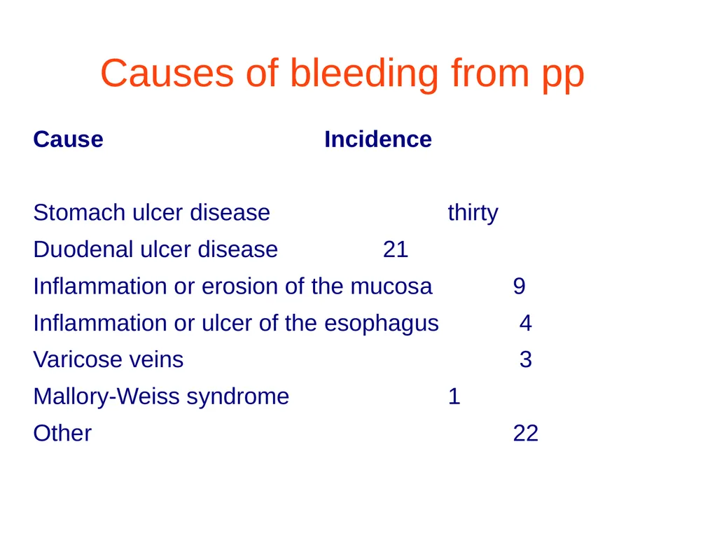
Causes of bleeding from pp Cause Incidence
Stomach ulcer disease thirty
Duodenal ulcer disease 21
Inflammation or erosion of the mucosa 9
Inflammation or ulcer of the esophagus 4
Varicose veins 3
Mallory-Weiss syndrome 1
Other 22<br>
Stomach ulcer disease thirty
Duodenal ulcer disease 21
Inflammation or erosion of the mucosa 9
Inflammation or ulcer of the esophagus 4
Varicose veins 3
Mallory-Weiss syndrome 1
Other 22<br>
12
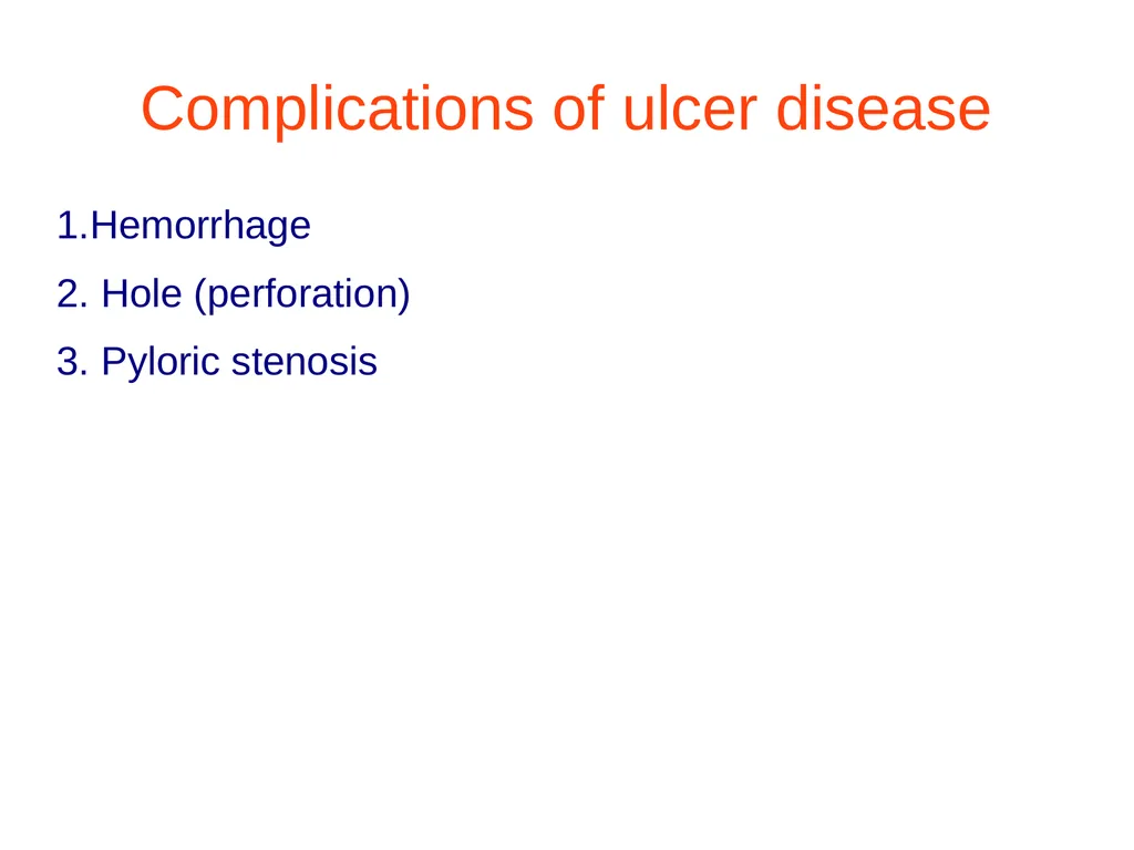
Complications of ulcer disease 1.Hemorrhage
2. Hole (perforation)
3. Pyloric stenosis<br>
2. Hole (perforation)
3. Pyloric stenosis<br>
13
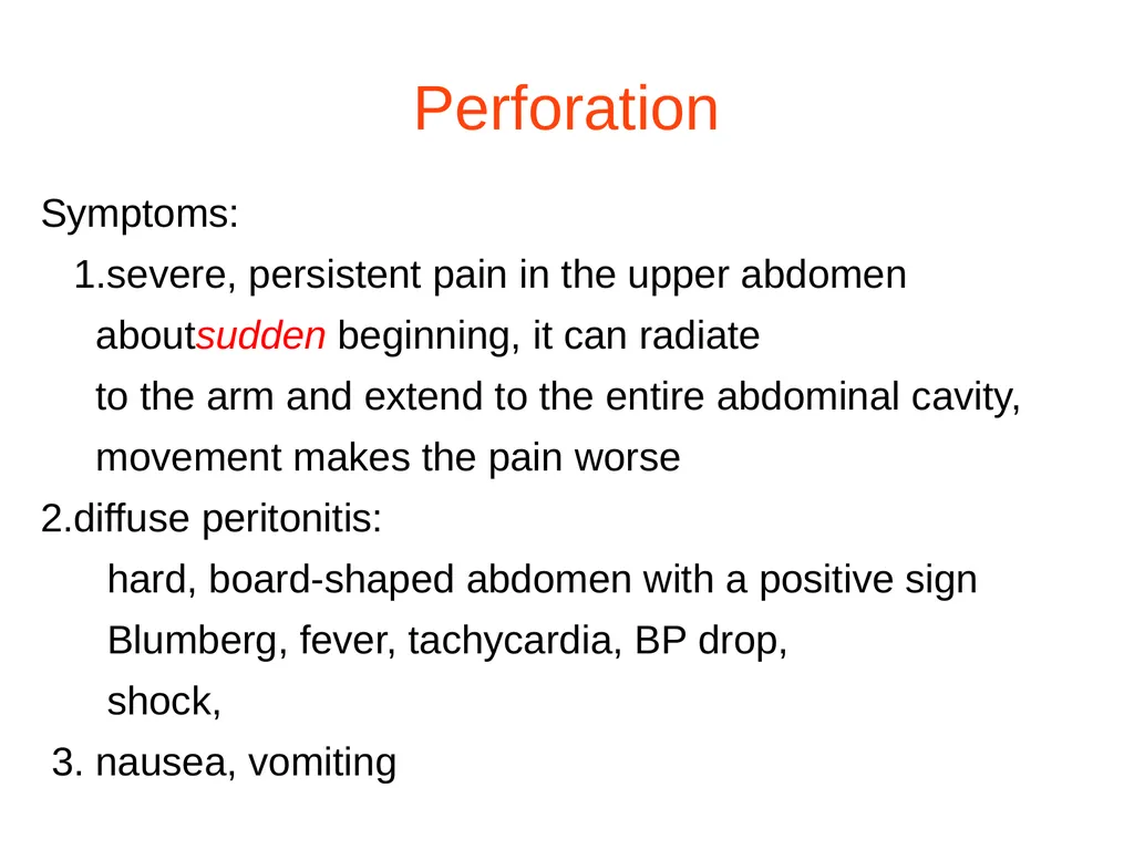
Perforation Symptoms:
1.severe, persistent pain in the upper abdomen
aboutsudden beginning, it can radiate
to the arm and extend to the entire abdominal cavity,
movement makes the pain worse
2.diffuse peritonitis:
hard, board-shaped abdomen with a positive sign
Blumberg, fever, tachycardia, BP drop,
shock,
3. nausea, vomiting<br>
1.severe, persistent pain in the upper abdomen
aboutsudden beginning, it can radiate
to the arm and extend to the entire abdominal cavity,
movement makes the pain worse
2.diffuse peritonitis:
hard, board-shaped abdomen with a positive sign
Blumberg, fever, tachycardia, BP drop,
shock,
3. nausea, vomiting<br>
14
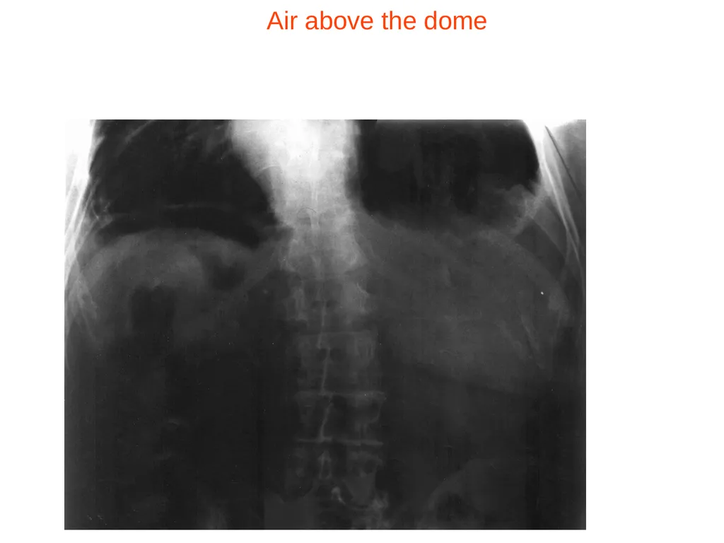
Air above the dome<br>
15
Complications of ulcer disease –bloody or tarry (black) stools - bleeding
–vomiting blood or coffee grounds - pyloric stenosisIt occurs in patients with recurrent ulcersin the pyloric canal or duodenal bulb
Symptoms:
retention of food, nausea and profuse vomiting<br>
–vomiting blood or coffee grounds - pyloric stenosisIt occurs in patients with recurrent ulcersin the pyloric canal or duodenal bulb
Symptoms:
retention of food, nausea and profuse vomiting<br>
16
Treatment Multiple treatment regimens for gastric ulcer:3 drugs:- proton pump blocking drug - omeprazole, lansoprazole 20-40 mg/day,- metronidazole - 3X 500 mg.- amoxicycline - 4X500mg or tetracycline.Treatment complications include diarrhea and pseudomembranous enteritis. Treatment should last up to 14 days.
H2 receptor antagonists inhibit gastric acid secretion.
Protective drugs - facilitate the healing of ulcers and protectits bottom before producing salt acid, pepsin.<br>
H2 receptor antagonists inhibit gastric acid secretion.
Protective drugs - facilitate the healing of ulcers and protectits bottom before producing salt acid, pepsin.<br>
17
Zollnger-Ellison syndrome In 60% of patients, the tumor is malignant, located in the pancreas, duodenum, speculum, and hepatoduodenal ligament. Characteristic treatment-resistant gastric and duodenal ulcers. The diagnosis is made by detecting an increased concentration of gastrin in the blood serum. Endoscopic ulcers, the tumor is diagnosed using endoscopic sonography, CT, RMI, scintigraphy, angiography. 30% of patients undergo radical surgery.<br>
18
Stomach cancer -The second most common cancer after lung cancer-It most often occurs in the 5th and 6th decade of life2x more common in men-In general, the incidence of disease has decreased in recent yearsexcept cancers of the proximal stomach-Symptoms: indigestion, weight loss, lossappetite.Diagnostics: X-ray, endoscopy, biopsy.Treatment: surgery, chemotherapy, radiotherapy.<br>
19
Malabsorption syndrome Typical symptoms:chronic, frequent, profuse diarrhea, steatorrhea, weight loss, deficiency symptoms. Fat content > 7g per day was indicative of ZZW.Etiology:-improper digestion:condition after stomach resection, pancreatic endocrine failure - chronic inflammationcystic fibrosis, biliary deficiency, cholestasis - Leśniewski-Croma's disease, ileal resection-abnormal absorption:diseases of the small intestine, gluten disease, intestinal parasites, intestinal lymphomas, intestinal blood supply disorders and others.<br>
20
Leśniewski-Croma disease Genetic basis, NOD2/CARD 15 gene mutation, intestinal wall obstruction.Symptoms:abdominal pain, diarrhea without blood, flatulence, weight loss, rarely occursfor perforation, fistula, perianal abscesses. Complication: colon cancer, amyloidosis. It runswith relapses after 1 and 2 years.Diagnosis-based on infusion, coloyleoscopy, biopsy of perianal abscesses and fistulas. Endoscopic treatment of strictures or fistulas. Indications for urgent surgical intervention: intestinal perforation, peritoneal blockage, obstruction.80% of patients relapsed.<br>
21
Ulcerative colitis -superficial inflammation of the mucous membrane of the rectum and/or colon.-red and swollen mucous membrane, bleeding when touched.- destruction of the mucous membrane, disappearance of its folds.Symptoms: mucous-bloody diarrhea, abdominal pain,diarrhea.- 70% have primary, sclerosing, bile duct obstruction, 15% skin lesions, joint problems.Diagnosis - rectoscopy, colonoscopy with intestinal biopsy, histopathological examination.<br>
22
Ulcerative colitis Treatment: aminosalicylates, glucosteroids, azathioprine, cyclosporine.Surgical procedure: incomplete colon resection leaving the colon or proctocolectomy. Mortality in emergencies 30%.Indications: fulminant colon with sepsis, perforation, severe hemorrhage.<br>
23
Colon and rectal cancer -Second after lung cancer in both women and menafter 50 years of age may occur in younger people. Most often the rectum (60%), esophageal colon (20%).Paths of spread -lymphatic vessels, chewed to the paraaortic nodes, pelvic walls, inguinal lymph nodes.- bloodstream - chewed to the lungs, liver.Symptoms are not very specific: some blood in the stool, a sudden change in the rhythm of bowel movements, especially in young people. Lymph node progression: abdominal pain, fever of unknown etiology, weight loss, chronic anemia. Late symptoms: a tumor palpable through the abdominal wall.<br>
24
Colon and rectal cancer Risk factors:- genetic factors - familial adenomatous polyposis, occurrence of colorectal cancer in closest relatives- diet - animal fats, high-protein diet- chronic intestinal diseases- age over 40- smoking tobacco- urinary-sigmoid anastomoses- Streptococcus bovis.<br>
25
Colon and rectal cancer Diagnosis:examination of the rectum, bad.x-ray, colonoscopywith taking the sample for examination by histopathologist.Assessment of the stage of advancement - imaging tests: ultrasound, CT scan, chest X-ray. Carcinoembryonic antigen - CEA is not used for early diagnosis, some cases are monitored after treatment. 15% of colon and rectal cancers can be detected by palpation through the rectum.Treatment- surgery, depending on the stage of the disease, chemotherapy, radiotherapy, immunotherapy. Advanced tumor - palliative treatment, bypass anastomosis, artificial anus, cryotherapy, laser therapy, - reduction of tumor mass. 70% recurrencefalls on the first two years after the first radical treatment.<br>
26
Colon and rectal cancer Five-year survival 75% in phase I, 5% in phase IV.
Prevention - after 45 years of age occult blood test, rectal test, colonoscopy every 5 years.<br>
Prevention - after 45 years of age occult blood test, rectal test, colonoscopy every 5 years.<br>
27
Acute pancreatic blockage Reversible, damaging pancreatic and tissue damage. peripancreatic in the form of edema, hemorrhagic necrosis, fatty necrosis.Pathogenesis- self-life of the pancreas, activation of proteolytic and subsequent enzymes. Effect - protein lysis, interstitial bleeding, vascular damage, necrosis of pancreatic parenchyma cells - acute pancreatic necrosis.Reasons: alcohol abuse, biliary tract diseases - urolithiasis, endoscopic retrograde cholangiopancreatography (ERCP), drugs azathioprine, sulfonamides, estrogens, vascular causes and inflammation of vessels, irritating duodenal ulcer, obstruction of the major papilla of the duodenum, bifid pancreas, pancreatic cancer, idiopathic inflammation. pancreas.<br>
28
Acute pancreatic blockage Symptoms:abdominal pain, located in the upper abdomenand around the navel, sometimes radiating to the back, chest.and lower abdomen, fetal position, vomiting and abdominal distension, fever, tachycardia, hypotension, shock rarely, 10-20% of patients have fluid in the abdomen, changes in the lungs.Tests-increased activity of pancreatic hormones - amylase, lipase, trypsin. Additional tests included leukocytosis, hyperglycemia, hypocalcemia.- Ultrasound, CT scan, chest x-ray, abdominal x-ray.85-90% is a self-limiting form of AP that disappears after a few days.Procedure:pain treatment, patient hydration, infection prevention, expression of gastric contents. Drugs: antibiotics, H2-blocking drugs, pronate pump inhibitors. Laparotomy with drainage and removal of necrotic tissue. nutritionextrajet in patientswith prolonged inflammation.<br>
29
Acute pancreatic blockage Complications: local - necrosis, fluid collections - abscess, cyst, hemorrhage, infection, gastrointestinal obstruction, mechanical jaundice;systemic: pulmonary - pleural effusion, pneumonia, acute respiratory distress syndrome;-cardiovascular - hypotension, sudden cardiac death, pericardial effusion;- hepatological - DIC;- renal - oliguria, thrombosis of an artery and/or veinrenal;-metabolic - hyperglycemia, hypocalcemia, encephalopathy;CNS- psychosis, fat embolism.<br>
30
Chronic pancreatic blockage A non-neoplastic disease, there are irreversible morphological changes, atrophy of glandular tissue with the development of connective tissue and tissue impairment. exocrine andendocrine.Reasons:in children, cystic fibrosis, in adults, alcoholism - drinking even less than 50g a day.Symptoms:constant or periodic transient abdominal pain, located in the epigastrium or diffuse, typical is continuous and does not go away with neutralizing drugs, relief is provided by opioid painkillers, weight loss, abnormal bowel movements - steatorrhea, symptoms of malabsorption, rarely exudate into pleura and/or peritoneum.<br>
31
Chronic pancreatic blockage Impaired exocrine function, often glucose intolerance - diabetes 30%patients, pancreatic triad: pancreatic calcifications, steatorrhea, diabetes, vitamin B12 malabsorption.Bad. abdominal examination, ultrasound, endosonography, CT, ERCP.Treatment: pain relief and treatment of malabsorption syndrome, complete alcohol abstinence, avoidance of fatty and hard-to-digest foods.Pharmacological supplementation of pancreatic enzymes, which reduces pain in 75% of people. Pain treatment - opioids. Endoscopic sphincterotomy of the pancreatic sphincters, dilation of the narrowed section of the pancreatic duct and removal of deposits. Patients who do not comply with the recommendations have a poor prognosis.<br>
32
Jaundice Indicators of liver cell damage:ALAT - alanine aminotransferaseASPAT - aspartate aminotransferaseGGTP-transferaseƳ-glutamylGLDH - glutamate dehydrogenaseIndicators of bile stasis:ALP- alkaline phosphataseLAP - leucine aminopeptidase GGTP5-nucleotidase<br>
33
Jaundice Hemolytic-pre-hepatic jaundice. Acute and chronic hemolytic anemia-Exceeded metabolic capacity of the liver due toexcessive productionbilirurbin from disintegrating erythrocytes, uncompressed (indirect) bilirurbin, no bilirurbin in urine, mild jaundice.Hemolytic jaundice of the newborn may cause CNS damage. Treatment - exchange transfusionand tototherapy.-Parenchymal hepatic jaundice- indirect biriurbin- Gilbert's syndrome - mild chronic hyperbilirubinemia. The pathogenesis is unclear, congenital defects in the uptake of bilirurbin by hepatocytes are detected in young people. The concentration of bilirurbin increases during periods of starvation and stress. Does not require treatment. The prognosis is good.<br>
34
Jaundice - Crigler-Najjar syndrome. Full type I, inherited in an autosomal recessive manner, lack of the glucuronyltransferase enzyme. Severe in patients hyperbilirubinemia. The prognosis is poor - death in the first year of life. Type II glucuronosyltransferase deficiency. The prognosis is good, with no CNS damage.-Syndromes associated with increased concentration of compressed birurbin: Dubin-Jonson syndrome, Rotor syndrome, infectious hepatitis, chronic hepatitis and cirrhosis, toxic liver damage, drug-induced hepatitis.Cholestatic jaundice (obstructive)-impediment outflow of bile in the section from the liver to the greater ampulla of the duodenum (Vater) may cause cholestasis (biliary stasis). Symptoms: jaundice, light discolored stools, dark urine, itchy skin.The presence of bilirurbin, bile acids and bile enzymes in the blood.<br>
35
Jaundice - Crigler-Najjar syndrome. Full type I, inherited in an autosomal recessive manner, lack of the glucuronyltransferase enzyme. Severe in patients hyperbilirubinemia. The prognosis is poor - death in the first year of life.Type II glucuronosyltransferase deficiency. The prognosis is good, with no CNS damage.-Syndromes associated with increased concentration of compressed birurbin: Dubin-Jonson syndrome, Rotor syndrome, infectious hepatitis, chronic hepatitis and cirrhosis, toxic liver damage, drug-induced hepatitis.Cholestatic jaundice (obstructive)-impediment outflow of bile in the section from the liver to the greater ampulla of the duodenum (Vater) may cause cholestasis (biliary stasis). Symptoms: jaundice, light discolored stools, dark urine, itchy skin.The presence of bilirurbin, bile acids and bile enzymes in the blood.<br>
36
Cholestatic jaundice Reasons:- cholelithiasis, pancreatitis, pancreatic tumor, hepatic hydatid disease, biliary tract inflammation and cancer, inflammatory and neoplastic lesions in the ampulla of Vater - rarely - hemobilia, congenital biliary atresia, biliary cysts, parasitic infections- spontaneous jaundice of pregnancy, a mild form, may occur in the third trimester of a normal pregnancy, skin rashes, signs of slight cholestasis,does not exceed 8.5mmol/l. Symptoms disappearafter termination.<br>
37
Jaundice Reasons: :- cholelithiasis, pancreatitis, pancreatic tumor, hepatic hydatid disease, biliary tract inflammation and cancer, inflammatory and neoplastic lesions in the ampulla of Vater - rarely - hemobilia, congenital biliary atresia, biliary cysts, parasitic infections- spontaneous jaundice of pregnancy, mild form, may occur in the third trimester of a normal pregnancy, skin lesions occur, signs of slight cholestasis, does not exceed 85atminor. Symptoms disappear after termination.<br>
38
Hepatitis A virus (HAV) There is an RNA virus, enteroviruses. It dies at 100 degrees Celsiusafter 20 minutes It spreads through the oral route. The source of infection is water and food products. Excreted in feces 2-3 weeks before the onset of jaundice and about 8 days after its onset.Clinical course: 15-49 days. Symptoms of indigestion predominate, and less frequently, flu-like symptoms. Jaundice may not occur. Serum ALT activity increases 2-14 days before the onset of hyperbilirubinemia. The liver is enlarged, splenomegaly is rare, and jaundice lasts for several days to several weeks. The inflammation subsides after 6 months and does not become a chronic process.Diagnosis: identification of the titer of specific IgM antibodies (in the early period up to 3-6 months)No treatment.Prevention-anti-HAV vaccine. It is recommended to administer two doses over a period of 6-12 months. Effectiveness 100%. immunization for several years.<br>
39
Hepatitis B virus (HBV) Double-stranded DNA virus. It is destroyed after 30 minutes in an autoclave and 60 minutes in dry air at 60 degrees Celsius. at room temperature and frozen for several years.Epidemiology:over 350 million. in the world, mainly South Asia, China, Africa. Carrier status develops throughout life and is a reservoir of the virus. In Poland, approximately 1.5%.Infection in the perinatal period, unsterilized medical equipment, sexual route, medical personnel in contact with blood, multiple recipients, immunosuppressive treatment.Clinical course-incubation period 28-280 days. Jaundice or no jaundice, liver enlarged, spleen rarely palpable, jaundice 4-6 weeks. 0.1-0.5% hyperacute liver disease with symptoms of hepatic coma. Cured in 90% of cases with development of immunity. 5-10% chronic liver inflammation It increases liver cancer by 100 times.<br>
40
Hepatitis B virus (HBV) Symptoms of the pancreas include inflammation, exudative pleurisy, hemolytic and aplastic anemia. HBV virus - hepatotropic, genetic material also detected in pancreatic cells, kidneys, T and B lymphocytes. Recovery 1-2 months.Diagnosis: -appearance of HBsAg protein in serum - 2-4 weeks before elevated aminotransferases- II HbeAg marker in serum - 3-9 weeks.- anti-HBc IgM antibodies (acute infection, virus reactivation)- IgG antibodies (after a few weeks) - past infection, combined with + Hbs antigen - chronic infection.<br>
41
Hepatitis B virus (HBV) Carrier: from an infected mother in the perinatal period, asymptomatic infection, after the acute phase of infection.Chronic viral hepatitis.The transition from ACS to chronic disease depends on the immune status.Symptoms:weakness, joint pain, lack of appetite, painin the right hypochondrium, asymptomatic, tender liverand enlarged.-Advanced forms - erythema of the hand, less often spider veins on the skin of the neck, forearm, face, chest. hypoalbuminemia, hypergammaglobulinemia-Jaundice, tendency to bruise, prolonged bleedingfrom minor wounds and mucous membranes indicate significant damage to the liver - cirrhosis.<br>
42
Hepatitis B virus (HBV) Treatment - antiviral treatment - interferon and nucleoside analogues - lamivudine - inhibition of the synthesis of nucleoid acids.Patients with cirrhosis - liver transplant.Prevention:-mandatory hospitalization of a patient with OWZ-disinfection in the patient's home- HbsAg determination for future health care workers- vaccinations - immunity up to several years, effectiveness 98%- adults - incidental exposure to infection with anti-HBs immunoglobulin.<br>
43
Hepatitis D virus (HDV) A defective RNA virus that replicates in HBV infection.Clinical course such as HBV zap. Sometimes hyperacute, fatal liver disease occurs. Progression to liver cirrhosis.<br>
44
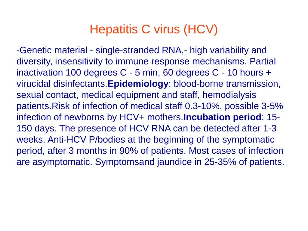
Hepatitis C virus (HCV) -Genetic material - single-stranded RNA,- high variability and diversity, insensitivity to immune response mechanisms. Partial inactivation 100 degrees C - 5 min, 60 degrees C - 10 hours + virucidal disinfectants.Epidemiology: blood-borne transmission, sexual contact, medical equipment and staff, hemodialysis patients.Risk of infection of medical staff 0.3-10%, possible 3-5% infection of newborns by HCV+ mothers.Incubation period: 15-150 days. The presence of HCV RNA can be detected after 1-3 weeks. Anti-HCV P/bodies at the beginning of the symptomatic period, after 3 months in 90% of patients. Most cases of infection are asymptomatic. Symptomsand jaundice in 25-35% of patients.<br>
45
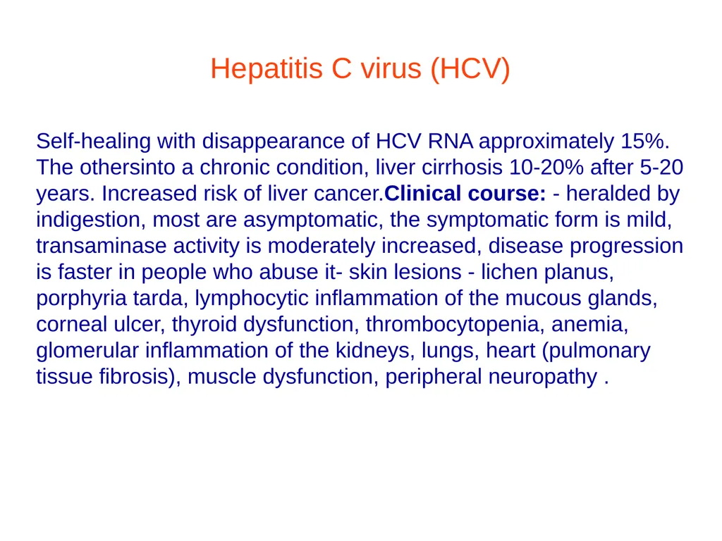
Hepatitis C virus (HCV) Self-healing with disappearance of HCV RNA approximately 15%. The othersinto a chronic condition, liver cirrhosis 10-20% after 5-20 years. Increased risk of liver cancer.Clinical course: - heralded by indigestion, most are asymptomatic, the symptomatic form is mild, transaminase activity is moderately increased, disease progression is faster in people who abuse it- skin lesions - lichen planus, porphyria tarda, lymphocytic inflammation of the mucous glands, corneal ulcer, thyroid dysfunction, thrombocytopenia, anemia, glomerular inflammation of the kidneys, lungs, heart (pulmonary tissue fibrosis), muscle dysfunction, peripheral neuropathy .<br>
46
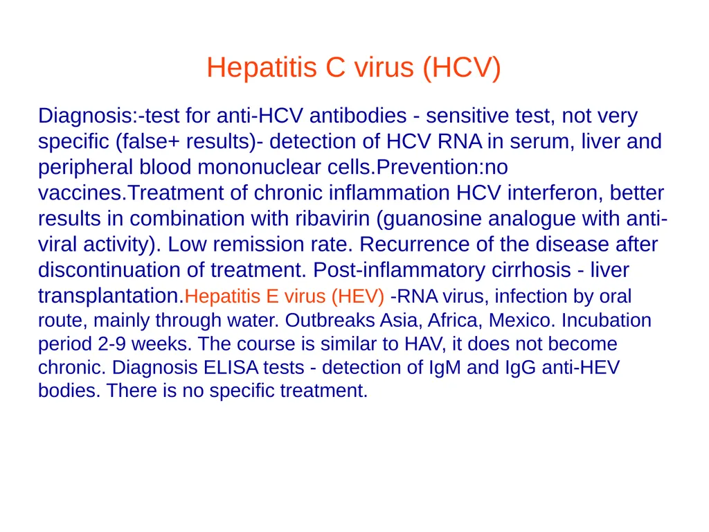
Hepatitis C virus (HCV) Diagnosis:-test for anti-HCV antibodies - sensitive test, not very specific (false+ results)- detection of HCV RNA in serum, liver and peripheral blood mononuclear cells.Prevention:no vaccines.Treatment of chronic inflammation HCV interferon, better results in combination with ribavirin (guanosine analogue with anti-viral activity). Low remission rate. Recurrence of the disease after discontinuation of treatment. Post-inflammatory cirrhosis - liver transplantation.Hepatitis E virus (HEV) -RNA virus, infection by oral route, mainly through water. Outbreaks Asia, Africa, Mexico. Incubation period 2-9 weeks. The course is similar to HAV, it does not become chronic. Diagnosis ELISA tests - detection of IgM and IgG anti-HEV bodies. There is no specific treatment.<br>
47
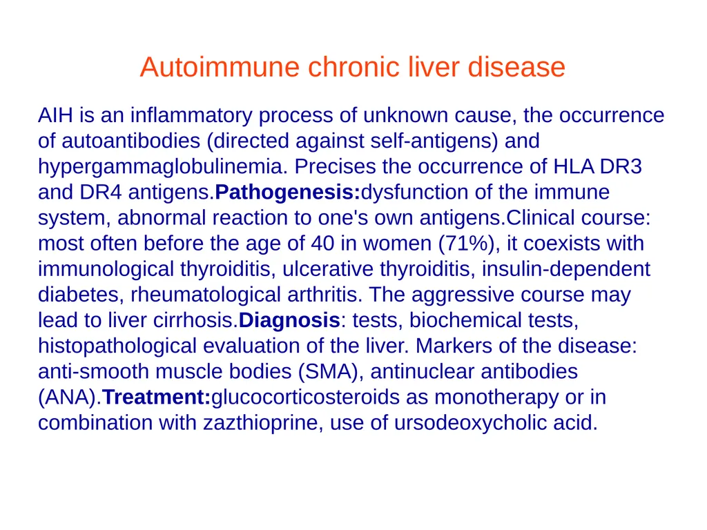
Autoimmune chronic liver disease AIH is an inflammatory process of unknown cause, the occurrence of autoantibodies (directed against self-antigens) and hypergammaglobulinemia. Precises the occurrence of HLA DR3 and DR4 antigens.Pathogenesis:dysfunction of the immune system, abnormal reaction to one's own antigens.Clinical course: most often before the age of 40 in women (71%), it coexists with immunological thyroiditis, ulcerative thyroiditis, insulin-dependent diabetes, rheumatological arthritis. The aggressive course may lead to liver cirrhosis.Diagnosis: tests, biochemical tests, histopathological evaluation of the liver. Markers of the disease: anti-smooth muscle bodies (SMA), antinuclear antibodies (ANA).Treatment:glucocorticosteroids as monotherapy or in combination with zazthioprine, use of ursodeoxycholic acid.<br>
48
Primary liver cirrhosis -90% of cases occur in women over 40 years of age, autoimmune, chronic diseasePathogenesis not explained, serum - p/mitochondrial antibodies (AMA). Against mitochondrial membrane antigens.Symptoms: -troubling skin itching before the onset of cholestatic jaundice- dark skin color, yellow eyelid tufts- the spleen is unknownly enlarged, sometimes the liver- advanced form - liver cirrhosis with symptoms of portal hypertension, liver failure.Bad.labor.: heightalkaline phosphatase, GGTP, increase in serum bilirubin, hypercholesterolemia, increase in IgMInserum.Treatment- no causal effect, good effect - ursodeoxycholic acid, replacement of vitamins A, D, E, K.<br>
49
Primary liver cirrhosis -Liver remodeling with post-inflammatory fibrosis, regenerative nodules, connective tissue septa.- a consequence of various chronic diseases, alcohol-related etiology, less often due to autoimmune diseases, metabolic diseases, vascular diseases, cardiac origin due to chronic blood stasis in the liver.Symptoms:weakness, limited mobility, lack of appetite, weight loss, feeling of fullness in the upper abdomen, erythema on the skin of the hands and legs, spider veins, sometimes jaundice, skin itching, impotence, gynecomastia, menstrual cycle disorders.Preliminary examination - initially enlarged liver, final stage - small with uneven surface.Bad.labor:affecting liver function, reducing the concentration of coagulation factors of the prothrombin system, increasing the INR, reducing antithrombin III, reducing albumin concentration.<br>
50
Secondary hypertension -BP value in the portal vein exceeds 12mmHg, collateral circulation between the portal basin and the circulatory system. inferior vena cava: portal-gastroesophageal junction, development of varicose veins of the esophagus and the bottom of the stomach, periumbilical connections, collateral connections of the mesentery with the development of hemorrhoids, gastrodiaphragmatic-adrenal or renal connections.Reasons: - prehepatic block, peripheral block (splenic vein thrombosis), central block (portal vein thrombosis)- intrahepatic block - presinusoidal block (primary biliary cirrhosis), extrasinusoidal block (liver cirrhosis)- extrahepatic block - hepatic vein obstructionin the course of thrombosis, tumor, congenital connective tissue septum.<br>
51
Secondary hypertension Symptoms:- visible vessels of collateral circulation, congestive splenomegaly and hypersplenism (thrompenia, leukocytopenia, anemia), ascites, bleeding from esophageal varices, gastric fundus.Treatment:treatment of the underlying disease, preventionand treatment of hemorrhages from esophageal varices, the bottom of the stomach, correction of coagulation disorders (fresh frozen plasma, vitamin K), prevention of hepatic coma after bleeding from esophageal varices, treatment of ascites (restriction of salt, fluids, diuretics), paracentesis, subsequent administration albumin intravenously, in cases of peritoneal inflammation - a broad-spectrum antibiotic.<br>
52
Hepatic encephalopathy and hepatic coma. Acute neuropsychiatric disorders (acute liver failure, viral infections, post-coital reactions, toxic damage)and chronic liver diseases.Reasons: bcancer of metabolic function and detoxification causes toxic substances to enter the brain through the circulation. Triggers increased ammonia production in the intestine as a result of gastrointestinal bleeding, esophageal varices, after richin meal protein, which penetrates the brain (alkalosis), intensive dehydration treatment, constipation.Trigger factors:Gastrointestinal bleedingInfection, sepsisHyponatremia, hypokalemia (electrolyte disturbances)DehydrationConstipationDiet with excess proteinAlcoholPsychotrop medicinesNon-steroidal anti-inflammatory drugs<br>
53
Degree ISHEN Neuropsychiatric condition Comments
0 - Latent form- No symptoms -Abnormal results of psychometric and neurophysiological tests. Lack of universal diagnostic criteria. Expert knowledge required
1- Poor concentration of attention - Making mistakes in simple mathematical operations (addition and subtraction). Alternating euphoria and discouragement. Sleep rhythm disturbances. Despite the preserved orientation to time and place, discreet behavioral disorders occur. Changing the handwriting.
2-Overt form- Lethargy and apathy. Slurred speech. Significant impairment of intellectual abilities. Personality changes. Behavior inappropriate to the situation. Clear asterixis. Dyspraxia. Disturbed orientation in time (at least 3 wrong answers about the day(the patient can be woken up). Strange behaviour.<br>
0 - Latent form- No symptoms -Abnormal results of psychometric and neurophysiological tests. Lack of universal diagnostic criteria. Expert knowledge required
1- Poor concentration of attention - Making mistakes in simple mathematical operations (addition and subtraction). Alternating euphoria and discouragement. Sleep rhythm disturbances. Despite the preserved orientation to time and place, discreet behavioral disorders occur. Changing the handwriting.
2-Overt form- Lethargy and apathy. Slurred speech. Significant impairment of intellectual abilities. Personality changes. Behavior inappropriate to the situation. Clear asterixis. Dyspraxia. Disturbed orientation in time (at least 3 wrong answers about the day(the patient can be woken up). Strange behaviour.<br>
54
3-Confusion and drowsiness. (the patient can be woken up). Strange behaviour. Confusion also concerns place (at least 3 wrong answers regarding country, region, city, place of stay). Current response to stimuli
4- Coma- Lack of response to painful stimuli
ISHEN – division according to the International Society of Hepatic Encephalopathy and Nitrogen Metabolism.
Stages of encelopathy:Stage I - drowsiness, poor concentration, confusion, slowness, mood swings, slurred speech, coarse-wave tremors, disturbancesin EEG.Stage II - more severe drowsiness, apathy, change in handwriting, coarse-wave tremors.Stage III - constant drowsiness, tremors, foetor hepanicus (smell of raw liver), abnormalities in the EEG recording.Stage IV - coma, deep sleep, does not respond to pain stimuli.<br>
4- Coma- Lack of response to painful stimuli
ISHEN – division according to the International Society of Hepatic Encephalopathy and Nitrogen Metabolism.
Stages of encelopathy:Stage I - drowsiness, poor concentration, confusion, slowness, mood swings, slurred speech, coarse-wave tremors, disturbancesin EEG.Stage II - more severe drowsiness, apathy, change in handwriting, coarse-wave tremors.Stage III - constant drowsiness, tremors, foetor hepanicus (smell of raw liver), abnormalities in the EEG recording.Stage IV - coma, deep sleep, does not respond to pain stimuli.<br>
55
The basic symptom of urolithiasis is biliary colic. Patients with cholelithiasis also have deposits in the common bile duct.Refers toapproximately 18% of women and 8% of men.Asymptomatic stones, "silent" stones constitute 75% of all stones.Symptomatic stone disease accounts for 25% of all cases.The basic symptom of urolithiasis is biliary colic5xF rulefemalefair (light skin)fat (obese)forts (forty years old)fecund (fertile) Cholelithiasis<br>
56
Symptoms Biliary colic is a typical example of visceral pain caused by muscle spasm of the gallbladder or sphincter of Oddi.
Location: right hypochondrium/epigastrium
(sometimes in the precordial area)
Radiation: to the right shoulder blade and arm
Character: begins suddenly, escalates quickly,
(within minutes), reaches a plateau, subsides
gradually (over a few hours) or
suddenly, comes back after days or months<br>
Location: right hypochondrium/epigastrium
(sometimes in the precordial area)
Radiation: to the right shoulder blade and arm
Character: begins suddenly, escalates quickly,
(within minutes), reaches a plateau, subsides
gradually (over a few hours) or
suddenly, comes back after days or months<br>
57
Acute cholecystitis Initially, it looks like biliary colic, but the pain lasts longer(over 3 hours). It is accompanied by nausea, vomiting and fever(37-38 degrees)
The most characteristic symptom of cholecystitis is tenderness and muscular defense of the gallbladder area.
In 30-40% of cases, an enlarged follicle is felt in the hypochondrium. Jaundice occurs in 15% of patients due to swelling of the tissues around the cystic duct. Generally, improvement occurs on the 2-3rd day of the disease.<br>
The most characteristic symptom of cholecystitis is tenderness and muscular defense of the gallbladder area.
In 30-40% of cases, an enlarged follicle is felt in the hypochondrium. Jaundice occurs in 15% of patients due to swelling of the tissues around the cystic duct. Generally, improvement occurs on the 2-3rd day of the disease.<br>
58
Acute cholecystitis A symptom may be present on physical examinationMurphy(sudden pain that appears when a patient with pressure on the right hypochondrium tries to take a deep breath) and the symptomChełmoński(pain triggered by hitting the hand placed in the right subcostal area.Acute complications1. Empyema2. Gangrenous inflammation, peritoneal inflammation, sepsis3. Perforation4. Cholelithiasis - mechanical jaundice, acute pancreatitis, ascending inflammation.Chronic complications1. Chronic inflammation of the gallbladder2. Bilioduodenal fistula3. Adenocarcinoma of the gallbladder.<br>
59
Charcot's triad in cholangitis:-pain in the right upper abdomen,-jaundice and fever- frequent relapsesDiagnostics:Ultrasound, endoscopic retrograde cholangiopancreatography (ERCP), plain X-ray.Invasive treatment - removal of the follicle with deposits laparoscopically - deposits > 3 cm.Lithotripsy (ESWL) development of deposits with an ultrasonic shock wave - deposits > 1.5-2 cm. With ERCP, smaller deposits >1.5 cm are removed using a special basket, and the greater papilla of the duodenum is incised - sphincterotomy.Conservative treatment- use of ursodeoxycholic acid derivatives. Low-fat, low-cholesterol, easily digestible diet. We stop an attack of biliary colic with antispasmodics and painkillers and hydrate the patient.<br>
60
Ductal stones The cause of extrahepatic mechanical jaundice - severe inflammation of the bile ducts, pancreas, chronic inflammation of the liver. Obstruction of bile flow, colonization with Gram-negative bacteria.Symptoms:Charcot's triad, increased concentration of bilirurbin - mainly compressed, alkaline phosphatase. Examination USG, RMI of the biliary tract, ERCP, percutaneous transhepatic cholangiography (PTC) - precise assessment of the deposit, removal, qualification for the proceduresurgical.<br>
61
Ductal stones The cause of extrahepatic mechanical jaundice - severe inflammation of the bile ducts, pancreas, chronic inflammation of the liver. Obstruction of bile flow, colonization with Gram-negative bacteria.Symptoms:Charcot's triad, increased concentration of bilirurbin - mainly compressed, alkaline phosphatase. Examination USG, RMI of the biliary tract, ERCP, percutaneous transhepatic cholangiography (PTC) - precise assessment of the deposit, removal, qualification for the proceduresurgical.<br>
62
Thank you for your attention.<br>