Electron microscope By Dr. Nelson Xess Department of Microbiology Bhanupratap Deo Govt. P.G College, Kanker, Chhattisgarh Electron microscope Because the wavelength of an electron beam is 100,000 times shorter than the wavelength of visible
"Electron microscope By Dr. Nelson Xess Department" is the property of its rightful owner. Permission is granted to
download and print the materials on this website for personal, non-commercial use only, and to display it
on your personal computer provided you do not modify the materials and that you retain all copyright
notices contained in the materials. By downloading content from our website, you accept the terms of this
agreement.
Presentation Transcript
01
Electron microscope By
Dr. Nelson Xess
Department of Microbiology
Bhanupratap Deo Govt. P.G College, Kanker, Chhattisgarh<br>
02
Electron microscope Because the wavelength of an electron beam is 100,000 times shorter than the wavelength of visible light photons, electron microscopes have far greater detection power than optical or light microscope . These microscopes can provide a structural image of the smallest objects with a resolution better than 50 picometers. Electron microscopes have a magnification of more than 10 million times, while optical microscopes have a resolution of 200 nanometers and a magnification of less than 2000 times due to the limitation of the diffraction phenomenon.<br>
03
Type of electron microscopes Transmission Electron Microscope (TEM)
Scanning Electron Microscope (SEM)<br>
04
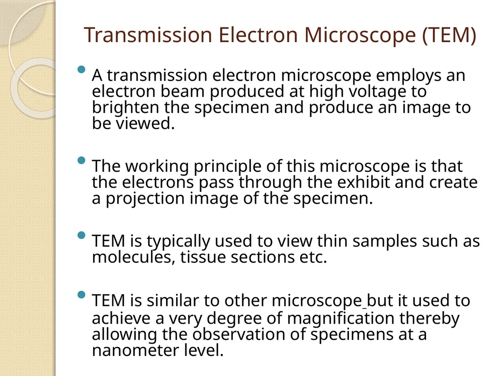
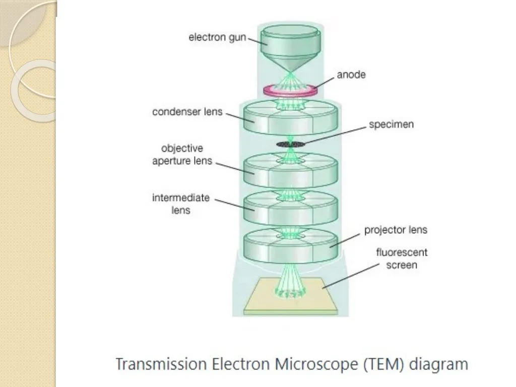
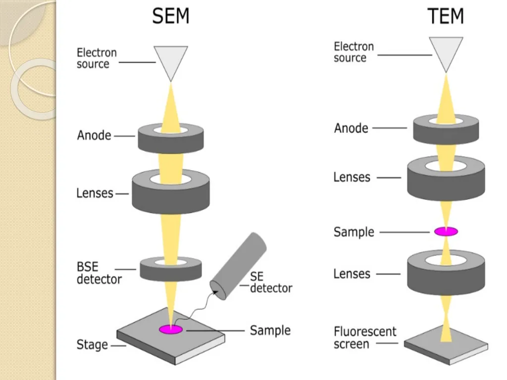
Transmission Electron Microscope (TEM) A transmission electron microscope employs an electron beam produced at high voltage to brighten the specimen and produce an image to be viewed.
The working principle of this microscope is that the electrons pass through the exhibit and create a projection image of the specimen.
TEM is typically used to view thin samples such as molecules, tissue sections etc.
TEM is similar to other microscope but it used to achieve a very degree of magnification thereby allowing the observation of specimens at a nanometer level.<br>
05
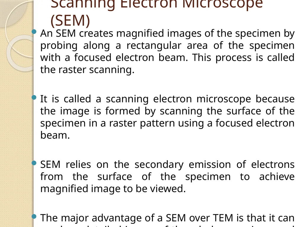
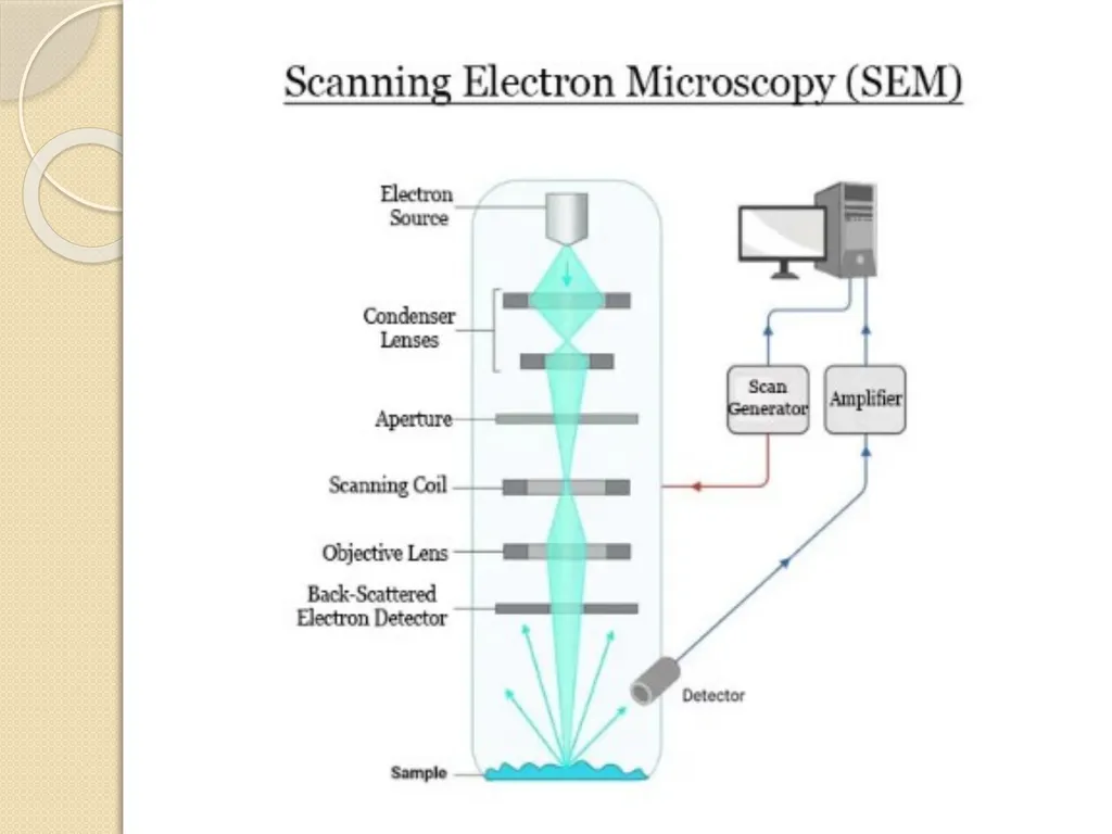
Scanning Electron Microscope (SEM) An SEM creates magnified images of the specimen by probing along a rectangular area of the specimen with a focused electron beam. This process is called the raster scanning.
It is called a scanning electron microscope because the image is formed by scanning the surface of the specimen in a raster pattern using a focused electron beam.
SEM relies on the secondary emission of electrons from the surface of the specimen to achieve magnified image to be viewed.
The major advantage of a SEM over TEM is that it can produce detailed image of the whole organisms and surfaces of the cells.<br>
06
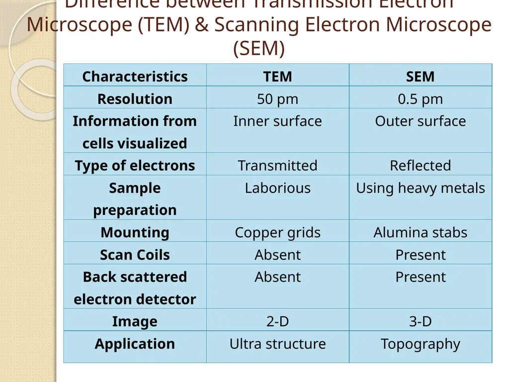
Difference between Transmission Electron Microscope (TEM) & Scanning Electron Microscope (SEM)<br>
07
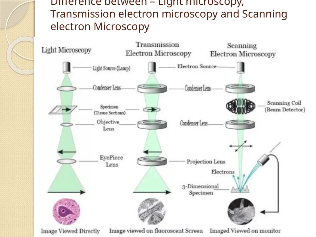
Difference between – Light microscopy, Transmission electron microscopy and Scanning electron Microscopy<br>