04
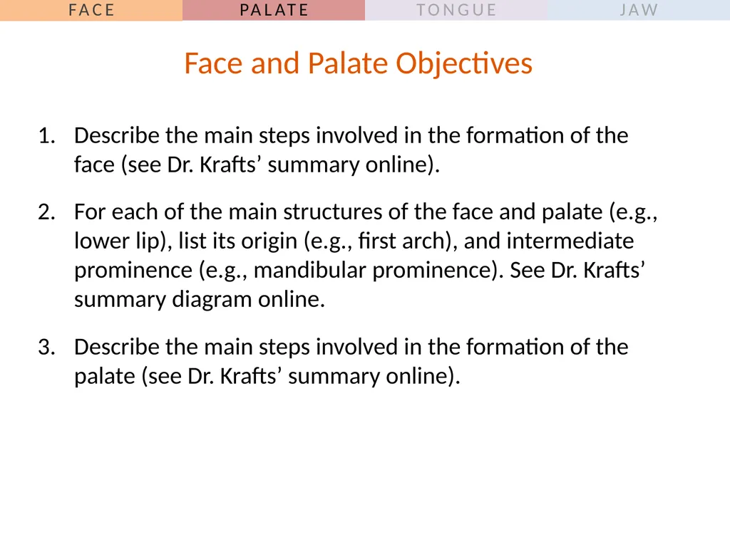
Describe the main steps involved in the formation of the face (see Dr. Krafts’ summary online).
For each of the main structures of the face and palate (e.g., lower lip), list its origin (e.g., first arch), and intermediate prominence (e.g., mandibular prominence). See Dr. Krafts’ summary diagram online.
Describe the main steps involved in the formation of the palate (see Dr. Krafts’ summary online). Face and Palate Objectives<br>
05
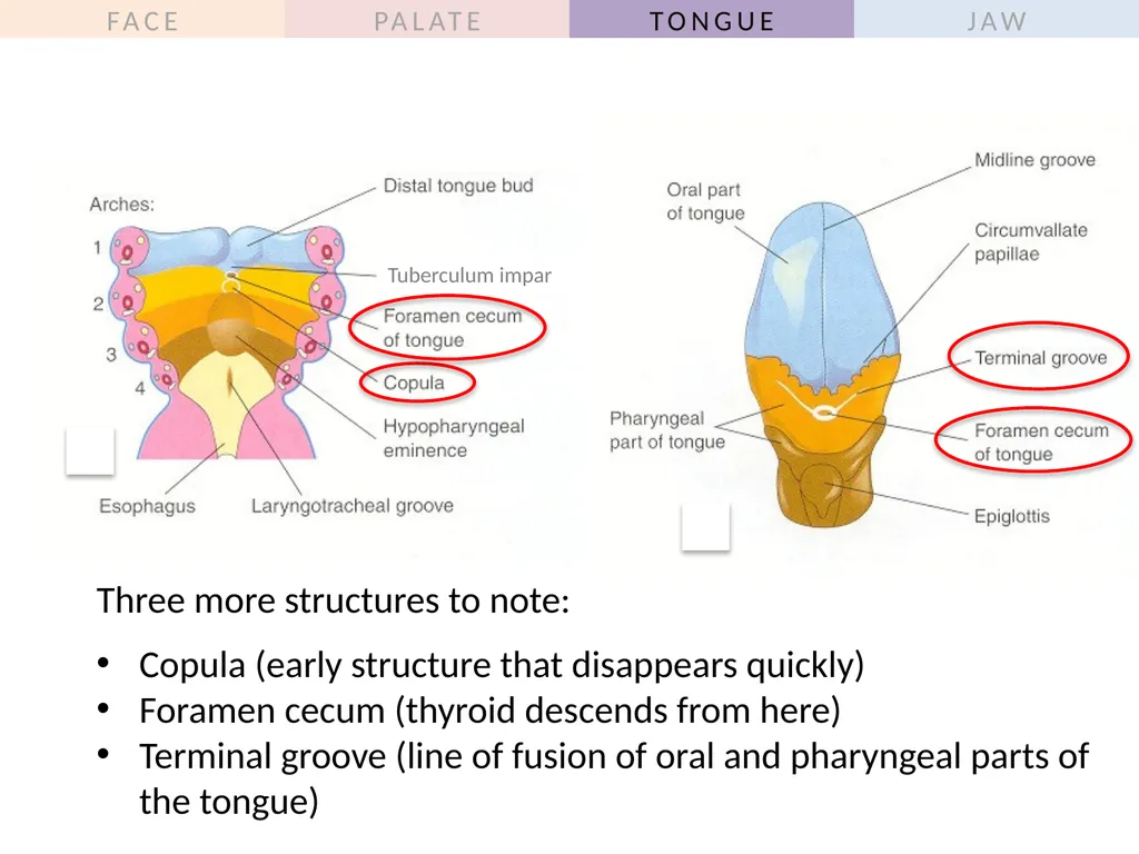
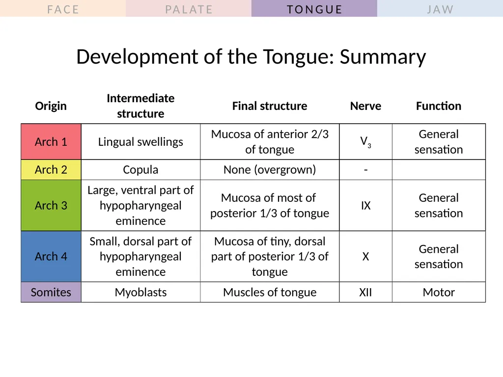
Describe how the mucosa of the tongue is formed from the first four arches. Include the contribution (if any!) of the following structures: lingual swellings, tuberculum impar, hypopharyngeal eminence, foramen cecum, and copula.
List the nerves supplying general sensation to the anterior 2/3 and posterior 1/3 of the mucosa of the tongue.
Describe the origin and motor innervation of the muscles of the tongue.
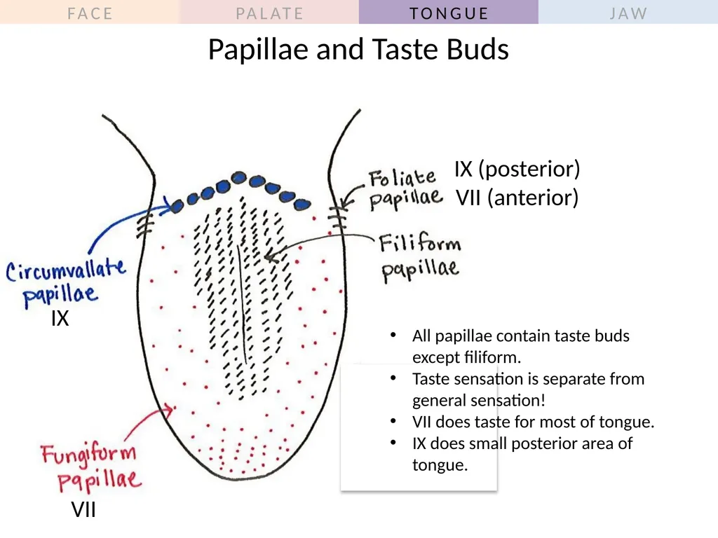
Describe the location of the four types of papillae, and list the nerves supplying taste sensation to the papillae containing taste buds. Tongue Objectives<br>
06
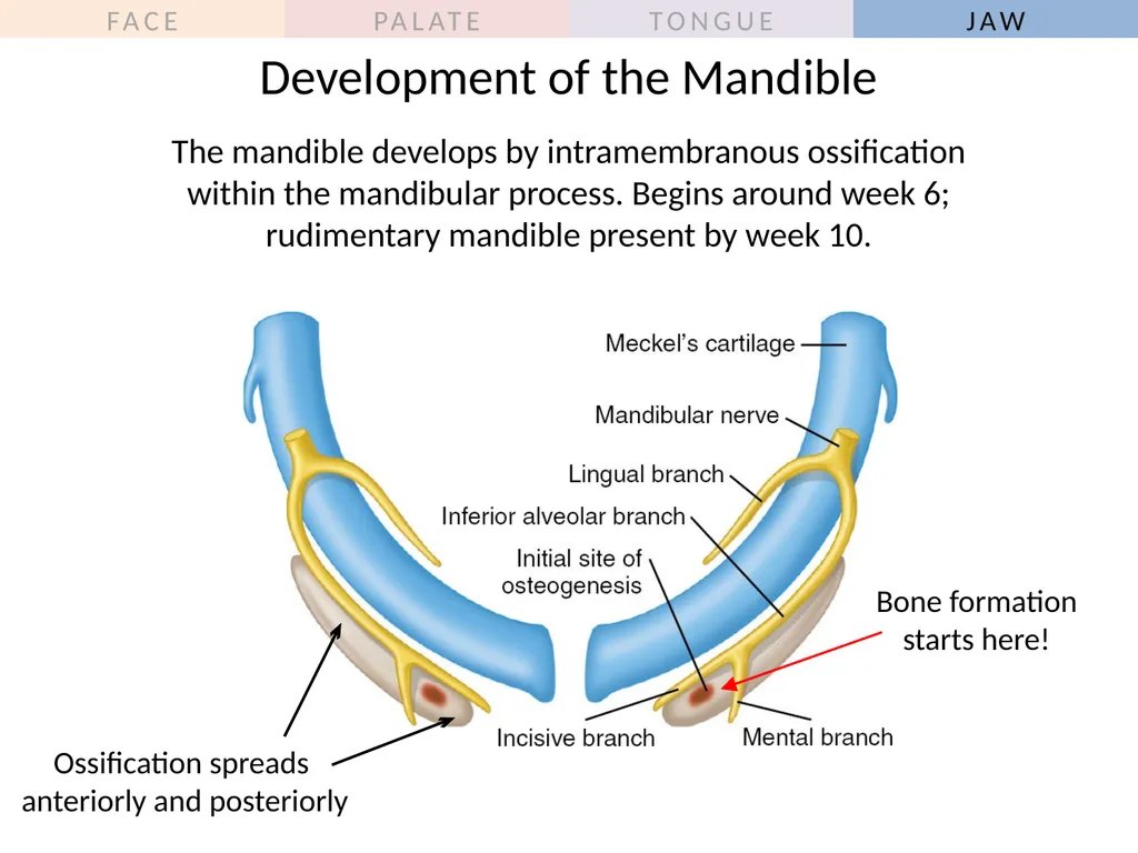
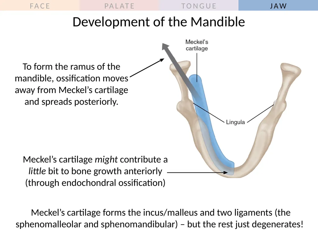
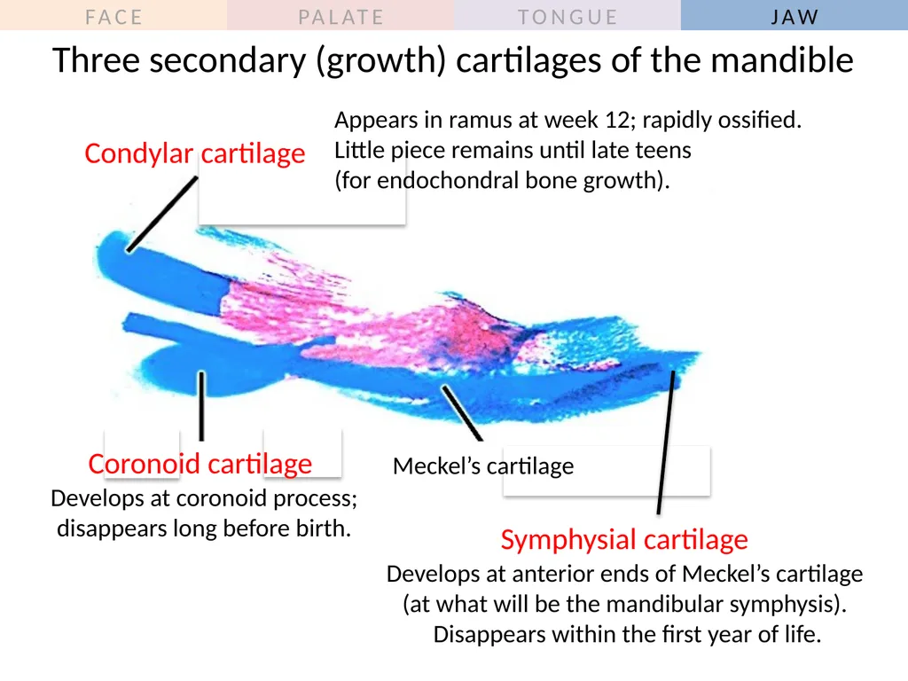
Describe how and when the mandible develops. Include the site of origin (between which nerve branches?), the role of Meckel’s cartilage, and the fate of the three secondary (growth cartilages).
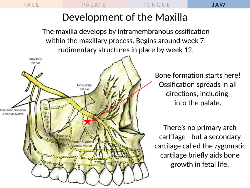
Describe how and when the maxilla develops. Include the site of origin of bone formation, and the contribution of secondary cartilages. Jaw Objectives<br>
07
Outline
General overview of prenatal development
Embryonic period phase 1
Formation of bilaminar disk
Formation of trilaminar disk (gastrulation)
Embryonic period phase 2
Formation of neural tube
Differentiation of mesoderm
Folding of embryo
Formation of pharyngeal arches
Development of the face and oral cavity
Face<br>
08
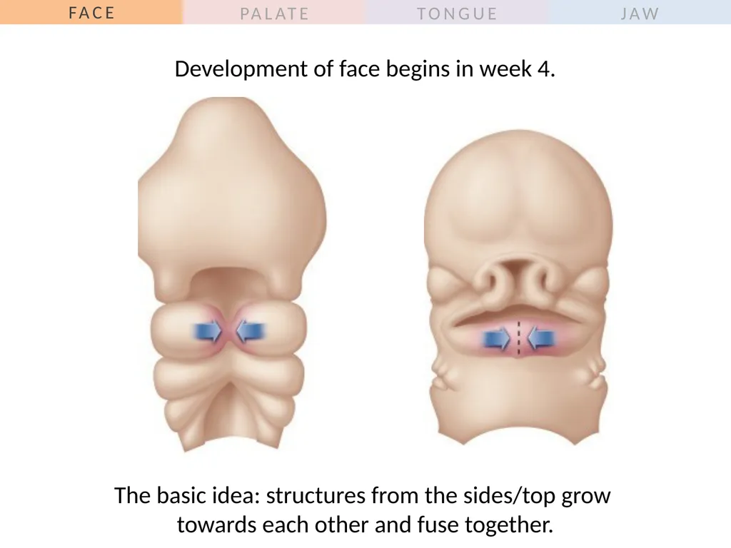
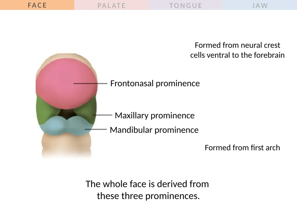
The basic idea: structures from the sides/top grow �towards each other and fuse together. Development of face begins in week 4.<br>
09
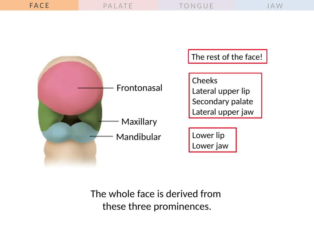
The whole face is derived from �these three prominences.<br>
10
The whole face is derived from �these three prominences.<br>
11
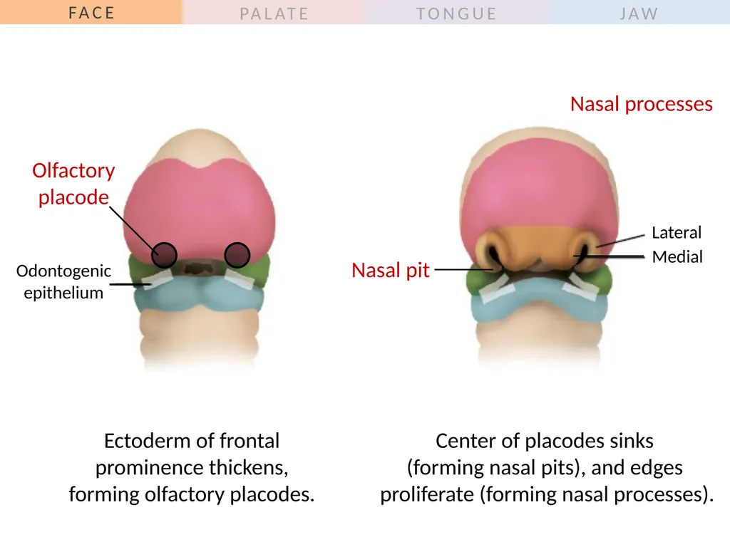
Lateral Medial Nasal processes Ectoderm of frontal �prominence thickens, �forming olfactory placodes. Nasal pit Odontogenic epithelium Olfactory�placode Center of placodes sinks �(forming nasal pits), and edges �proliferate (forming nasal processes).<br>
12
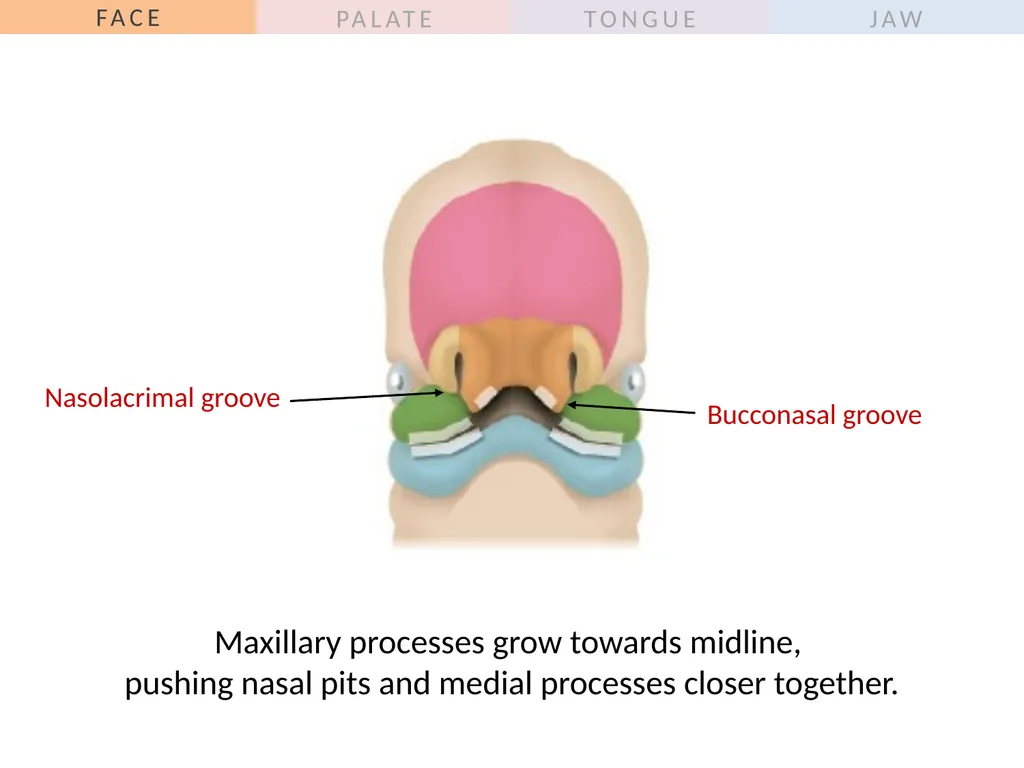
Maxillary processes grow towards midline, �pushing nasal pits and medial processes closer together. Bucconasal groove Nasolacrimal groove<br>
13
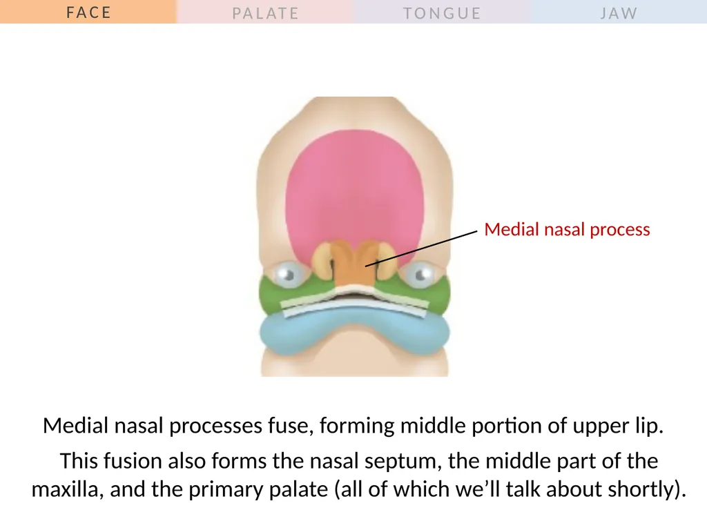
Medial nasal process Medial nasal processes fuse, forming middle portion of upper lip. This fusion also forms the nasal septum, the middle part of the maxilla, and the primary palate (all of which we’ll talk about shortly).<br>
14
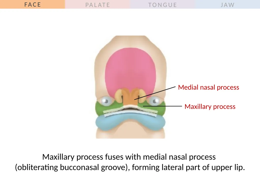
Maxillary process fuses with medial nasal process �(obliterating bucconasal groove), forming lateral part of upper lip. Medial nasal process Maxillary process<br>
15
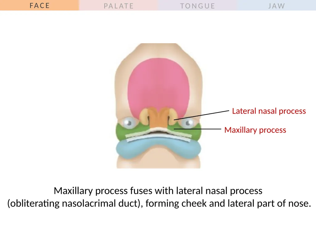
Maxillary process fuses with lateral nasal process �(obliterating nasolacrimal duct), forming cheek and lateral part of nose. Maxillary process Lateral nasal process<br>
16
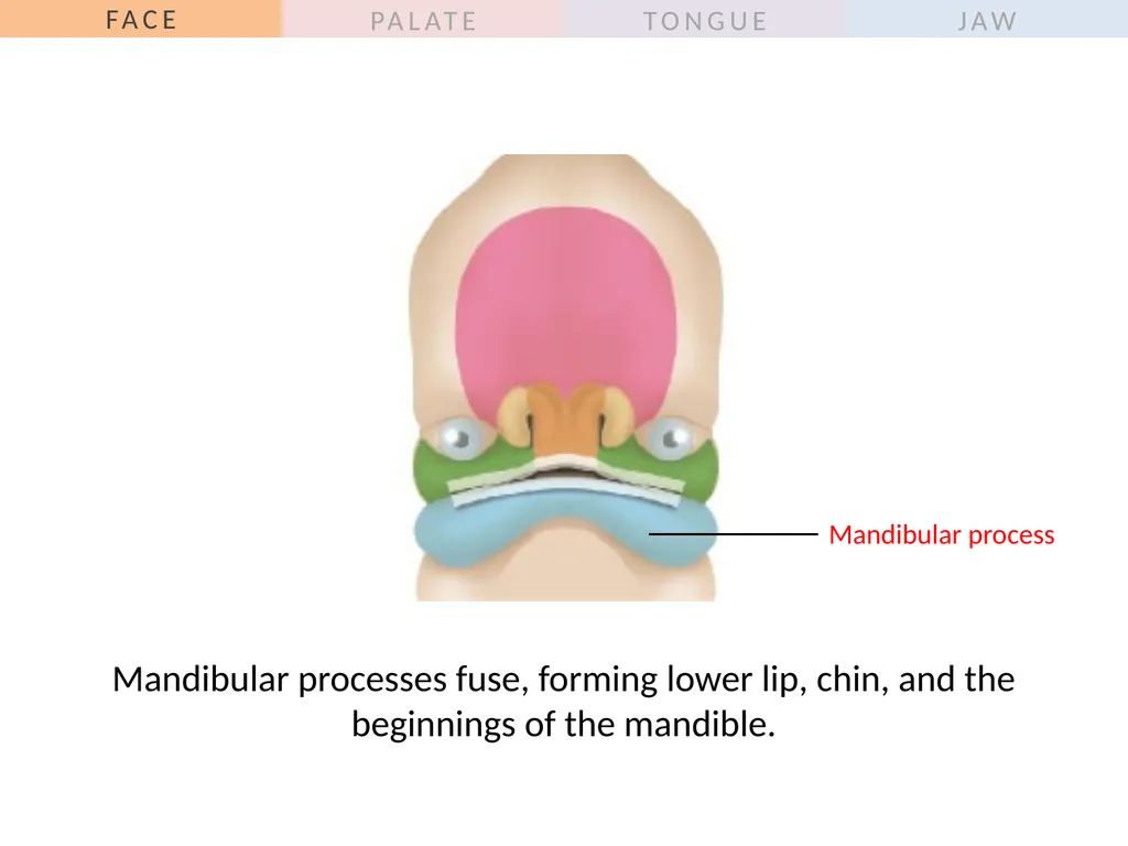
Mandibular processes fuse, forming lower lip, chin, and the beginnings of the mandible. Mandibular process<br>
17
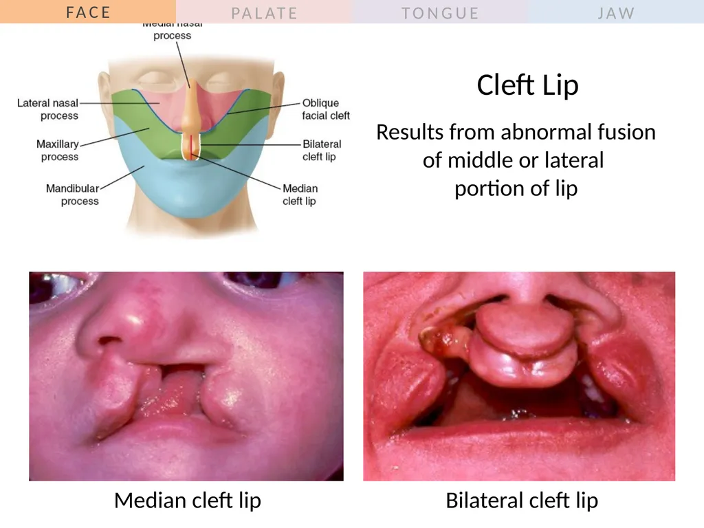
Cleft Lip Results from abnormal fusion of middle or lateral �portion of lip Median cleft lip Bilateral cleft lip<br>
18
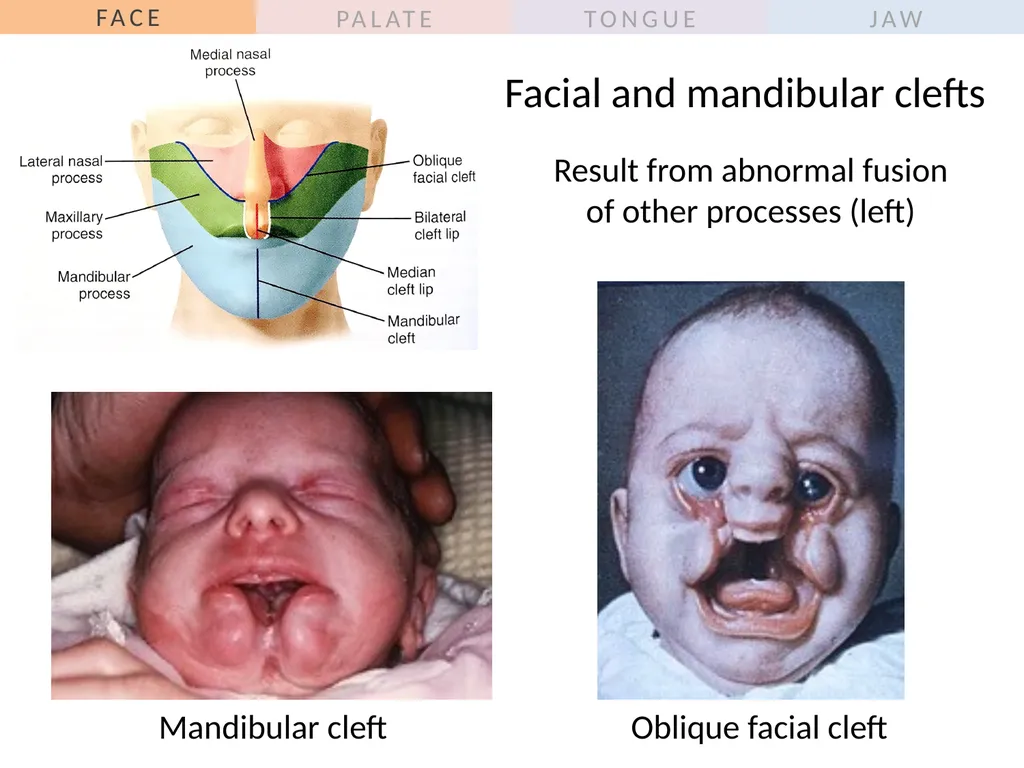
Facial and mandibular clefts Result from abnormal fusion of other processes (left) Mandibular cleft Oblique facial cleft<br>
19
Outline
General overview of prenatal development
Embryonic period phase 1
Formation of bilaminar disk
Formation of trilaminar disk (gastrulation)
Embryonic period phase 2
Formation of neural tube
Differentiation of mesoderm
Folding of embryo
Formation of pharyngeal arches
Development of head, face and oral cavity
Face
Palate<br>
20
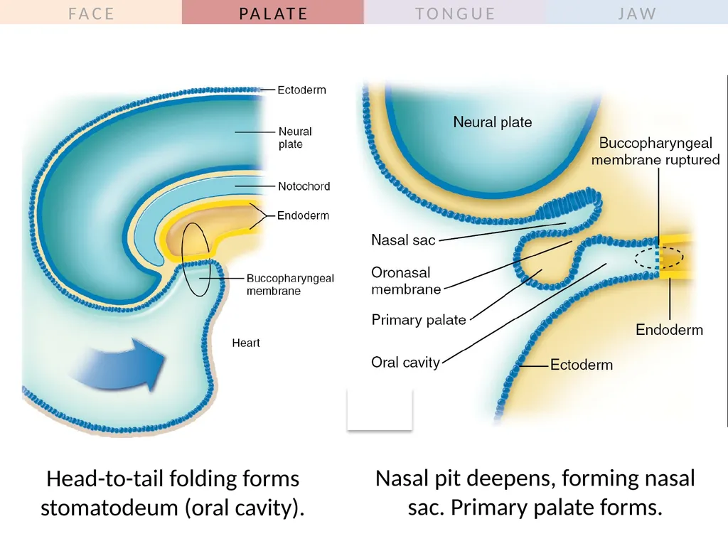
Head-to-tail folding forms stomatodeum (oral cavity). Nasal pit deepens, forming nasal sac. Primary palate forms.<br>
21
Nasal cavity forms, �oronasal membrane ruptures. Head lifts away from thorax. �Jaw and tongue move down and forward.<br>
22
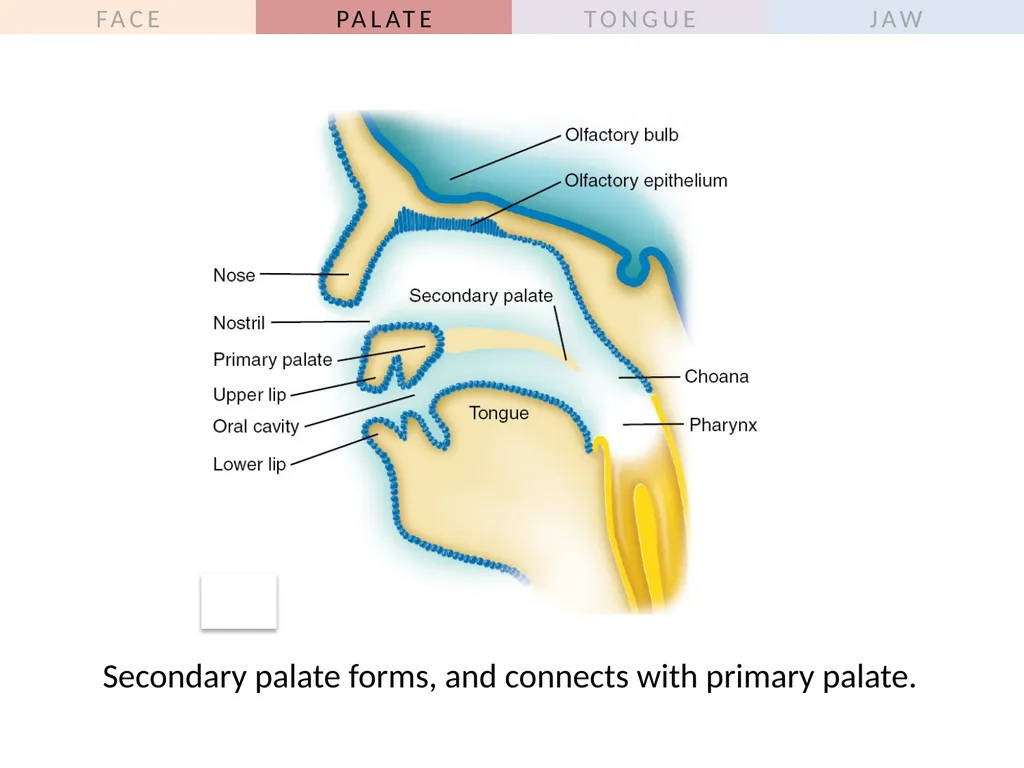
Secondary palate forms, and connects with primary palate.<br>
23
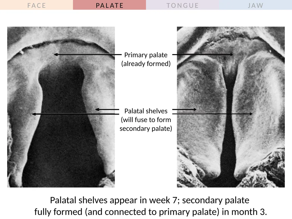
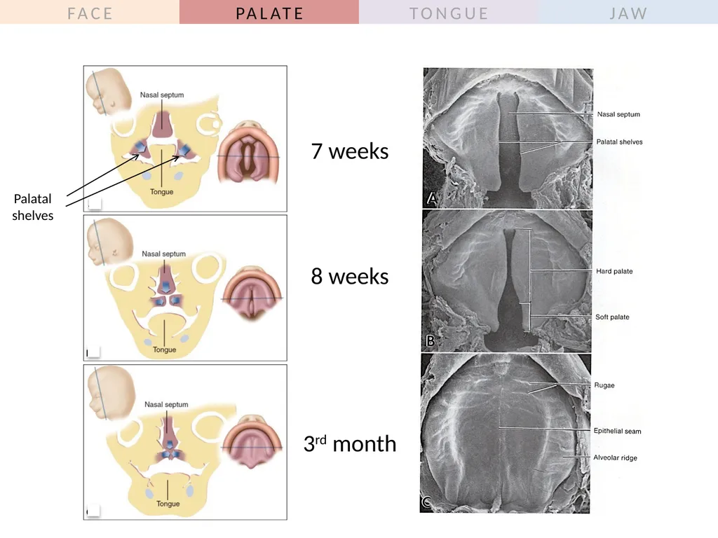
Palatal shelves appear in week 7; secondary palate �fully formed (and connected to primary palate) in month 3. Primary palate
(already formed) Palatal shelves
(will fuse to form secondary palate)<br>
24
7 weeks 8 weeks 3rd month Palatal shelves<br>
26
Outline
General overview of prenatal development
Embryonic period phase 1
Formation of bilaminar disk
Formation of trilaminar disk (gastrulation)
Embryonic period phase 2
Formation of neural tube
Differentiation of mesoderm
Folding of embryo
Formation of pharyngeal arches
Development of head, face and oral cavity
Face
Palate
Tongue<br>
27
Pharyngeal arches fuse in midline, form three swellings:
Tuberculum impar appears and remains small
Lingual swellings form anterior 2/3 of tongue
Hypopharyngeal eminence forms posterior 1/3 of tongue Arch 1 Arch 2 Arch 3 Arch 4 Tuberculum impar Lingual swellings Hypopharyngeal eminence General sensation: V3 Here’s how the mucosal surface of the tongue forms. General sensation: Mostly IX, tiny bit X<br>
28
Three more structures to note:
Copula (early structure that disappears quickly)
Foramen cecum (thyroid descends from here)
Terminal groove (line of fusion of oral and pharyngeal parts of the tongue)<br>
29
The muscles of the tongue are derived from occipital somites, which migrate into the floor of mouth,
carrying with them their nerve supply (XII).<br>
30
Development of the Tongue: Summary<br>
31
Papillae and Taste Buds IX VII IX (posterior)
VII (anterior) All papillae contain taste buds except filiform.
Taste sensation is separate from general sensation!
VII does taste for most of tongue.
IX does small posterior area of tongue.<br>
32
Outline
General overview of prenatal development
Embryonic period phase 1
Formation of bilaminar disk
Formation of trilaminar disk (gastrulation)
Embryonic period phase 2
Formation of neural tube
Differentiation of mesoderm
Folding of embryo
Formation of pharyngeal arches
Development of head, face and oral cavity
Face
Palate
Tongue
Jaw<br>
33
The mandible develops by intramembranous ossification within the mandibular process. Begins around week 6; rudimentary mandible present by week 10. Development of the Mandible Bone formation starts here! Ossification spreads �anteriorly and posteriorly<br>
34
Development of the Mandible To form the ramus of the mandible, ossification moves away from Meckel’s cartilage and spreads posteriorly. Meckel’s cartilage forms the incus/malleus and two ligaments (the sphenomalleolar and sphenomandibular) – but the rest just degenerates! Meckel’s cartilage might contribute a little bit to bone growth anteriorly (through endochondral ossification)<br>
35
Develops at coronoid process; disappears long before birth. Condylar cartilage Coronoid cartilage Meckel’s cartilage Symphysial cartilage Appears in ramus at week 12; rapidly ossified. �Little piece remains until late teens �(for endochondral bone growth). Develops at anterior ends of Meckel’s cartilage �(at what will be the mandibular symphysis). �Disappears within the first year of life. Three secondary (growth) cartilages of the mandible<br>
36
The maxilla develops by intramembranous ossification within the maxillary process. Begins around week 7; rudimentary structures in place by week 12. Development of the Maxilla There’s no primary arch cartilage - but a secondary cartilage called the zygomatic cartilage briefly aids bone growth in fetal life. Bone formation starts here! Ossification spreads in all directions, including �into the palate.<br>
37
Outline
General overview of prenatal development
Embryonic period phase 1
Formation of bilaminar disk
Formation of trilaminar disk (gastrulation)
Embryonic period phase 2
Formation of neural tube
Differentiation of mesoderm
Folding of embryo
Formation of pharyngeal arches
Development of head, face and oral cavity
Face (bones and muscles)
Palate
Tongue
Jaw<br>