
Endocrine Emergencies CHAPTER 24 Introduction
KB
Published · 102 slides · 0 views

1 / 1







Description
Endocrine Emergencies CHAPTER 24 Introduction Endocrine system influences almost every cell, organ, and function of the body. Disorders often display a broad range of signs and symptoms. Assess thoroughly and treat immediately to prevent
Related Topics
Share
Embed code
Download this presentation From Below
"Endocrine Emergencies CHAPTER 24 Introduction" is the property of its rightful owner. Permission is granted to download and print the materials on this website for personal, non-commercial use only, and to display it on your personal computer provided you do not modify the materials and that you retain all copyright notices contained in the materials. By downloading content from our website, you accept the terms of this agreement.
Presentation Transcript
01
Endocrine Emergencies CHAPTER 24<br>
02
Introduction Endocrine system influences almost every cell, organ, and function of the body.
Disorders often display a broad range of signs and symptoms.
Assess thoroughly and treat immediately to prevent life threats.<br>
Disorders often display a broad range of signs and symptoms.
Assess thoroughly and treat immediately to prevent life threats.<br>
03
Components of the Endocrine System (1 of 13) The endocrine system controls and regulates all the systems in the body.
Works with nervous system to maintain homeostasis
Coordinates responses to environmental changes and stress
Hormones: Chemical messengers<br>
Works with nervous system to maintain homeostasis
Coordinates responses to environmental changes and stress
Hormones: Chemical messengers<br>
04
Components of the Endocrine System (2 of 13) Hypothalamus
Small region of brain, containing control centers for body functions and emotions
Primary link between the endocrine and nervous systems
Hypothalamus and pituitary gland are intimately related through the vascular system.<br>
Small region of brain, containing control centers for body functions and emotions
Primary link between the endocrine and nervous systems
Hypothalamus and pituitary gland are intimately related through the vascular system.<br>
05
Components of the Endocrine System (3 of 13) Pituitary gland
Secretions control activity of other endocrine glands.
Known as “tropic” hormones
Hormones control other body functions
ADH, oxytocin<br>
Secretions control activity of other endocrine glands.
Known as “tropic” hormones
Hormones control other body functions
ADH, oxytocin<br>
06
Components of the Endocrine System (4 of 13) Thyroid gland
Secretes thyroxine (T4) when body’s metabolic rate decreases
Body’s major metabolic hormone
Stimulates energy production in cells
Cannot be produced without proper iodine intake
Production regulated by negative feedback mechanism<br>
Secretes thyroxine (T4) when body’s metabolic rate decreases
Body’s major metabolic hormone
Stimulates energy production in cells
Cannot be produced without proper iodine intake
Production regulated by negative feedback mechanism<br>
07
Components of the Endocrine System (5 of 13) Thyroid gland (cont’d)
Secretes calcitonin
Helps maintain calcium levels in the blood
Stimulates bone-building cells to absorb excess calcium
Stimulates kidneys to absorb and excrete excess calcium<br>
Secretes calcitonin
Helps maintain calcium levels in the blood
Stimulates bone-building cells to absorb excess calcium
Stimulates kidneys to absorb and excrete excess calcium<br>
08
Components of the Endocrine System (6 of 13) Parathyroid glands
Assist in the regulation of calcium
Parathyroid hormone
Secreted when calcium blood levels are low
Stimulates bone-dissolving cells to break down bone and release calcium into bloodstream
Decreases amount of calcium released in the urine<br>
Assist in the regulation of calcium
Parathyroid hormone
Secreted when calcium blood levels are low
Stimulates bone-dissolving cells to break down bone and release calcium into bloodstream
Decreases amount of calcium released in the urine<br>
09
Components of the Endocrine System (7 of 13) Thymus gland
Helps immune system identify and destroy foreign intruders<br>
Helps immune system identify and destroy foreign intruders<br>
10
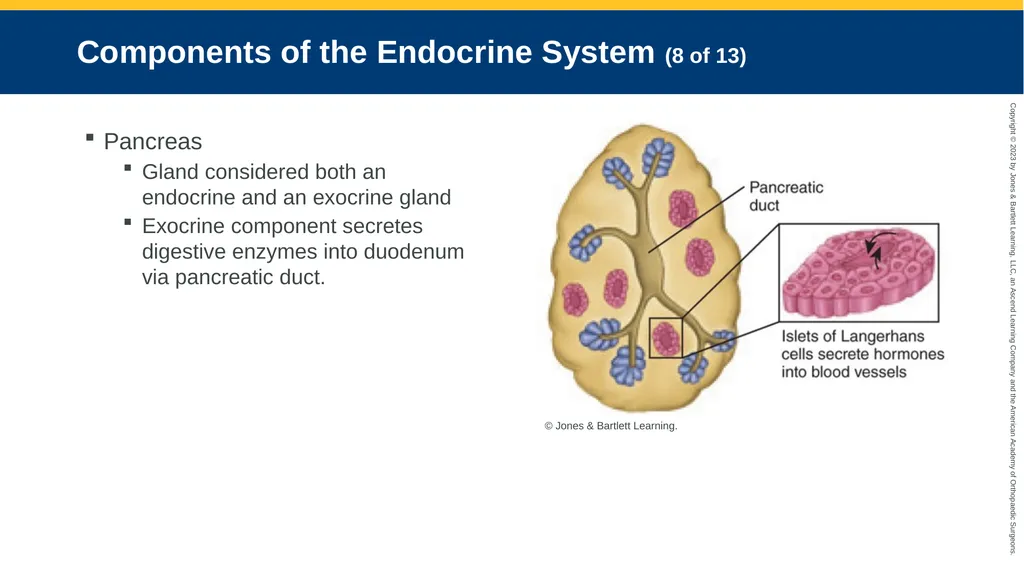
Components of the Endocrine System (8 of 13) Pancreas
Gland considered both an endocrine and an exocrine gland
Exocrine component secretes digestive enzymes into duodenum via pancreatic duct. © Jones & Bartlett Learning.<br>
Gland considered both an endocrine and an exocrine gland
Exocrine component secretes digestive enzymes into duodenum via pancreatic duct. © Jones & Bartlett Learning.<br>
11
Components of the Endocrine System (9 of 13) Pancreas (cont’d)
Endocrine component comprises the islets of Langerhans.
Cell groups within the pancreas that act like “an organ within an organ”
Alpha cells secrete glucagon.
Beta cells secrete insulin.
Delta cells secrete somatostatin.<br>
Endocrine component comprises the islets of Langerhans.
Cell groups within the pancreas that act like “an organ within an organ”
Alpha cells secrete glucagon.
Beta cells secrete insulin.
Delta cells secrete somatostatin.<br>
12
Components of the Endocrine System (10 of 13) Pancreas (cont’d)
When blood glucose level falls, glucagon is secreted to raise it.
When blood glucose level rises, insulin is secreted.<br>
When blood glucose level falls, glucagon is secreted to raise it.
When blood glucose level rises, insulin is secreted.<br>
13
Components of the Endocrine System (11 of 13) Adrenal glands
Located on superior aspect of each kidney
Divided into two distinct sections, the adrenal cortex and the adrenal medulla
Adrenal cortex secretes aldosterone
When stimulated by hypothalamus, adrenal medulla secretes norepinephrine and epinephrine.<br>
Located on superior aspect of each kidney
Divided into two distinct sections, the adrenal cortex and the adrenal medulla
Adrenal cortex secretes aldosterone
When stimulated by hypothalamus, adrenal medulla secretes norepinephrine and epinephrine.<br>
14
Components of the Endocrine System (12 of 13) Gonads
Promote sexual maturation
Fulfill reproductive needs
Testes in men
Produce hormones called androgens
Regulate changes of puberty
Most important is testosterone<br>
Promote sexual maturation
Fulfill reproductive needs
Testes in men
Produce hormones called androgens
Regulate changes of puberty
Most important is testosterone<br>
15
Components of the Endocrine System (13 of 13) Ovaries in women
Secrete estrogen and progesterone
Estrogen signals secretion of luteinizing hormone when an egg is developing.
Estrogen supports development of secondary sex characteristics.
Both assist in the regulation of the menstrual cycle.
Progesterone prepares uterus for implantation of fertilized egg.<br>
Secrete estrogen and progesterone
Estrogen signals secretion of luteinizing hormone when an egg is developing.
Estrogen supports development of secondary sex characteristics.
Both assist in the regulation of the menstrual cycle.
Progesterone prepares uterus for implantation of fertilized egg.<br>
16
Patient Assessment Endocrine emergencies tend to affect many organ systems.
Do not take them lightly.<br>
Do not take them lightly.<br>
17
Scene Size-Up Address hazards.
Follow standard precautions.
Check the home for medications.<br>
Follow standard precautions.
Check the home for medications.<br>
18
Primary Survey (1 of 5) ABCDE
Identify and manage life threats.
Form a general impression.
Signs and symptoms depend on affected hormone.
Is patient alert or is there a change in mental status?
Diaphoresis is a sign of severe distress.<br>
Identify and manage life threats.
Form a general impression.
Signs and symptoms depend on affected hormone.
Is patient alert or is there a change in mental status?
Diaphoresis is a sign of severe distress.<br>
19
Primary Survey (2 of 5) Check for signs and symptoms of specific conditions.
“Buffalo hump” and “moon face”
Mottled skin
Enlarged body parts
Underweight or overweight
Exophthalmos<br>
“Buffalo hump” and “moon face”
Mottled skin
Enlarged body parts
Underweight or overweight
Exophthalmos<br>
20
Primary Survey (3 of 5) Airway and breathing
Ensure patent airway.
Ensure there are no obstructions.
Assess breathing effort.<br>
Ensure patent airway.
Ensure there are no obstructions.
Assess breathing effort.<br>
21
Primary Survey (4 of 5) Circulation
Assess skin color, moisture, and temperature.
Obtain blood pressure.
If necessary:
Administer IV fluid.<br>
Assess skin color, moisture, and temperature.
Obtain blood pressure.
If necessary:
Administer IV fluid.<br>
22
Primary Survey (5 of 5) Transport decision
Many patients should be transported to a specialty facility.
Provide rapid transport to the closest facility if the patient is unstable.<br>
Many patients should be transported to a specialty facility.
Provide rapid transport to the closest facility if the patient is unstable.<br>
23
History Taking (1 of 2) Especially useful in diabetic emergencies
Investigate chief complaint/present illness.
If patient is unresponsive:
Obtain blood glucose level
Consider signs and symptoms.
Undiagnosed or poorly managed diabetes may cause:
Polyphagia
Polyuria
Polydipsia<br>
Investigate chief complaint/present illness.
If patient is unresponsive:
Obtain blood glucose level
Consider signs and symptoms.
Undiagnosed or poorly managed diabetes may cause:
Polyphagia
Polyuria
Polydipsia<br>
24
History Taking (2 of 2) Hyperthyroidism and thyrotoxicosis may cause cardiac dysrhythmias.
Ascertain any allergies prior to administering medication.
Document all medications being taken.<br>
Ascertain any allergies prior to administering medication.
Document all medications being taken.<br>
25
Secondary Assessment (1 of 3) Physical exam
Observe appearance and position.
Identify atypical findings.
Finer abnormalities will help determine treatment.
Condition of skin<br>
Observe appearance and position.
Identify atypical findings.
Finer abnormalities will help determine treatment.
Condition of skin<br>
26
Secondary Assessment (2 of 3) Goals with comatose patients:
Determine level of consciousness.
Look for cause of coma.<br>
Determine level of consciousness.
Look for cause of coma.<br>
27
Secondary Assessment (3 of 3) Vital signs
Look for hypertension and bradycardia.
Be alert for abnormal respiratory patterns.
Look for respiratory-related motions.<br>
Look for hypertension and bradycardia.
Be alert for abnormal respiratory patterns.
Look for respiratory-related motions.<br>
28
Reassessment Continually reassess the patient.
Be prepared to manage the airway.
Obtain blood specimens early in patients with diabetes.
Provide emotional support.
Monitor cardiac rhythm.
Recheck vital signs, pupils, and level of consciousness.<br>
Be prepared to manage the airway.
Obtain blood specimens early in patients with diabetes.
Provide emotional support.
Monitor cardiac rhythm.
Recheck vital signs, pupils, and level of consciousness.<br>
29
Emergency Medical Care (1 of 2) If patient has altered mental status:
Establish IV with 0.9% (NS) or a saline lock.
Initiate treatment for reading <60 mg/dL.
Give D50 at a dose per local protocols.
If condition does not improve after dextrose and you suspect narcotic overdose, consider naloxone.<br>
Establish IV with 0.9% (NS) or a saline lock.
Initiate treatment for reading <60 mg/dL.
Give D50 at a dose per local protocols.
If condition does not improve after dextrose and you suspect narcotic overdose, consider naloxone.<br>
30
Emergency Medical Care (2 of 2) Transport for comatose patients
Intubated: supine with cervical collar
Not intubated: lateral recumbent or recovery position
Increasing intracranial pressure, posturing, or unequal pupils: head elevated to 30–45 degrees and midline
Keep mouth and pharynx free of secretions.<br>
Intubated: supine with cervical collar
Not intubated: lateral recumbent or recovery position
Increasing intracranial pressure, posturing, or unequal pupils: head elevated to 30–45 degrees and midline
Keep mouth and pharynx free of secretions.<br>
31
Glucose Metabolic Derangements (1 of 2) Endocrine disorders are caused by:
Hypersecretion of a gland
Insufficient secretion of a gland
Glucose metabolic derangements
Caused by dysfunction of the pancreas<br>
Hypersecretion of a gland
Insufficient secretion of a gland
Glucose metabolic derangements
Caused by dysfunction of the pancreas<br>
32
Glucose Metabolic Derangements (2 of 2) Most endocrine emergencies result in:
Compromise of the ABCs
Improper fluid balance
Deteriorating mental status
Abnormal vital signs and blood glucose levels<br>
Compromise of the ABCs
Improper fluid balance
Deteriorating mental status
Abnormal vital signs and blood glucose levels<br>
33
Diabetes A group of complex metabolic disorders
Diabetes mellitus
Gestational diabetes
Hypoglycemia/hyperglycemia
Diabetic ketoacidosis
Hyperosmolar hyperglycemic nonketotic syndrome<br>
Diabetes mellitus
Gestational diabetes
Hypoglycemia/hyperglycemia
Diabetic ketoacidosis
Hyperosmolar hyperglycemic nonketotic syndrome<br>
34
Diabetes Mellitus (1 of 3) Metabolic disorder in which body’s ability to metabolize simple carbohydrates is impaired.
Characterized by:
Polyphagia
Polydipsia
Polyuria<br>
Characterized by:
Polyphagia
Polydipsia
Polyuria<br>
35
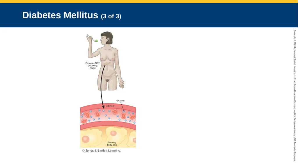
Diabetes Mellitus (2 of 3) Glucose: Primary fuel for cellular metabolism
Body cannot metabolize glucose.
Pancreas does not produce enough insulin
Cells do not respond to insulin<br>
Body cannot metabolize glucose.
Pancreas does not produce enough insulin
Cells do not respond to insulin<br>
36
Diabetes Mellitus (3 of 3) © Jones & Bartlett Learning.<br>
37
Life-Altering Complications from Diabetes (1 of 3) Kidney failure
Glomeruli become sclerotic.
Heart disease
Lipolysis raises fat level in the blood.
Microangiopathy restricts blood flow.
Increased risk for silent MI<br>
Glomeruli become sclerotic.
Heart disease
Lipolysis raises fat level in the blood.
Microangiopathy restricts blood flow.
Increased risk for silent MI<br>
38
Life-Altering Complications from Diabetes (2 of 3) Cerebrovascular disease, stroke, and hypertension
Peripheral artery disease
Kidney disease
Eyes
Primary cause of blindness
Cataracts
Retinal detachment<br>
Peripheral artery disease
Kidney disease
Eyes
Primary cause of blindness
Cataracts
Retinal detachment<br>
39
Life-Altering Complications from Diabetes (3 of 3) Neuropathy
Loss of sensation and function
Many conditions can be delayed or prevented with lifestyle changes.<br>
Loss of sensation and function
Many conditions can be delayed or prevented with lifestyle changes.<br>
40
Type 1 Diabetes (1 of 3) Pathophysiology
Generally affects children
Environmental factors may be part of the cause.
Pancreatic cells do not produce insulin.
Daily insulin is required by injection or pump.
Strict dietary control must be observed.<br>
Generally affects children
Environmental factors may be part of the cause.
Pancreatic cells do not produce insulin.
Daily insulin is required by injection or pump.
Strict dietary control must be observed.<br>
41
Type 1 Diabetes (2 of 3) Assessment
Assess compliance with disease management.
With altered mental status, suspect low blood glucose level.
Consider additional chronic conditions.
Look for sores or infections.<br>
Assess compliance with disease management.
With altered mental status, suspect low blood glucose level.
Consider additional chronic conditions.
Look for sores or infections.<br>
42
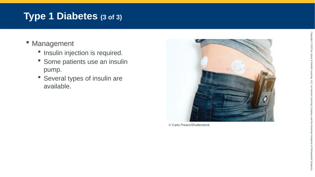
Type 1 Diabetes (3 of 3) Management
Insulin injection is required.
Some patients use an insulin pump.
Several types of insulin are available. © Carlo Prearo/Shutterstock.<br>
Insulin injection is required.
Some patients use an insulin pump.
Several types of insulin are available. © Carlo Prearo/Shutterstock.<br>
43
Type 2 Diabetes (1 of 3) Pathophysiology
Most common form of diabetes
Blood glucose levels are elevated.
Often, pancreas produces enough insulin, but body cannot effectively use it.
Development is associated with obesity and physical inactivity.<br>
Most common form of diabetes
Blood glucose levels are elevated.
Often, pancreas produces enough insulin, but body cannot effectively use it.
Development is associated with obesity and physical inactivity.<br>
44
Type 2 Diabetes (2 of 3) Assessment
Symptoms may include:
Frequent urination
Thirst
Blurred vision
Frequent infections
Cranky, confused, or shaky
Unresponsiveness
Seizure<br>
Symptoms may include:
Frequent urination
Thirst
Blurred vision
Frequent infections
Cranky, confused, or shaky
Unresponsiveness
Seizure<br>
45
Type 2 Diabetes (3 of 3) Management
Weight loss helps to control disease.
Food intake must be spread throughout the day.
Medication/insulin required daily.<br>
Weight loss helps to control disease.
Food intake must be spread throughout the day.
Medication/insulin required daily.<br>
46
Prediabetes (1 of 2) Blood glucose levels or hemoglobin A1c levels are above normal levels.
Not high enough to be diagnosed as diabetes
Affects 1 out of 3 US adults, and 84% are unaware of status.
Without intervention, many develop type 2 diabetes.<br>
Not high enough to be diagnosed as diabetes
Affects 1 out of 3 US adults, and 84% are unaware of status.
Without intervention, many develop type 2 diabetes.<br>
47
Prediabetes (2 of 2) Risk factors
Being overweight
Age greater than 45 years
Parent or sibling with type 2 diabetes
Physical inactivity
Gestational diabetes
Polycystic ovary syndrome<br>
Being overweight
Age greater than 45 years
Parent or sibling with type 2 diabetes
Physical inactivity
Gestational diabetes
Polycystic ovary syndrome<br>
48
Gestational Diabetes (1 of 2) Pathophysiology
Form of glucose intolerance during pregnancy
Increases risk of type 2 diabetes
Resolves before delivery for most women
Does not produce birth defects<br>
Form of glucose intolerance during pregnancy
Increases risk of type 2 diabetes
Resolves before delivery for most women
Does not produce birth defects<br>
49
Gestational Diabetes (2 of 2) Assessment
Oral glucose tolerance test is used.
Hemoglobin A1c test can be used during the first pregnancy visit.
Management
Stabilize blood glucose levels.
Diet, exercise, blood glucose testing
May require insulin injections<br>
Oral glucose tolerance test is used.
Hemoglobin A1c test can be used during the first pregnancy visit.
Management
Stabilize blood glucose levels.
Diet, exercise, blood glucose testing
May require insulin injections<br>
50
Hypoglycemia (1 of 9) Low blood glucose level
45 mg/dL or less
Experienced by patients with type 1 and type 2 diabetes
Requires close monitoring and control of diabetes to prevent long-term complications
Severe hypoglycemia requires intervention and treatment.<br>
45 mg/dL or less
Experienced by patients with type 1 and type 2 diabetes
Requires close monitoring and control of diabetes to prevent long-term complications
Severe hypoglycemia requires intervention and treatment.<br>
51
Hypoglycemia (2 of 9) Pathophysiology
Often results from:
Too much insulin
Too little food
Both
Counter-regulation is body’s natural defense to maintain blood glucose at appropriate level.<br>
Often results from:
Too much insulin
Too little food
Both
Counter-regulation is body’s natural defense to maintain blood glucose at appropriate level.<br>
52
Hypoglycemia (3 of 9) Pathophysiology (cont’d)
Body’s first line of defense
Reduce insulin production by the pancreas
Increase glucagon production by alpha cells
Second line of defense
Secretion of catecholamines
Last defense
Stimulation of autonomic nervous system<br>
Body’s first line of defense
Reduce insulin production by the pancreas
Increase glucagon production by alpha cells
Second line of defense
Secretion of catecholamines
Last defense
Stimulation of autonomic nervous system<br>
53
Hypoglycemia (4 of 9) Pathophysiology (cont’d)
In type 1 diabetes, islets of Langerhans do not make insulin.
Body’s first line of defense against hypoglycemia is lost.
In type 2 diabetes, the pancreas generates insulin, but:
Body may be insulin resistant.
Pancreas may not produce enough insulin.<br>
In type 1 diabetes, islets of Langerhans do not make insulin.
Body’s first line of defense against hypoglycemia is lost.
In type 2 diabetes, the pancreas generates insulin, but:
Body may be insulin resistant.
Pancreas may not produce enough insulin.<br>
54
Hypoglycemia (5 of 9) Assessment
Common signs and symptoms include:
Trembling, rapid pulse, sweat, and hunger
Blood glucose <60 mg/dL
Unexplainable agitation, irritability, combative behavior
Altered mentation or confusion
Nausea
Weakness
Cool, clammy skin<br>
Common signs and symptoms include:
Trembling, rapid pulse, sweat, and hunger
Blood glucose <60 mg/dL
Unexplainable agitation, irritability, combative behavior
Altered mentation or confusion
Nausea
Weakness
Cool, clammy skin<br>
55
Hypoglycemia (6 of 9) Assessment (cont’d)
Additional signs and symptoms include:
Headache
Memory loss
Incoordination
Slurred speech
Dilated pupils
Seizures and coma in severe cases<br>
Additional signs and symptoms include:
Headache
Memory loss
Incoordination
Slurred speech
Dilated pupils
Seizures and coma in severe cases<br>
56
Hypoglycemia (7 of 9) Assessment (cont’d)
Hypoglycemia develops rapidly.
Suspect in any diabetic patient with:
Bizarre behavior
Neurologic signs
Coma<br>
Hypoglycemia develops rapidly.
Suspect in any diabetic patient with:
Bizarre behavior
Neurologic signs
Coma<br>
57
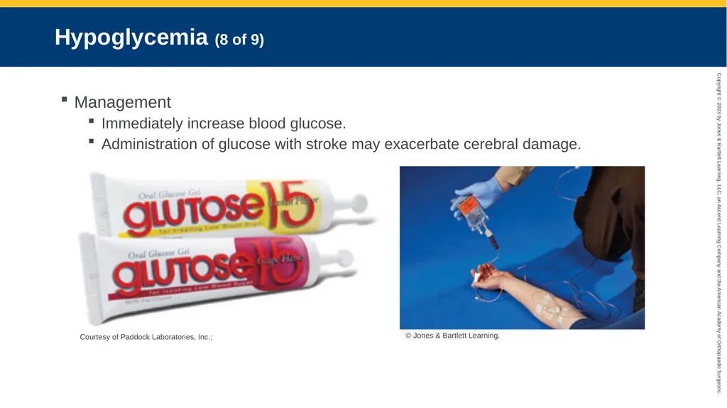
Hypoglycemia (8 of 9) Management
Immediately increase blood glucose.
Administration of glucose with stroke may exacerbate cerebral damage. Courtesy of Paddock Laboratories, Inc.; © Jones & Bartlett Learning.<br>
Immediately increase blood glucose.
Administration of glucose with stroke may exacerbate cerebral damage. Courtesy of Paddock Laboratories, Inc.; © Jones & Bartlett Learning.<br>
58
Hypoglycemia (9 of 9) Management (cont’d)
Rule out hypoglycemia with a field glucose test.
Administer glucose tablets or sugar if patient is alert and able to swallow.
If patient has altered LOC or difficult airway:
Aspiration may result with administration of oral substances.
If patient has decreased LOC and obtaining a patent IV line is difficult:
Administer glucagon IM or IN in adults.<br>
Rule out hypoglycemia with a field glucose test.
Administer glucose tablets or sugar if patient is alert and able to swallow.
If patient has altered LOC or difficult airway:
Aspiration may result with administration of oral substances.
If patient has decreased LOC and obtaining a patent IV line is difficult:
Administer glucagon IM or IN in adults.<br>
59
Hyperglycemia and Diabetic Ketoacidosis (1 of 9) Pathophysiology
Hyperglycemia
Classic symptom of diabetes
Early signs: Excessive thirst and urination
Occurs when blood glucose exceeds 120 mg/dL
Onset may be rapid or gradual.
Can be caused by:
Excessive food, insufficient insulin, infection or illness, injury, surgery, emotional stress<br>
Hyperglycemia
Classic symptom of diabetes
Early signs: Excessive thirst and urination
Occurs when blood glucose exceeds 120 mg/dL
Onset may be rapid or gradual.
Can be caused by:
Excessive food, insufficient insulin, infection or illness, injury, surgery, emotional stress<br>
60
Hyperglycemia and Diabetic Ketoacidosis (2 of 9) Other causes of hyperglycemia
Dawn phenomenon
Somogyi effect
Patients with type 2 diabetes may go undiagnosed for several years.<br>
Dawn phenomenon
Somogyi effect
Patients with type 2 diabetes may go undiagnosed for several years.<br>
61
Hyperglycemia and Diabetic Ketoacidosis (3 of 9) Untreated hyperglycemia will progress to DKA.
Occurs when certain acids accumulate because insulin is not available © Jones & Bartlett Learning.<br>
Occurs when certain acids accumulate because insulin is not available © Jones & Bartlett Learning.<br>
62
Hyperglycemia and Diabetic Ketoacidosis (4 of 9) Pathophysiology (cont’d)
Rising blood glucose leads to massive osmotic diuresis.
Deficiency of insulin prevents cells from taking up the extra glucose.
Ketones in bloodstream cause a decrease in the blood’s pH and result in acidosis.
In type 2 diabetes, DKA is rare because insulin is still present.<br>
Rising blood glucose leads to massive osmotic diuresis.
Deficiency of insulin prevents cells from taking up the extra glucose.
Ketones in bloodstream cause a decrease in the blood’s pH and result in acidosis.
In type 2 diabetes, DKA is rare because insulin is still present.<br>
63
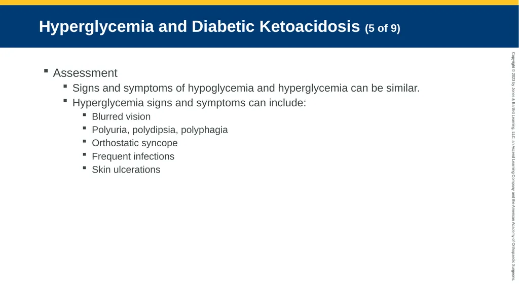
Hyperglycemia and Diabetic Ketoacidosis (5 of 9) Assessment
Signs and symptoms of hypoglycemia and hyperglycemia can be similar.
Hyperglycemia signs and symptoms can include:
Blurred vision
Polyuria, polydipsia, polyphagia
Orthostatic syncope
Frequent infections
Skin ulcerations<br>
Signs and symptoms of hypoglycemia and hyperglycemia can be similar.
Hyperglycemia signs and symptoms can include:
Blurred vision
Polyuria, polydipsia, polyphagia
Orthostatic syncope
Frequent infections
Skin ulcerations<br>
64
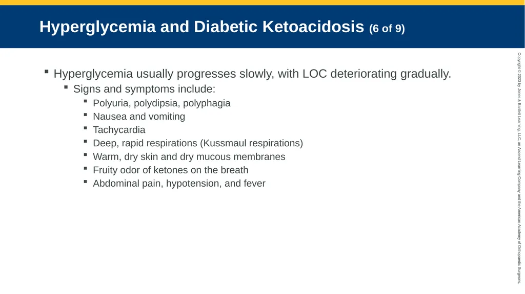
Hyperglycemia and Diabetic Ketoacidosis (6 of 9) Hyperglycemia usually progresses slowly, with LOC deteriorating gradually.
Signs and symptoms include:
Polyuria, polydipsia, polyphagia
Nausea and vomiting
Tachycardia
Deep, rapid respirations (Kussmaul respirations)
Warm, dry skin and dry mucous membranes
Fruity odor of ketones on the breath
Abdominal pain, hypotension, and fever<br>
Signs and symptoms include:
Polyuria, polydipsia, polyphagia
Nausea and vomiting
Tachycardia
Deep, rapid respirations (Kussmaul respirations)
Warm, dry skin and dry mucous membranes
Fruity odor of ketones on the breath
Abdominal pain, hypotension, and fever<br>
65
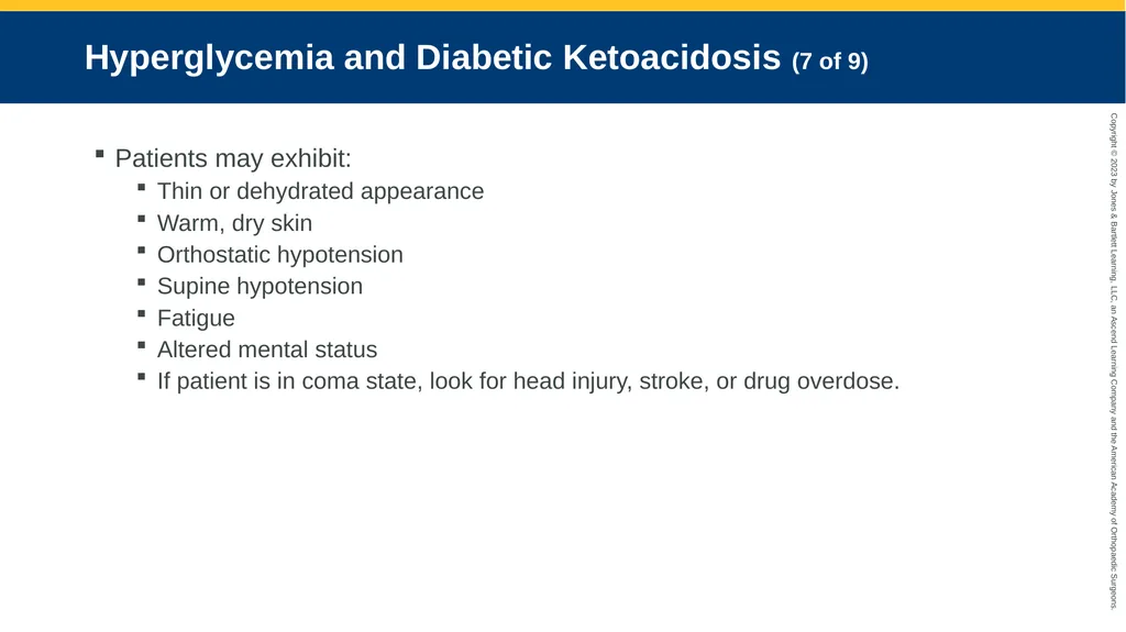
Hyperglycemia and Diabetic Ketoacidosis (7 of 9) Patients may exhibit:
Thin or dehydrated appearance
Warm, dry skin
Orthostatic hypotension
Supine hypotension
Fatigue
Altered mental status
If patient is in coma state, look for head injury, stroke, or drug overdose.<br>
Thin or dehydrated appearance
Warm, dry skin
Orthostatic hypotension
Supine hypotension
Fatigue
Altered mental status
If patient is in coma state, look for head injury, stroke, or drug overdose.<br>
66
Hyperglycemia and Diabetic Ketoacidosis (8 of 9) © Jones & Bartlett Learning.<br>
67
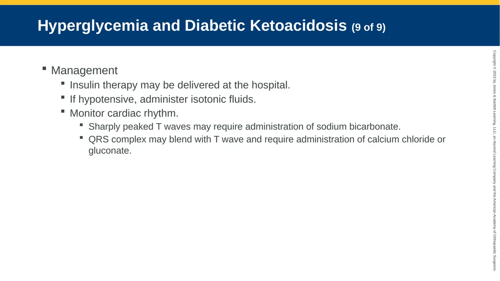
Hyperglycemia and Diabetic Ketoacidosis (9 of 9) Management
Insulin therapy may be delivered at the hospital.
If hypotensive, administer isotonic fluids.
Monitor cardiac rhythm.
Sharply peaked T waves may require administration of sodium bicarbonate.
QRS complex may blend with T wave and require administration of calcium chloride or gluconate.<br>
Insulin therapy may be delivered at the hospital.
If hypotensive, administer isotonic fluids.
Monitor cardiac rhythm.
Sharply peaked T waves may require administration of sodium bicarbonate.
QRS complex may blend with T wave and require administration of calcium chloride or gluconate.<br>
68
Hyperosmolar Hyperglycemic Nonketotic Syndrome (HHNS) (1 of 5) Pathophysiology
Formerly called hyperosmolar nonketotic coma (HONK)
Occurs primarily with type 2 diabetes
Characterized by:
Hyperglycemia
Altered mental status, drowsiness, and lethargy
Visual or sensory deficit
Partial paralysis or muscle weakness<br>
Formerly called hyperosmolar nonketotic coma (HONK)
Occurs primarily with type 2 diabetes
Characterized by:
Hyperglycemia
Altered mental status, drowsiness, and lethargy
Visual or sensory deficit
Partial paralysis or muscle weakness<br>
69
Hyperosmolar Hyperglycemic Nonketotic Syndrome (HHNS) (2 of 5) Pathophysiology (cont’d)
Few patients present in comatose state.
Most have severe dehydration and focal or global neurologic deficits.
Clinical features of HHNS and DKA overlap.
Often develops in patients with diabetes who have a secondary illness that leads to reduced fluid intake<br>
Few patients present in comatose state.
Most have severe dehydration and focal or global neurologic deficits.
Clinical features of HHNS and DKA overlap.
Often develops in patients with diabetes who have a secondary illness that leads to reduced fluid intake<br>
70
Hyperosmolar Hyperglycemic Nonketotic Syndrome (HHNS) (3 of 5) Assessment
Patients do not experience ketoacidosis.
Onset can take several weeks.
Most have a history of diabetes.
Blood glucose levels substantially higher
Neurologic changes are possible:
Drowsiness and lethargy
Delirium and coma
Focal or generalized seizures<br>
Patients do not experience ketoacidosis.
Onset can take several weeks.
Most have a history of diabetes.
Blood glucose levels substantially higher
Neurologic changes are possible:
Drowsiness and lethargy
Delirium and coma
Focal or generalized seizures<br>
71
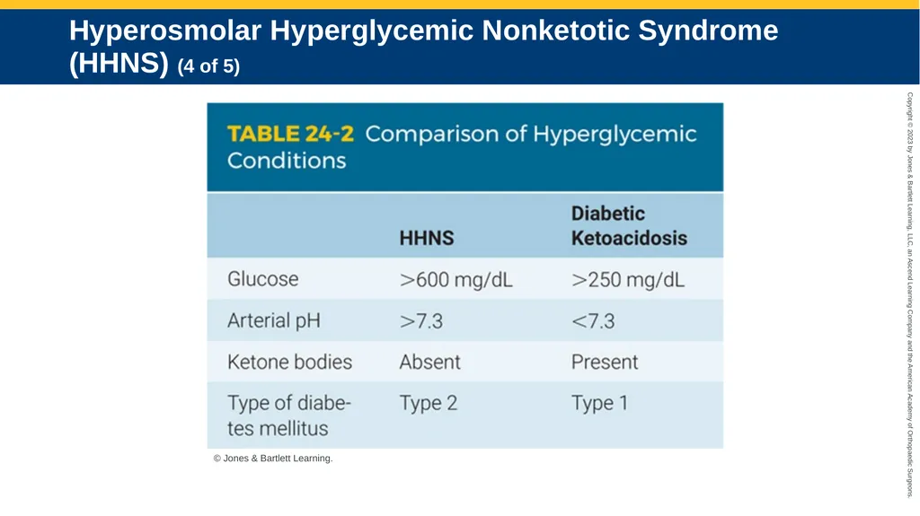
Hyperosmolar Hyperglycemic Nonketotic Syndrome (HHNS) (4 of 5) © Jones & Bartlett Learning.<br>
72
Hyperosmolar Hyperglycemic Nonketotic Syndrome (HHNS) (5 of 5) Management
Address dehydration and altered mental status.
Manage airway.
A bolus of NS is appropriate for nearly all who are clinically dehydrated.<br>
Address dehydration and altered mental status.
Manage airway.
A bolus of NS is appropriate for nearly all who are clinically dehydrated.<br>
73
Pancreatitis (1 of 3) Pathophysiology
Inflammation of the pancreas
Acute form is a medical emergency.
Chronic form is a progressive disease.<br>
Inflammation of the pancreas
Acute form is a medical emergency.
Chronic form is a progressive disease.<br>
74
Pancreatitis (2 of 3) Assessment
Patient may present with:
Flank and/or epigastric pain
Tachycardia
Nausea and vomiting
Abdominal distention
Organ failure may develop.<br>
Patient may present with:
Flank and/or epigastric pain
Tachycardia
Nausea and vomiting
Abdominal distention
Organ failure may develop.<br>
75
Pancreatitis (3 of 3) Management
Most are managed with supportive care.
Transport patients.
Pain management may be considered.
Lifestyle changes are critical for chronic pancreatitis.<br>
Most are managed with supportive care.
Transport patients.
Pain management may be considered.
Lifestyle changes are critical for chronic pancreatitis.<br>
76
Adrenal Insufficiency Decreased function of the adrenal cortex
Underproduction of cortisol and aldosterone
Results in:
Weakness, dehydration, inability to maintain blood pressure
Usually well tolerated<br>
Underproduction of cortisol and aldosterone
Results in:
Weakness, dehydration, inability to maintain blood pressure
Usually well tolerated<br>
77
Primary Adrenal Insufficiency (Addison Disease) (1 of 4) Pathophysiology
Both adrenal glands atrophied or destroyed
Rare disease
Usually result of idiopathic atrophy
Occurs when 90% of the adrenal cortex has been destroyed<br>
Both adrenal glands atrophied or destroyed
Rare disease
Usually result of idiopathic atrophy
Occurs when 90% of the adrenal cortex has been destroyed<br>
78
Primary Adrenal Insufficiency (Addison Disease) (2 of 4) Assessment
Signs of chronic disease include:
Fatigue
Anorexia
Salt craving
Muscle, joint pain
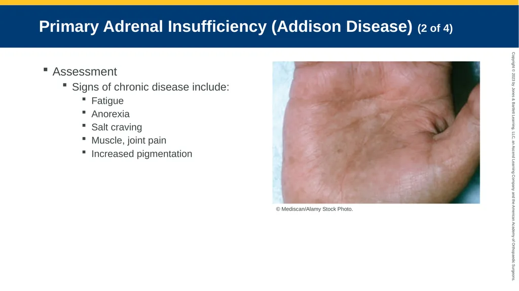
Increased pigmentation © Mediscan/Alamy Stock Photo.<br>
Signs of chronic disease include:
Fatigue
Anorexia
Salt craving
Muscle, joint pain
Increased pigmentation © Mediscan/Alamy Stock Photo.<br>
79
Primary Adrenal Insufficiency (Addison Disease) (3 of 4) Assessment (cont’d)
Body improperly regulates sodium, potassium, and water in body fluids.
Blood volume and pressure fall.
Sodium concentration of the blood falls.
Blood potassium rises.<br>
Body improperly regulates sodium, potassium, and water in body fluids.
Blood volume and pressure fall.
Sodium concentration of the blood falls.
Blood potassium rises.<br>
80
Primary Adrenal Insufficiency (Addison Disease) (4 of 4) Management
Assess and manage ABCs.
Initiate aggressive fluid replacement.
Hydrocortisone is indicated in the acute management of a crisis.
Consider electrolyte imbalances.<br>
Assess and manage ABCs.
Initiate aggressive fluid replacement.
Hydrocortisone is indicated in the acute management of a crisis.
Consider electrolyte imbalances.<br>
81
Secondary Adrenal Insufficiency (1 of 3) Pathophysiology
Characterized by a lack of ACTH secretion from pituitary gland
May result if a patient abruptly stops taking corticosteroids<br>
Characterized by a lack of ACTH secretion from pituitary gland
May result if a patient abruptly stops taking corticosteroids<br>
82
Secondary Adrenal Insufficiency (2 of 3) Assessment
May appear suddenly (addisonian crisis)
Chief manifestation is shock.
Symptoms may also include:
Weakness/lethargy
Confusion
Low blood pressure
Severe pain and/or vomiting<br>
May appear suddenly (addisonian crisis)
Chief manifestation is shock.
Symptoms may also include:
Weakness/lethargy
Confusion
Low blood pressure
Severe pain and/or vomiting<br>
83
Secondary Adrenal Insufficiency (3 of 3) Management
Maintain ABCs and have suction ready.
Rehydrate and correct abnormalities.
Check glucose level and cardiac rhythm.<br>
Maintain ABCs and have suction ready.
Rehydrate and correct abnormalities.
Check glucose level and cardiac rhythm.<br>
84
Cushing Syndrome (1 of 3) Pathophysiology
Caused by excess cortisol production or use of corticosteroid hormones
Characteristic changes:
Blood glucose level rises.
Protein synthesis is impaired.
Bones become weaker.<br>
Caused by excess cortisol production or use of corticosteroid hormones
Characteristic changes:
Blood glucose level rises.
Protein synthesis is impaired.
Bones become weaker.<br>
85
Cushing Syndrome (2 of 3) Assessment
Signs and symptoms include:
Weakness and fatigue
Increased thirst and urination
High blood glucose
Weight gain
Thinning and/or darkening of the skin<br>
Signs and symptoms include:
Weakness and fatigue
Increased thirst and urination
High blood glucose
Weight gain
Thinning and/or darkening of the skin<br>
86
Cushing Syndrome (3 of 3) Management
Assess and manage ABCs.
Prehospital treatment is generally supportive.
Obtain blood glucose level.<br>
Assess and manage ABCs.
Prehospital treatment is generally supportive.
Obtain blood glucose level.<br>
87
Adrenal Gland Tumor Pheochromocytoma
Tumor in adrenal gland, usually in the medulla
Causes excessive release of hormones
Combination of symptoms is common.
Frequent but sporadic
May increase in frequency, duration, and severity<br>
Tumor in adrenal gland, usually in the medulla
Causes excessive release of hormones
Combination of symptoms is common.
Frequent but sporadic
May increase in frequency, duration, and severity<br>
88
Growth Hormone Pathologies Anterior pituitary gland secretes growth hormone.
Problems involve oversecretion or undersecretion.
Oversecretion results in acromegaly.
Undersecretion is characterized by delayed development and growth.<br>
Problems involve oversecretion or undersecretion.
Oversecretion results in acromegaly.
Undersecretion is characterized by delayed development and growth.<br>
89
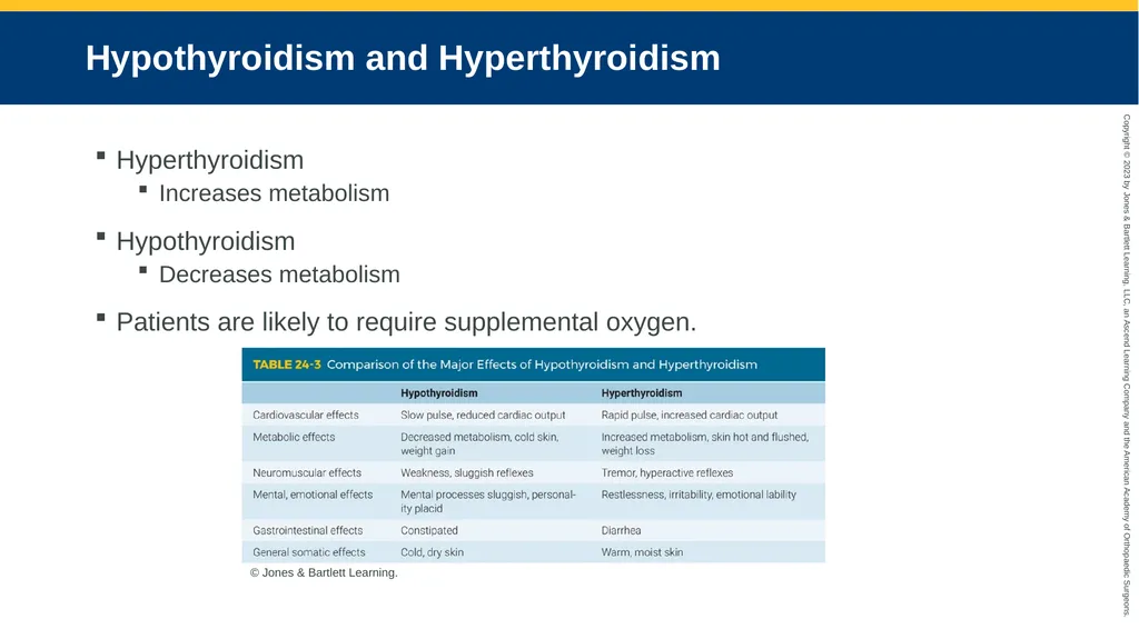
Hypothyroidism and Hyperthyroidism Hyperthyroidism
Increases metabolism
Hypothyroidism
Decreases metabolism
Patients are likely to require supplemental oxygen. © Jones & Bartlett Learning.<br>
Increases metabolism
Hypothyroidism
Decreases metabolism
Patients are likely to require supplemental oxygen. © Jones & Bartlett Learning.<br>
90
Graves Disease Most common cause of hyperthyroidism
10 times more common in women
Autoimmune disorder in which the thyroid gland hypertrophies as its activity increases
Produces a visible mass (goiter) in the neck
Excessive amount of thyroxine is secreted<br>
10 times more common in women
Autoimmune disorder in which the thyroid gland hypertrophies as its activity increases
Produces a visible mass (goiter) in the neck
Excessive amount of thyroxine is secreted<br>
91
Hashimoto Disease Cause of hyperthyroidism
Result of the infiltration of T lymphocytes and plasma cells
Autoimmune disorder that affects TSH receptors
Antibodies destroy follicles and cause hypothyroidism.<br>
Result of the infiltration of T lymphocytes and plasma cells
Autoimmune disorder that affects TSH receptors
Antibodies destroy follicles and cause hypothyroidism.<br>
92
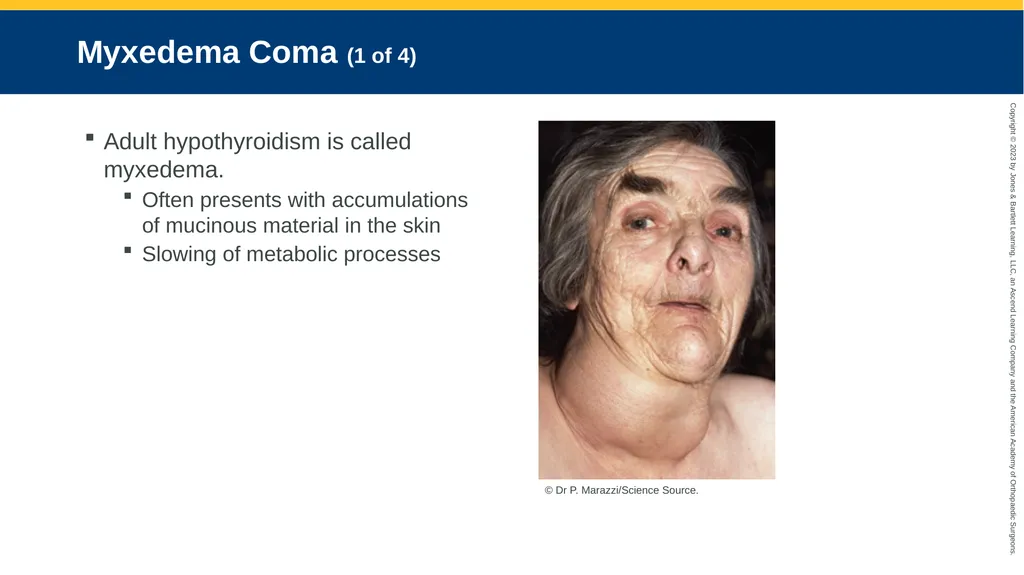
Myxedema Coma (1 of 4) Adult hypothyroidism is called myxedema.
Often presents with accumulations of mucinous material in the skin
Slowing of metabolic processes © Dr P. Marazzi/Science Source.<br>
Often presents with accumulations of mucinous material in the skin
Slowing of metabolic processes © Dr P. Marazzi/Science Source.<br>
93
Myxedema Coma (2 of 4) Symptoms
Severity is consistent with degree of deficiency.
May include:
Fatigue
Feeling cold
Weight gain
Dry skin
Sleepiness<br>
Severity is consistent with degree of deficiency.
May include:
Fatigue
Feeling cold
Weight gain
Dry skin
Sleepiness<br>
94
Myxedema Coma (3 of 4) Dropping hormone levels may lead to myxedema coma.
Often precipitated by triggers
Hallmark is deterioration of mental status.
Consistent finding is hypothermia.
Hypothyroidism decreases intestinal motility.<br>
Often precipitated by triggers
Hallmark is deterioration of mental status.
Consistent finding is hypothermia.
Hypothyroidism decreases intestinal motility.<br>
95
Myxedema Coma (4 of 4) Management
Supplemental oxygen for hypoxia
Intubation and ventilation may be indicated.
Monitor cardiac status.
Passive rewarming for hypothermia
Avoid sedatives, narcotics, anesthetics.<br>
Supplemental oxygen for hypoxia
Intubation and ventilation may be indicated.
Monitor cardiac status.
Passive rewarming for hypothermia
Avoid sedatives, narcotics, anesthetics.<br>
96
Thyrotoxicosis Caused by excessive levels of circulating thyroid hormone
Causes may include:
Hyperthyroidism
Goiters
Autoimmune disorders
Thyroid cancer<br>
Causes may include:
Hyperthyroidism
Goiters
Autoimmune disorders
Thyroid cancer<br>
97
Thyroid Storm Rare, life threatening
Usually triggered by:
Stressful event or increased volume of thyroid hormones in the circulation
Signs and symptoms may include:
Normal signs, symptoms of hyperthyroidism
Fever
Severe tachycardia
Vomiting<br>
Usually triggered by:
Stressful event or increased volume of thyroid hormones in the circulation
Signs and symptoms may include:
Normal signs, symptoms of hyperthyroidism
Fever
Severe tachycardia
Vomiting<br>
98
Hyperparathyroidism (1 of 2) Increased parathyroid hormone level
Primary causes result from the gland.
Secondary causes occur elsewhere.
Most common cause is benign tumor (adenoma).<br>
Primary causes result from the gland.
Secondary causes occur elsewhere.
Most common cause is benign tumor (adenoma).<br>
99
Hyperparathyroidism (2 of 2) Signs and symptoms
Can be vague
May include fatigue, weakness, vomiting
Definitive management is surgery to remove gland.
Manage ABCs; provide supportive care.<br>
Can be vague
May include fatigue, weakness, vomiting
Definitive management is surgery to remove gland.
Manage ABCs; provide supportive care.<br>
100
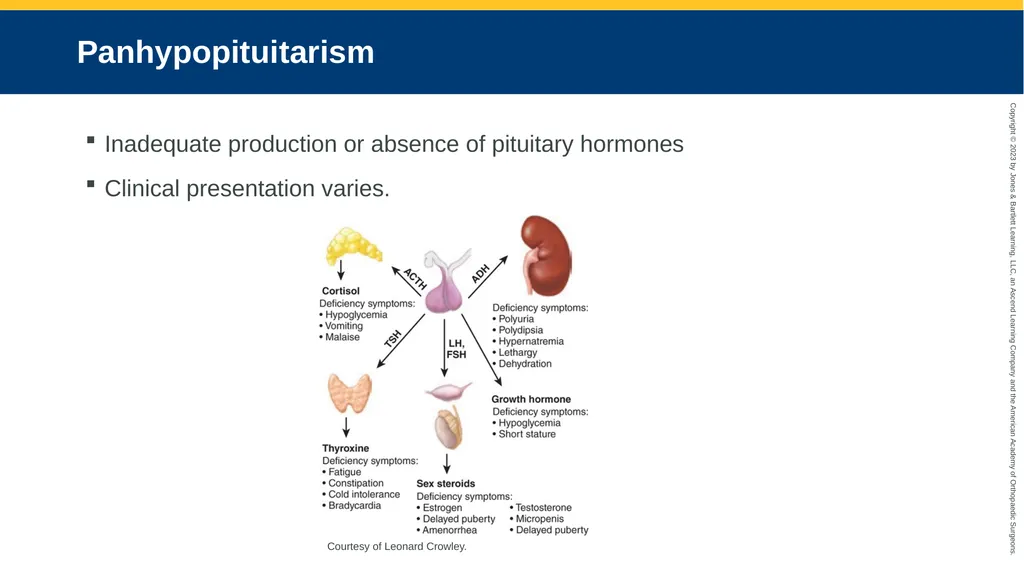
Panhypopituitarism Inadequate production or absence of pituitary hormones
Clinical presentation varies. Courtesy of Leonard Crowley.<br>
Clinical presentation varies. Courtesy of Leonard Crowley.<br>
101
Diabetes Insipidus and SIADH (1 of 2) Unable to regulate fluid due to:
Lack of ADH
Kidneys unable to respond appropriately
Management may include synthetic ADH.
In SIADH, excess of ADH results in a decrease in urinary output.
Systemic fluid overload<br>
Lack of ADH
Kidneys unable to respond appropriately
Management may include synthetic ADH.
In SIADH, excess of ADH results in a decrease in urinary output.
Systemic fluid overload<br>
102
Diabetes Insipidus and SIADH (2 of 2) © Jones & Bartlett Learning.<br>