
General Bacteriology MORPHOLOGY Gram positive


Description: General Bacteriology MORPHOLOGY Gram positive bacteria Morphology of Gram negative bacteria Different morphology of bacteria and Gram staining property Structure of Bacterial Cell Bacterial cell wall The cell wall has following functions:
Related Topics
Download Presentation
"General Bacteriology MORPHOLOGY Gram positive" is the property of its rightful owner. Permission is granted to download and print the materials on this website for personal, non-commercial use only, and to display it on your personal computer provided you do not modify the materials and that you retain all copyright notices contained in the materials. By downloading content from our website, you accept the terms of this agreement.
Presentation Transcript
slide1. General Bacteriology<br>
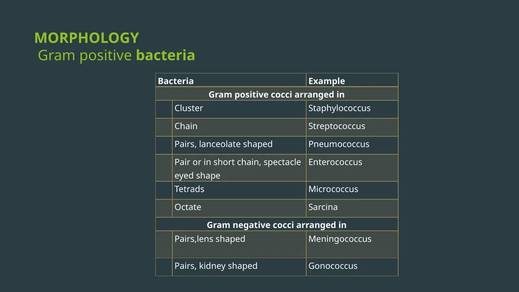
slide2. MORPHOLOGY Gram positive bacteria<br>
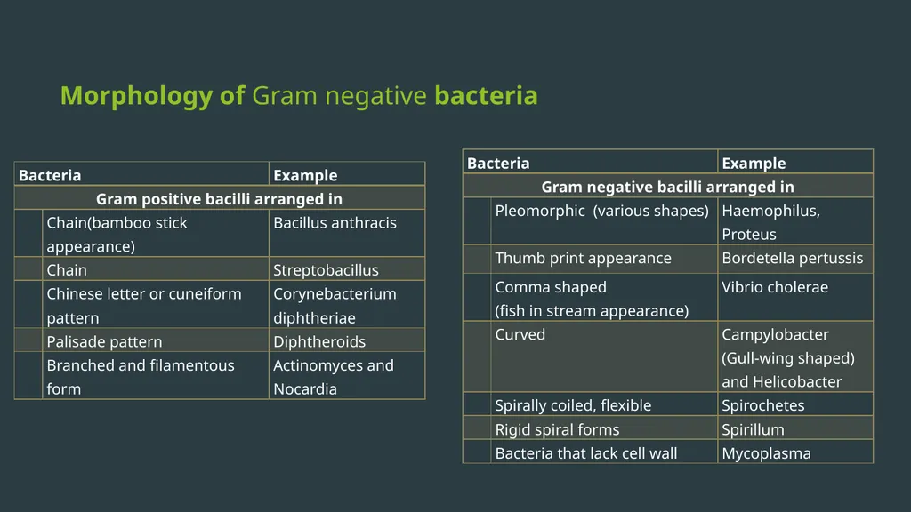
slide3. Morphology of Gram negative bacteria<br>
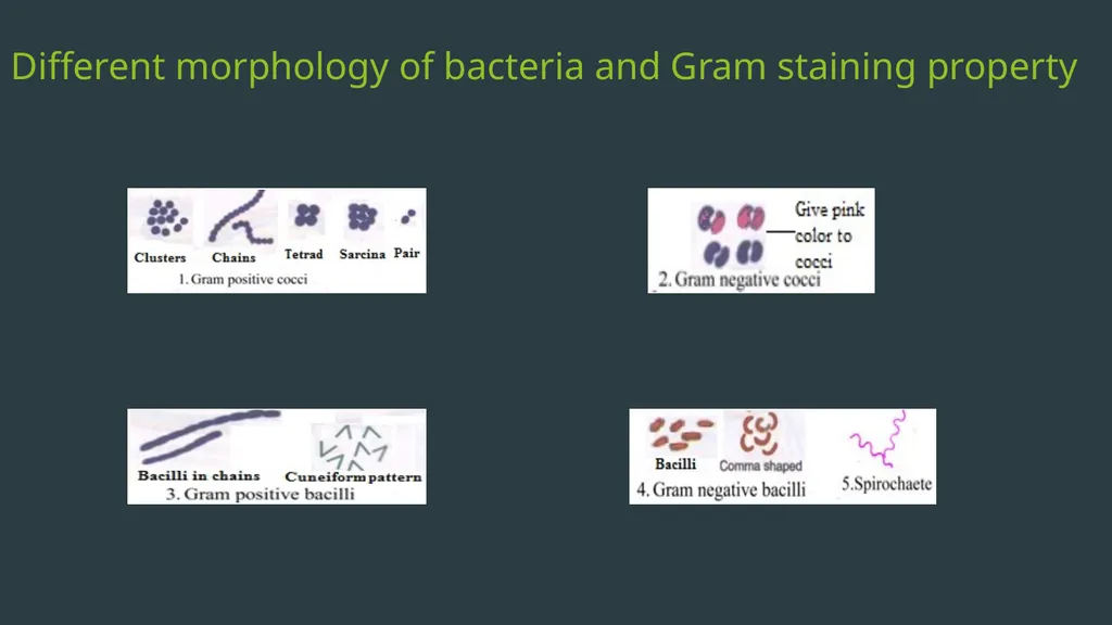
slide4. Different morphology of bacteria and Gram staining property<br>
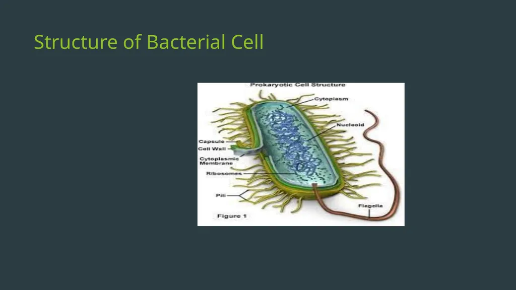
slide5. Structure of Bacterial Cell<br>
slide6. Bacterial cell wall The cell wall has following functions:
Provides protection to the cell against osmotic lysis.
Confers rigidity
Accounts for the shape of the cell.
Takes part in cell division.
Protect a cell from toxic substances and is the site of action of several antibiotics.
Virulence factors-Bacterial cell wall contains certain virulence factors (e.g. endotoxin), which contribute to their pathogenicity
Immunity: Antibody raised against specific cell wall antigens (e.g. antibody to LPS) may provide immunity against some bacterial infection.<br>
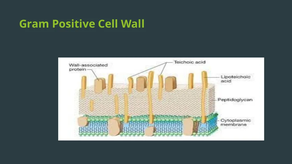
slide7. Gram Positive Cell Wall<br>
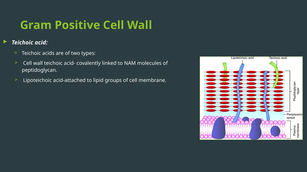
slide8. Gram Positive Cell Wall Teichoic acid:
Teichoic acids are of two types:
Cell wall teichoic acid- covalently linked to NAM molecules of peptidoglycan.
Lipoteichoic acid-attached to lipid groups of cell membrane.<br>
slide9. Gram-negative cell wall Peptidoglycan layer :
Very thin (1-2 layer, 2nm thick).
Composed of a mucopeptide chain similar to that of Gram positive cell wall, and consists of alternate NAM and NAG molecules.<br>
slide10. Gram-negative cell wall Outer membrane:
Phospholipid layer which lies outside the thin peptidoglycan layer.
Serves as a protective barrier to the cell.
Outer membrane proteins (OMP) or porin proteins<br>
slide11. Gram-negative cell wall Lipopolysaccharide (LPS): consists of:
Lipid A or the endotoxin
Core polysaccharide
O side chain (or O antigen)
Periplasmic space: It is the space between the inner cell membrane and outer membrane. It encompasses the peptidoglycan layer<br>
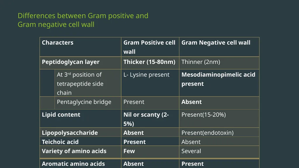
slide12. Differences between Gram positive and Gram negative cell wall<br>
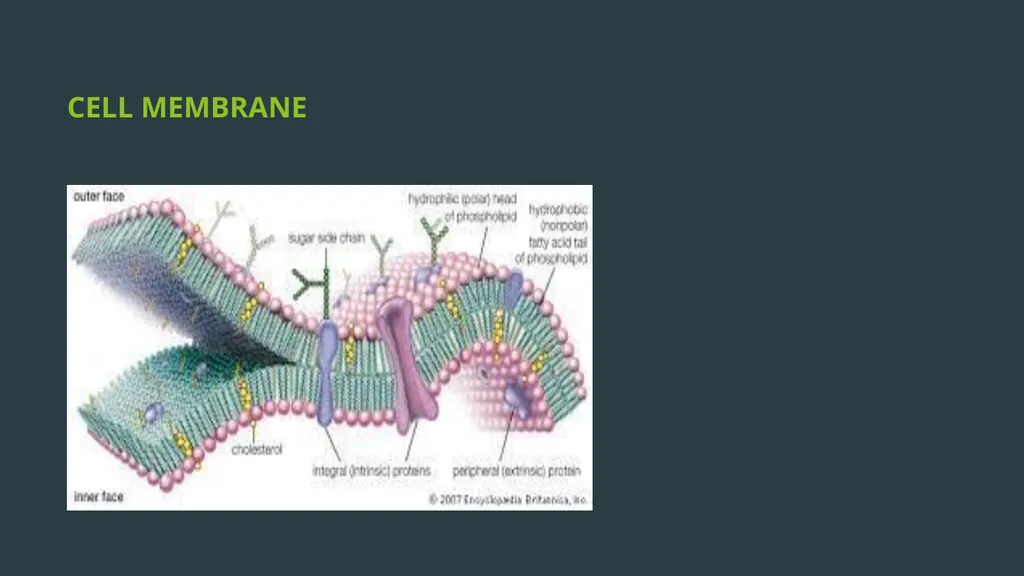
slide13. CELL MEMBRANE<br>
slide14. Functions of cell membrane Semi permeable membrane - osmotic barrier
Transport system
Site for metabolic processes
Special receptor molecules located in the membrane help the bacteria to detect and respond to chemicals in their surroundings.<br>
slide15. CYTOPLASMIC MATRIX Bacterial cytoplasm, lacks membrane-bound organelles.
Composed of water and is packed with ribosomes, storage granules called inclusions and cell membrane invaginations called mesosomes.
Lack true cytoskeleton, but do have a cytoskeleton-like system of proteins.
The plasma membrane and everything within it is called as the protoplast.<br>
slide16. Ribosomes Ribosomes are the sites for protein synthesis.
Composed of ribosomal RNA (rRNA) and ribosomal proteins.
Ribosomes are integrated in linear strands of mRNA to form polysomes.
At this site, the genetic codons of the mRNA are translated into peptide sequences.
Each 70 S unit consists of a 30 S and a 50S subunits.<br>
slide17. Intracytoplasmic inclusions Storage sites of nutrients/ energy present in some bacteria.
Formed by the bacteria under nutritional deficiency conditions
There are two types of inclusions-
Organic inclusion bodies
Inorganic inclusion bodies<br>
slide18. Mesosomes Mesosomes are invaginations of the plasma membrane in the shape of vesicles, tubules, or lamellae
Mesosomes often are found next to septa in dividing bacteria or sometimes seen attached to the bacterial chromosome.
Involved in-
Site of bacterial respiration
Cell wall formation during division
Chromosome replication and distribution to daughter cells.<br>
slide19. NUCLEOID Bacteria possess a single haploid chromosome
Comprises of super coiled circular double stranded DNA
Bacterial DNA lacks basic proteins.
Divides by simple binary fission.
Nucleoid demonstrated by electron microscopy or staining with the Feulgen stain
Bacteria possess extra-chromosomal DNA called plasmids.<br>
slide20. CELL WALL APPENDAGES Capsule and slime layer
S layer
Flagella
Fimbriae or pili
Spore<br>
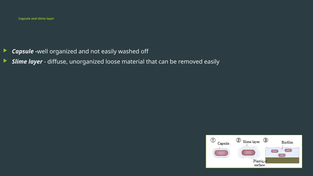
slide21. Capsule and slime layer Capsule -well organized and not easily washed off
Slime layer - diffuse, unorganized loose material that can be removed easily<br>
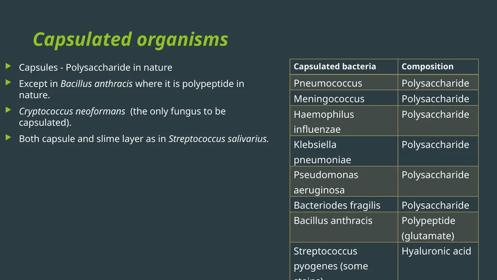
slide22. Capsulated organisms Capsules - Polysaccharide in nature
Except in Bacillus anthracis where it is polypeptide in nature.
Cryptococcus neoformans (the only fungus to be capsulated).
Both capsule and slime layer as in Streptococcus salivarius.<br>
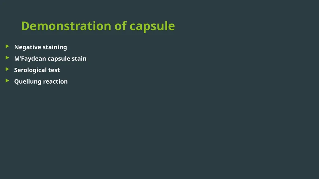
slide23. Demonstration of capsule Negative staining
M’Faydean capsule stain
Serological test
Quellung reaction<br>
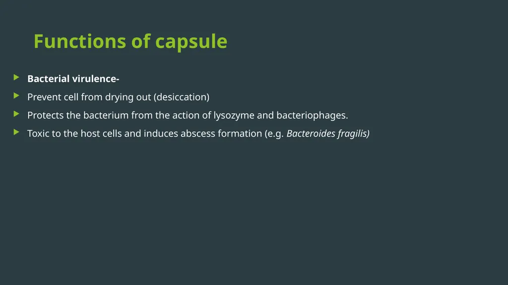
slide24. Functions of capsule Bacterial virulence-
Prevent cell from drying out (desiccation)
Protects the bacterium from the action of lysozyme and bacteriophages.
Toxic to the host cells and induces abscess formation (e.g. Bacteroides fragilis)<br>
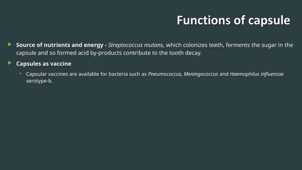
slide25. Source of nutrients and energy - Streptococcus mutans, which colonizes teeth, ferments the sugar in the capsule and so formed acid by-products contribute to the tooth decay.
Capsules as vaccine
Capsular vaccines are available for bacteria such as Pneumococcus, Meningococcus and Haemophilus influenzae serotype-b.<br>
slide26. Biofilm formation and adhesion Made of millions of adherent bacterial cells embedded within a self-produced matrix of extracellular polymeric substance
Capable of adherence to damaged tissues and plastic surfaces
Adhesion is a first step in colonization and sometimes leads to disease<br>
slide27. S layer Protect the cell against ion and pH fluctuations, osmotic stress, enzymes, and also helps to maintain the shape and envelope rigidity.
Prevents bacteria from phagocytosis and cell adhesion.<br>
slide28. Flagella Thread-like appendages, protruding from the cell wall, composed of protein subunits called flagellin.
Organs of locomotion, confer motility to the bacteria.<br>
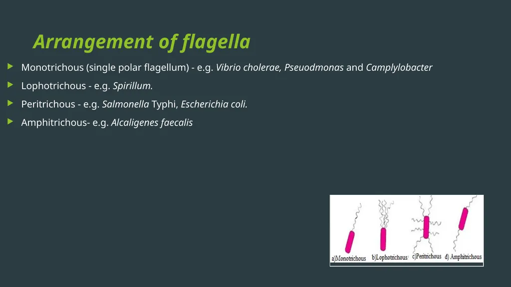
slide29. Arrangement of flagella Monotrichous (single polar flagellum) - e.g. Vibrio cholerae, Pseuodmonas and Camplylobacter
Lophotrichous - e.g. Spirillum.
Peritrichous - e.g. Salmonella Typhi, Escherichia coli.
Amphitrichous- e.g. Alcaligenes faecalis<br>
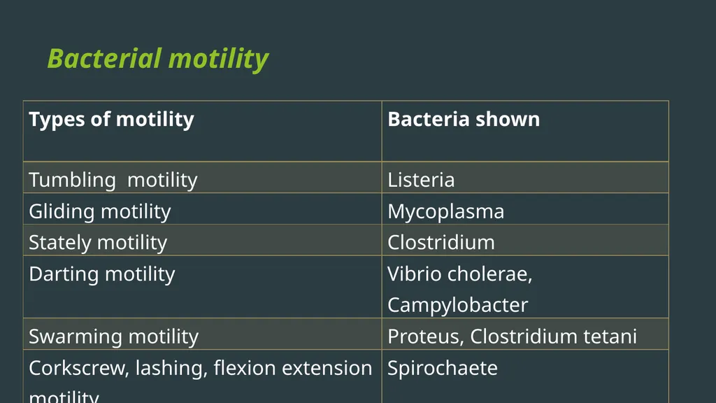
slide30. Bacterial motility<br>
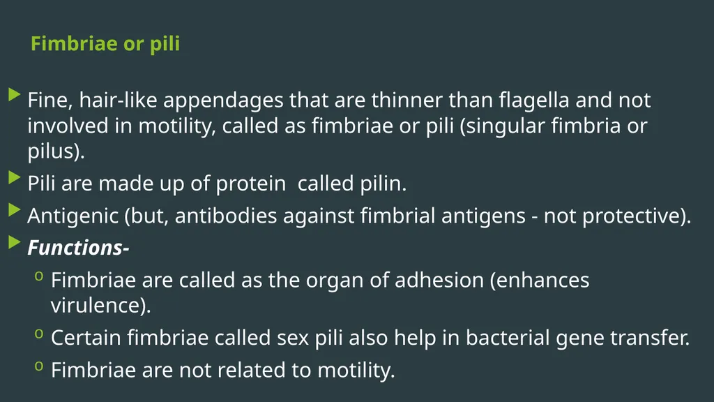
slide31. Fimbriae or pili Fine, hair-like appendages that are thinner than flagella and not involved in motility, called as fimbriae or pili (singular fimbria or pilus).
Pili are made up of protein called pilin.
Antigenic (but, antibodies against fimbrial antigens - not protective).
Functions-
Fimbriae are called as the organ of adhesion (enhances virulence).
Certain fimbriae called sex pili also help in bacterial gene transfer.
Fimbriae are not related to motility.<br>
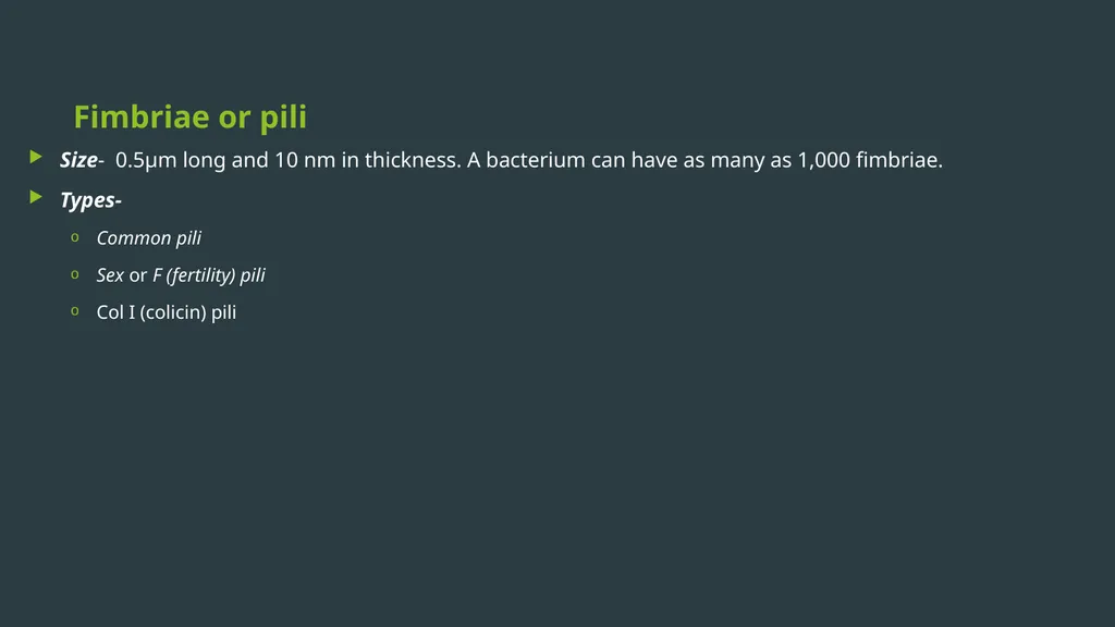
slide32. Fimbriae or pili Size- 0.5µm long and 10 nm in thickness. A bacterium can have as many as 1,000 fimbriae.
Types-
Common pili
Sex or F (fertility) pili
Col I (colicin) pili<br>
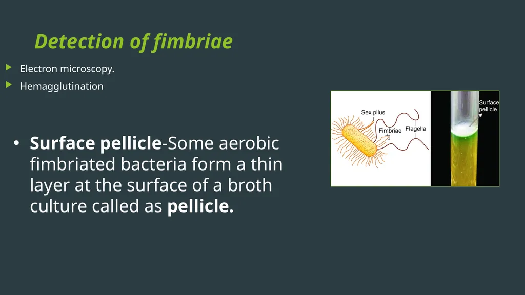
slide33. Detection of fimbriae Electron microscopy.
Hemagglutination Surface pellicle-Some aerobic fimbriated bacteria form a thin layer at the surface of a broth culture called as pellicle.<br>
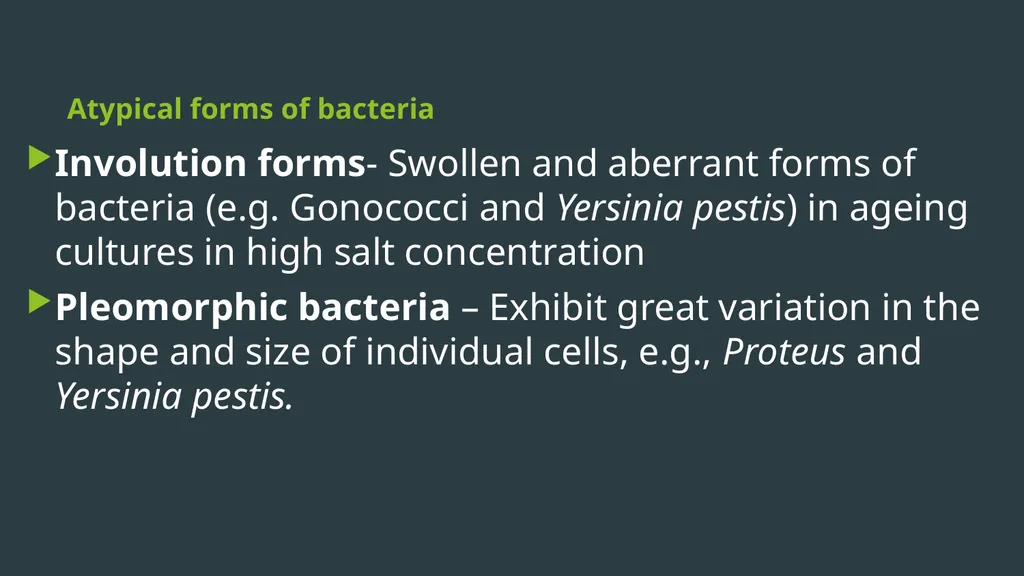
slide34. Atypical forms of bacteria Involution forms- Swollen and aberrant forms of bacteria (e.g. Gonococci and Yersinia pestis) in ageing cultures in high salt concentration
Pleomorphic bacteria – Exhibit great variation in the shape and size of individual cells, e.g., Proteus and Yersinia pestis.<br>
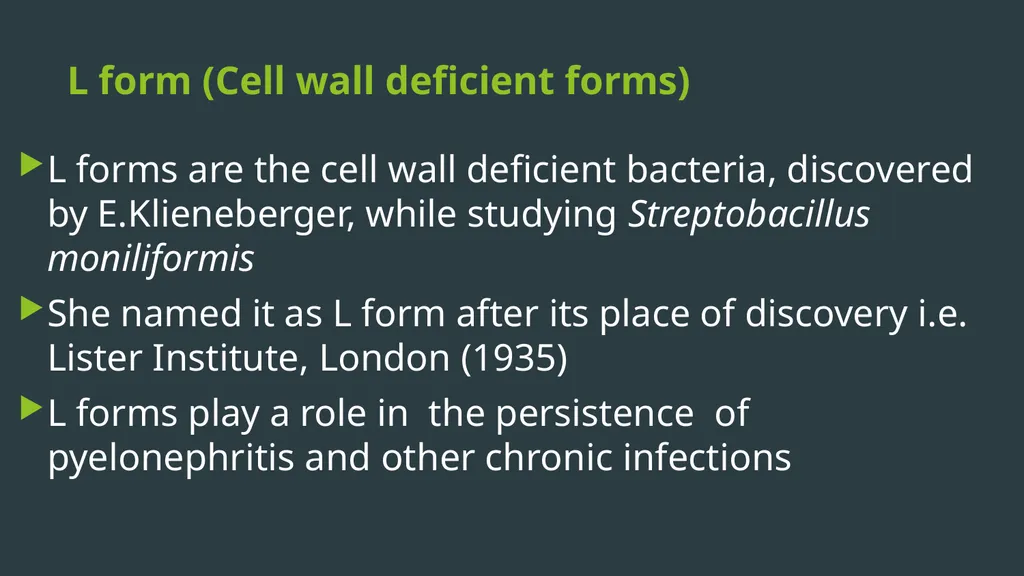
slide35. L form (Cell wall deficient forms) L forms are the cell wall deficient bacteria, discovered by E.Klieneberger, while studying Streptobacillus moniliformis
She named it as L form after its place of discovery i.e. Lister Institute, London (1935)
L forms play a role in the persistence of pyelonephritis and other chronic infections<br>
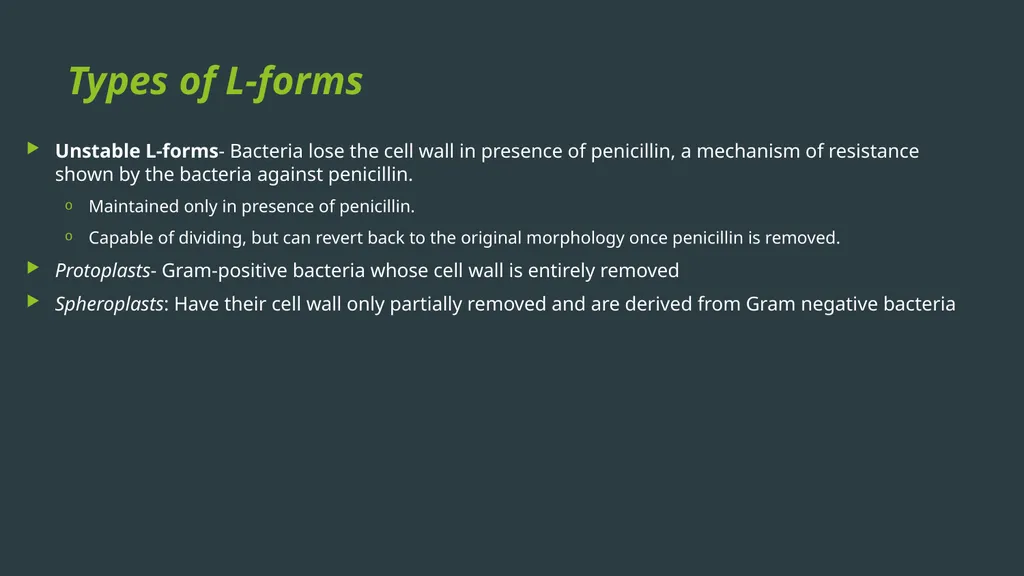
slide36. Types of L-forms Unstable L-forms- Bacteria lose the cell wall in presence of penicillin, a mechanism of resistance shown by the bacteria against penicillin.
Maintained only in presence of penicillin.
Capable of dividing, but can revert back to the original morphology once penicillin is removed.
Protoplasts- Gram-positive bacteria whose cell wall is entirely removed
Spheroplasts: Have their cell wall only partially removed and are derived from Gram negative bacteria<br>
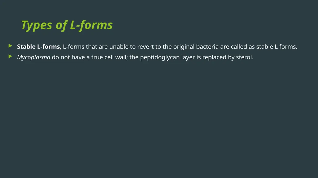
slide37. Types of L-forms Stable L-forms, L-forms that are unable to revert to the original bacteria are called as stable L forms.
Mycoplasma do not have a true cell wall; the peptidoglycan layer is replaced by sterol.<br>
slide38. REFERENCES Textbook of Medical Microbiology by Ananthnarayan, Paniker
Textbook of Medical Microbiology by C.P Baweja
Textbook of Medical Microbiology by S. Bhat, A.S.Sastry
Textbook of Medical Microbiology by D.R.Arora, Brij bala Arora<br>
slide2. MORPHOLOGY Gram positive bacteria<br>
slide3. Morphology of Gram negative bacteria<br>
slide4. Different morphology of bacteria and Gram staining property<br>
slide5. Structure of Bacterial Cell<br>
slide6. Bacterial cell wall The cell wall has following functions:
Provides protection to the cell against osmotic lysis.
Confers rigidity
Accounts for the shape of the cell.
Takes part in cell division.
Protect a cell from toxic substances and is the site of action of several antibiotics.
Virulence factors-Bacterial cell wall contains certain virulence factors (e.g. endotoxin), which contribute to their pathogenicity
Immunity: Antibody raised against specific cell wall antigens (e.g. antibody to LPS) may provide immunity against some bacterial infection.<br>
slide7. Gram Positive Cell Wall<br>
slide8. Gram Positive Cell Wall Teichoic acid:
Teichoic acids are of two types:
Cell wall teichoic acid- covalently linked to NAM molecules of peptidoglycan.
Lipoteichoic acid-attached to lipid groups of cell membrane.<br>
slide9. Gram-negative cell wall Peptidoglycan layer :
Very thin (1-2 layer, 2nm thick).
Composed of a mucopeptide chain similar to that of Gram positive cell wall, and consists of alternate NAM and NAG molecules.<br>
slide10. Gram-negative cell wall Outer membrane:
Phospholipid layer which lies outside the thin peptidoglycan layer.
Serves as a protective barrier to the cell.
Outer membrane proteins (OMP) or porin proteins<br>
slide11. Gram-negative cell wall Lipopolysaccharide (LPS): consists of:
Lipid A or the endotoxin
Core polysaccharide
O side chain (or O antigen)
Periplasmic space: It is the space between the inner cell membrane and outer membrane. It encompasses the peptidoglycan layer<br>
slide12. Differences between Gram positive and Gram negative cell wall<br>
slide13. CELL MEMBRANE<br>
slide14. Functions of cell membrane Semi permeable membrane - osmotic barrier
Transport system
Site for metabolic processes
Special receptor molecules located in the membrane help the bacteria to detect and respond to chemicals in their surroundings.<br>
slide15. CYTOPLASMIC MATRIX Bacterial cytoplasm, lacks membrane-bound organelles.
Composed of water and is packed with ribosomes, storage granules called inclusions and cell membrane invaginations called mesosomes.
Lack true cytoskeleton, but do have a cytoskeleton-like system of proteins.
The plasma membrane and everything within it is called as the protoplast.<br>
slide16. Ribosomes Ribosomes are the sites for protein synthesis.
Composed of ribosomal RNA (rRNA) and ribosomal proteins.
Ribosomes are integrated in linear strands of mRNA to form polysomes.
At this site, the genetic codons of the mRNA are translated into peptide sequences.
Each 70 S unit consists of a 30 S and a 50S subunits.<br>
slide17. Intracytoplasmic inclusions Storage sites of nutrients/ energy present in some bacteria.
Formed by the bacteria under nutritional deficiency conditions
There are two types of inclusions-
Organic inclusion bodies
Inorganic inclusion bodies<br>
slide18. Mesosomes Mesosomes are invaginations of the plasma membrane in the shape of vesicles, tubules, or lamellae
Mesosomes often are found next to septa in dividing bacteria or sometimes seen attached to the bacterial chromosome.
Involved in-
Site of bacterial respiration
Cell wall formation during division
Chromosome replication and distribution to daughter cells.<br>
slide19. NUCLEOID Bacteria possess a single haploid chromosome
Comprises of super coiled circular double stranded DNA
Bacterial DNA lacks basic proteins.
Divides by simple binary fission.
Nucleoid demonstrated by electron microscopy or staining with the Feulgen stain
Bacteria possess extra-chromosomal DNA called plasmids.<br>
slide20. CELL WALL APPENDAGES Capsule and slime layer
S layer
Flagella
Fimbriae or pili
Spore<br>
slide21. Capsule and slime layer Capsule -well organized and not easily washed off
Slime layer - diffuse, unorganized loose material that can be removed easily<br>
slide22. Capsulated organisms Capsules - Polysaccharide in nature
Except in Bacillus anthracis where it is polypeptide in nature.
Cryptococcus neoformans (the only fungus to be capsulated).
Both capsule and slime layer as in Streptococcus salivarius.<br>
slide23. Demonstration of capsule Negative staining
M’Faydean capsule stain
Serological test
Quellung reaction<br>
slide24. Functions of capsule Bacterial virulence-
Prevent cell from drying out (desiccation)
Protects the bacterium from the action of lysozyme and bacteriophages.
Toxic to the host cells and induces abscess formation (e.g. Bacteroides fragilis)<br>
slide25. Source of nutrients and energy - Streptococcus mutans, which colonizes teeth, ferments the sugar in the capsule and so formed acid by-products contribute to the tooth decay.
Capsules as vaccine
Capsular vaccines are available for bacteria such as Pneumococcus, Meningococcus and Haemophilus influenzae serotype-b.<br>
slide26. Biofilm formation and adhesion Made of millions of adherent bacterial cells embedded within a self-produced matrix of extracellular polymeric substance
Capable of adherence to damaged tissues and plastic surfaces
Adhesion is a first step in colonization and sometimes leads to disease<br>
slide27. S layer Protect the cell against ion and pH fluctuations, osmotic stress, enzymes, and also helps to maintain the shape and envelope rigidity.
Prevents bacteria from phagocytosis and cell adhesion.<br>
slide28. Flagella Thread-like appendages, protruding from the cell wall, composed of protein subunits called flagellin.
Organs of locomotion, confer motility to the bacteria.<br>
slide29. Arrangement of flagella Monotrichous (single polar flagellum) - e.g. Vibrio cholerae, Pseuodmonas and Camplylobacter
Lophotrichous - e.g. Spirillum.
Peritrichous - e.g. Salmonella Typhi, Escherichia coli.
Amphitrichous- e.g. Alcaligenes faecalis<br>
slide30. Bacterial motility<br>
slide31. Fimbriae or pili Fine, hair-like appendages that are thinner than flagella and not involved in motility, called as fimbriae or pili (singular fimbria or pilus).
Pili are made up of protein called pilin.
Antigenic (but, antibodies against fimbrial antigens - not protective).
Functions-
Fimbriae are called as the organ of adhesion (enhances virulence).
Certain fimbriae called sex pili also help in bacterial gene transfer.
Fimbriae are not related to motility.<br>
slide32. Fimbriae or pili Size- 0.5µm long and 10 nm in thickness. A bacterium can have as many as 1,000 fimbriae.
Types-
Common pili
Sex or F (fertility) pili
Col I (colicin) pili<br>
slide33. Detection of fimbriae Electron microscopy.
Hemagglutination Surface pellicle-Some aerobic fimbriated bacteria form a thin layer at the surface of a broth culture called as pellicle.<br>
slide34. Atypical forms of bacteria Involution forms- Swollen and aberrant forms of bacteria (e.g. Gonococci and Yersinia pestis) in ageing cultures in high salt concentration
Pleomorphic bacteria – Exhibit great variation in the shape and size of individual cells, e.g., Proteus and Yersinia pestis.<br>
slide35. L form (Cell wall deficient forms) L forms are the cell wall deficient bacteria, discovered by E.Klieneberger, while studying Streptobacillus moniliformis
She named it as L form after its place of discovery i.e. Lister Institute, London (1935)
L forms play a role in the persistence of pyelonephritis and other chronic infections<br>
slide36. Types of L-forms Unstable L-forms- Bacteria lose the cell wall in presence of penicillin, a mechanism of resistance shown by the bacteria against penicillin.
Maintained only in presence of penicillin.
Capable of dividing, but can revert back to the original morphology once penicillin is removed.
Protoplasts- Gram-positive bacteria whose cell wall is entirely removed
Spheroplasts: Have their cell wall only partially removed and are derived from Gram negative bacteria<br>
slide37. Types of L-forms Stable L-forms, L-forms that are unable to revert to the original bacteria are called as stable L forms.
Mycoplasma do not have a true cell wall; the peptidoglycan layer is replaced by sterol.<br>
slide38. REFERENCES Textbook of Medical Microbiology by Ananthnarayan, Paniker
Textbook of Medical Microbiology by C.P Baweja
Textbook of Medical Microbiology by S. Bhat, A.S.Sastry
Textbook of Medical Microbiology by D.R.Arora, Brij bala Arora<br>