
Laboratory tests Objectives Identify the main






Description: Laboratory tests Objectives Identify the main causes of outbreak in the work zone Know the different types of samples and diagnostic tests that can be conducted, based on the infection dynamics Review the role of the laboratory coordinated
Related Topics
Download Presentation
"Laboratory tests Objectives Identify the main" is the property of its rightful owner. Permission is granted to download and print the materials on this website for personal, non-commercial use only, and to display it on your personal computer provided you do not modify the materials and that you retain all copyright notices contained in the materials. By downloading content from our website, you accept the terms of this agreement.
Presentation Transcript
slide1. Laboratory tests<br>
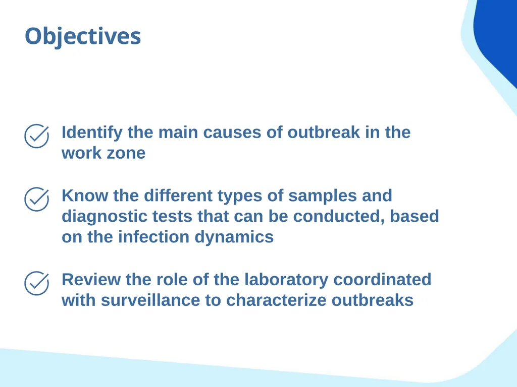
slide2. Objectives Identify the main causes of outbreak in the work zone Know the different types of samples and diagnostic tests that can be conducted, based on the infection dynamics Review the role of the laboratory coordinated with surveillance to characterize outbreaks<br>
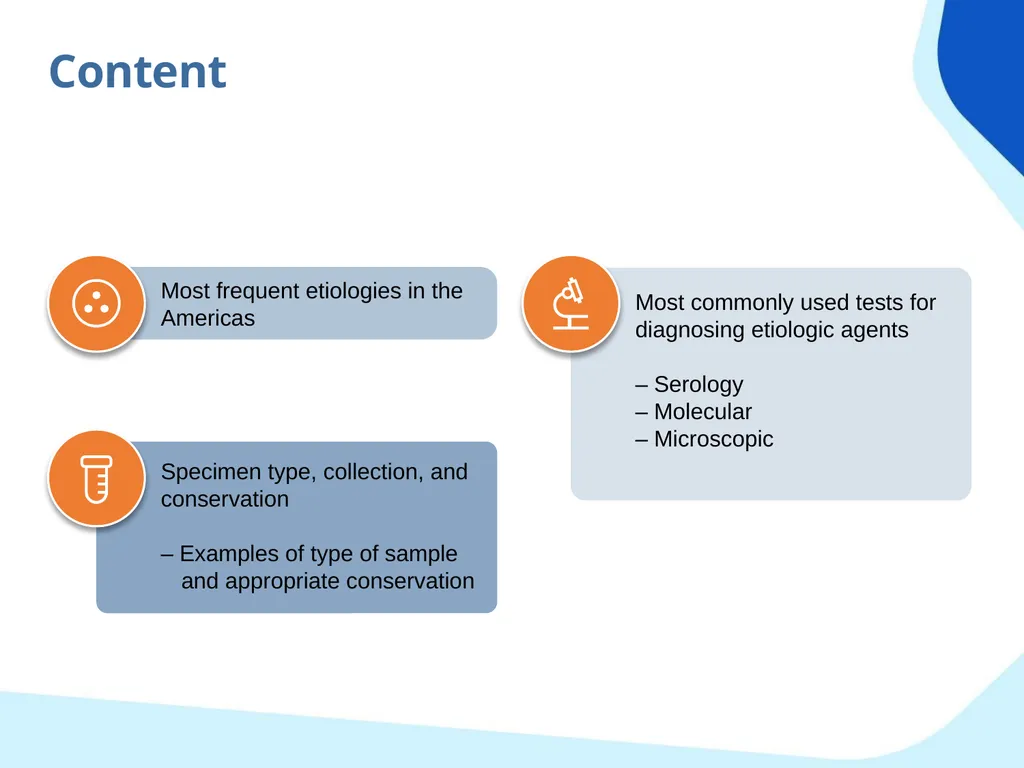
slide3. Content Most frequent etiologies in the Americas Most commonly used tests for diagnosing etiologic agents
– Serology
– Molecular
– Microscopic Specimen type, collection, and conservation
– Examples of type of sample and appropriate conservation<br>
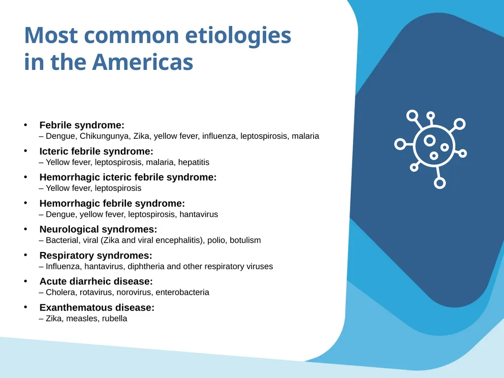
slide4. Most common etiologies
in the Americas Febrile syndrome:
– Dengue, Chikungunya, Zika, yellow fever, influenza, leptospirosis, malaria
Icteric febrile syndrome:
– Yellow fever, leptospirosis, malaria, hepatitis
Hemorrhagic icteric febrile syndrome:
– Yellow fever, leptospirosis
Hemorrhagic febrile syndrome:
– Dengue, yellow fever, leptospirosis, hantavirus
Neurological syndromes:
– Bacterial, viral (Zika and viral encephalitis), polio, botulism
Respiratory syndromes:
– Influenza, hantavirus, diphtheria and other respiratory viruses
Acute diarrheic disease:
– Cholera, rotavirus, norovirus, enterobacteria
Exanthematous disease:
– Zika, measles, rubella<br>
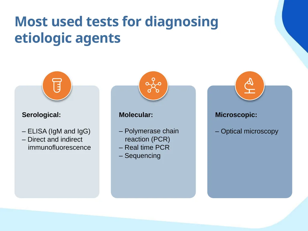
slide5. Most used tests for diagnosing etiologic agents Serological:
– ELISA (IgM and IgG)
– Direct and indirect immunofluorescence Molecular:
– Polymerase chain reaction (PCR)
– Real time PCR
– Sequencing Microscopic:
– Optical microscopy<br>
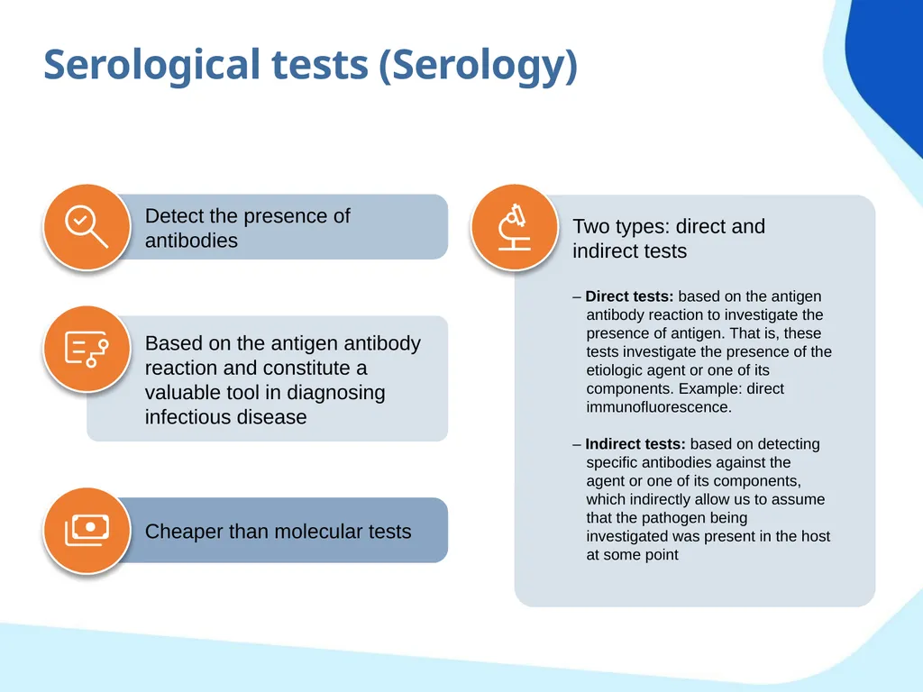
slide6. Serological tests (Serology) Two types: direct and indirect tests
– Direct tests: based on the antigen antibody reaction to investigate the presence of antigen. That is, these tests investigate the presence of the etiologic agent or one of its components. Example: direct immunofluorescence.
– Indirect tests: based on detecting specific antibodies against the agent or one of its components, which indirectly allow us to assume that the pathogen being investigated was present in the host at some point Detect the presence of antibodies Cheaper than molecular tests Based on the antigen antibody reaction and constitute a valuable tool in diagnosing infectious disease<br>
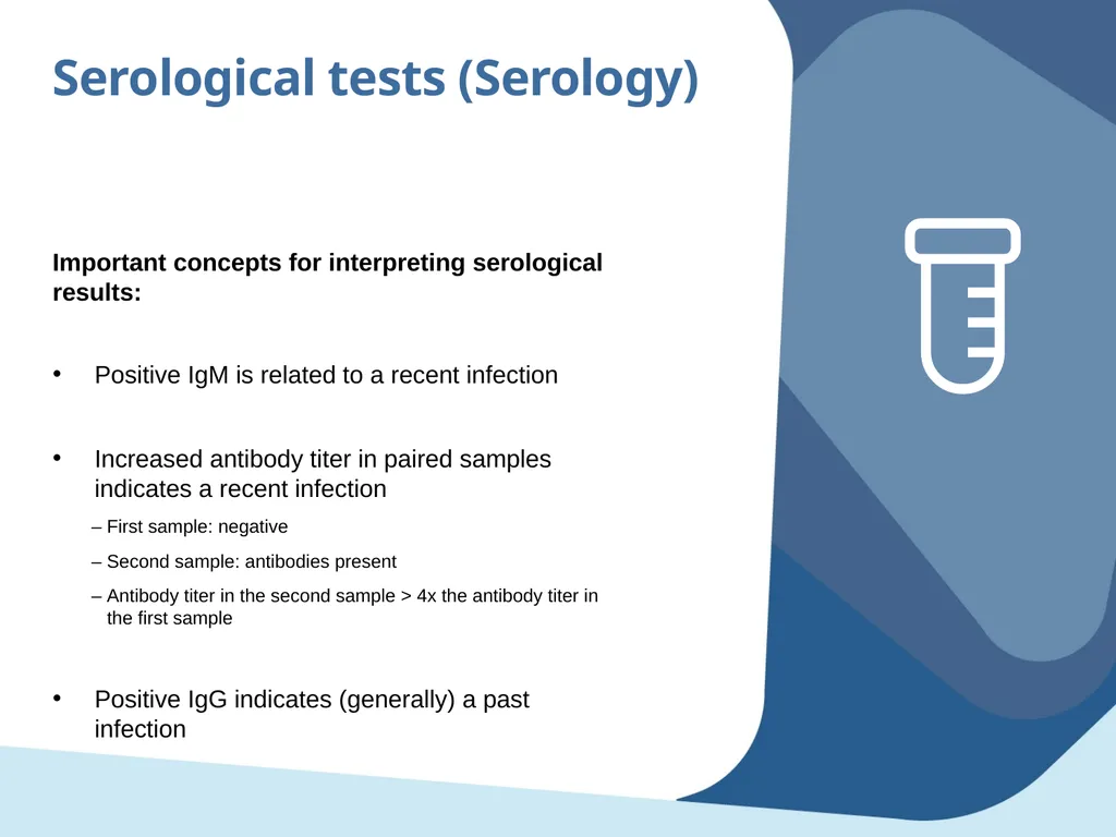
slide7. Serological tests (Serology) Important concepts for interpreting serological results: Positive IgM is related to a recent infection
Increased antibody titer in paired samples indicates a recent infection
– First sample: negative
– Second sample: antibodies present
– Antibody titer in the second sample > 4x the antibody titer in the first sample
Positive IgG indicates (generally) a past infection<br>
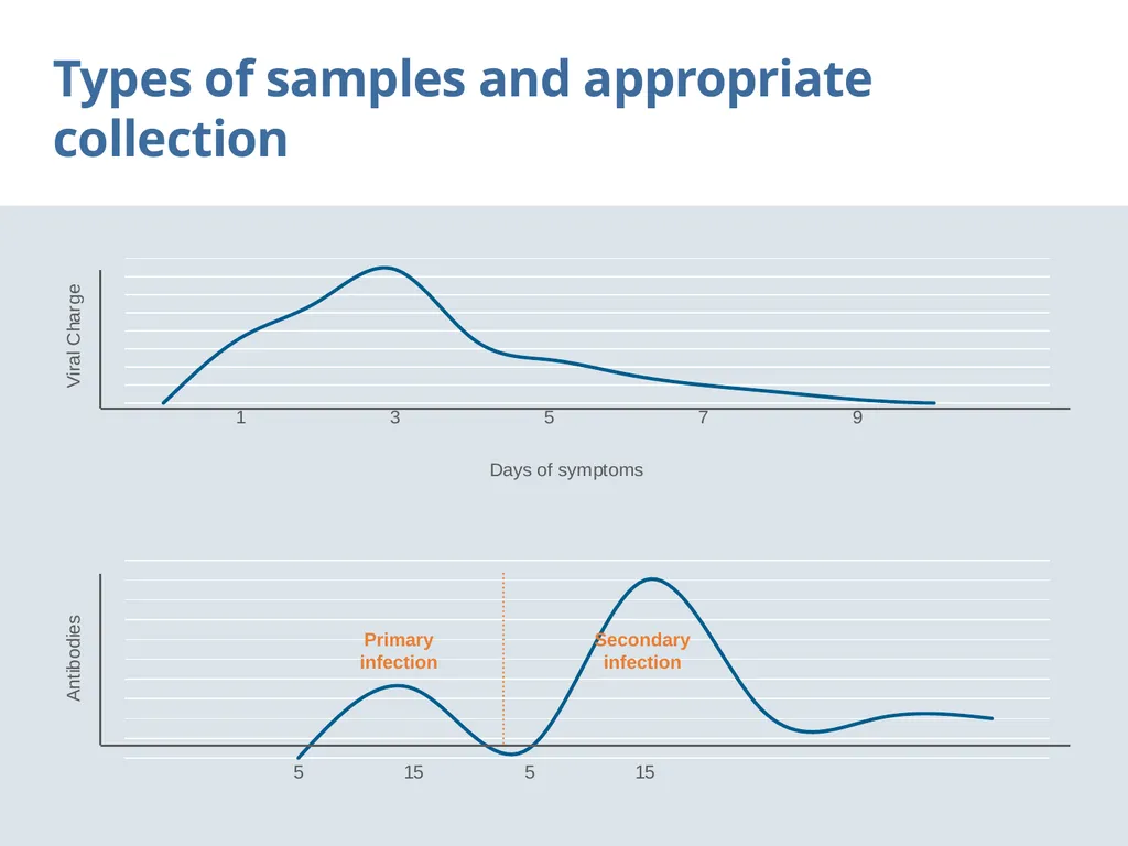
slide8. Types of samples and appropriate collection Primary infection Secondary infection<br>
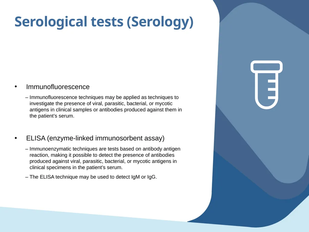
slide9. Serological tests (Serology) Immunofluorescence
– Immunofluorescence techniques may be applied as techniques to investigate the presence of viral, parasitic, bacterial, or mycotic antigens in clinical samples or antibodies produced against them in the patient’s serum.
ELISA (enzyme-linked immunosorbent assay)
– Immunoenzymatic techniques are tests based on antibody antigen reaction, making it possible to detect the presence of antibodies produced against viral, parasitic, bacterial, or mycotic antigens in clinical specimens in the patient’s serum.
– The ELISA technique may be used to detect IgM or IgG.<br>
slide10. Molecular tests Are based on detecting the etiologic agent’s genome.
Make it possible to detect the etiologic agent’s RNA or DNA with high sensitivity and specificity.
Are more costly than serology.
Make it possible to confirm the presence of the etiologic agent in clinical samples.
Make it possible to detect RNA or DNA even in very small sample volumes.<br>
slide11. Molecular tests Polymerase chain reaction (end point PCR)
– The end point PCR is based on amplifying and visualizing the complementary DNA or ADNA (cDNA). This technique consists of amplifying and visualizing a specific DNA fragment from the etiologic agent.
Real time PCR
– Also known as quantitative PCR, this test is a variant of the polymerase chain reaction (PCR).
– Real time PCR makes it possible to amplify and at the same time absolutely quantify specific DNA or cDNA molecules<br>
slide12. Microscopic Consists of using special instruments such as magnifying glasses and microscopes to magnify the size of structures that are invisible to the naked human eye, such as viruses, bacteria, and parasites.
Microscopic observation is necessary in the field of parasitology.
Modes of observation vary based on strategies employed and range from micrometric to nanometric observation.
Therefore, various types of microscopes are required, such as photonic and electronic (scanning, transmission, and atomic energy).<br>
slide13. Microscopic Optical microscopy technique allows us to visualize bacteria and parasites simply by examining the sample with a microscope.
For bacteria, a Gram stain (a purple stain) is often performed first. Bacteria is classified as follows:
– Gram positive (appear blue because they retain the Gram stain)
– Gram negative (appear red because they do not capture the stain)
A blood swab is performed to detect parasites found in the blood, such as filariasis, malaria, or babesiosis. For this test, a drop of blood is placed on a microscope slide, stained, and examined under the microscope to visualize the parasite. Optical microscopy<br>
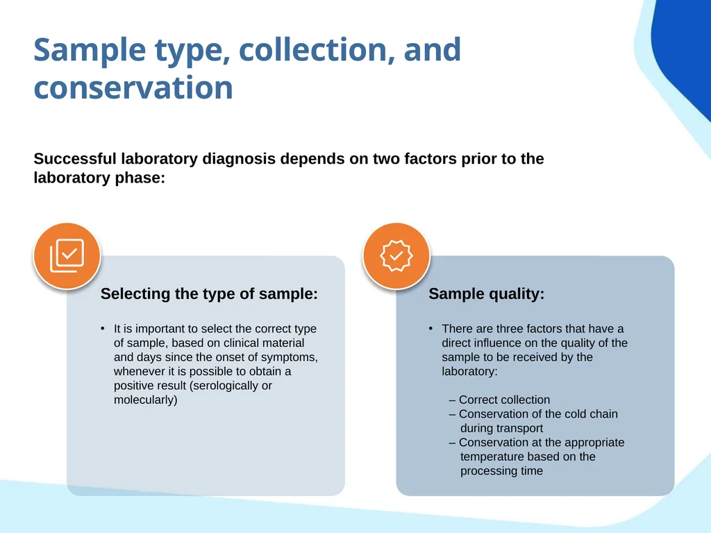
slide14. Sample type, collection, and conservation Successful laboratory diagnosis depends on two factors prior to the laboratory phase: Selecting the type of sample:
It is important to select the correct type of sample, based on clinical material and days since the onset of symptoms, whenever it is possible to obtain a positive result (serologically or molecularly) Sample quality:
There are three factors that have a direct influence on the quality of the sample to be received by the laboratory:
– Correct collection
– Conservation of the cold chain during transport
– Conservation at the appropriate temperature based on the processing time<br>
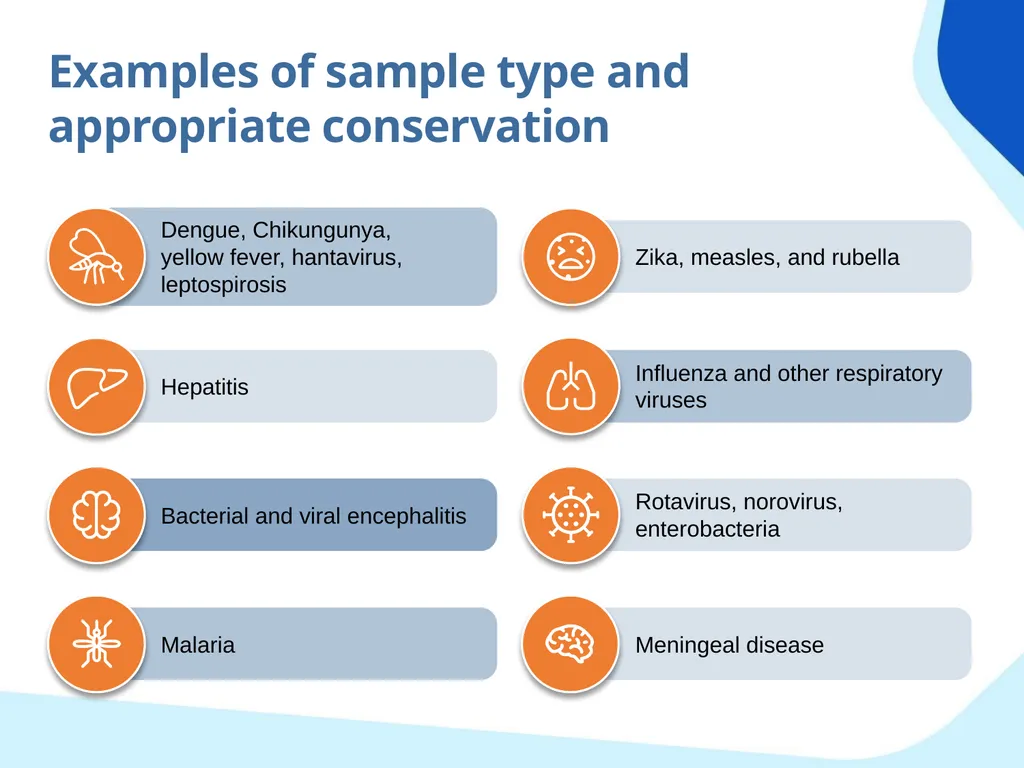
slide15. Examples of sample type and appropriate conservation Dengue, Chikungunya, yellow fever, hantavirus, leptospirosis Zika, measles, and rubella Hepatitis Influenza and other respiratory viruses Bacterial and viral encephalitis Rotavirus, norovirus, enterobacteria Malaria Meningeal disease<br>
slide16. Dengue, Chikungunya, yellow fever, hantavirus, leptospirosis Type of sample: serum
Quantity: 3 to 7 mL
Transport medium: no additives
Transport conditions: 2 to 8°C
Conservation: -20°C (up to 1 week) / -70°C (period greater than 1 week)
Laboratory diagnosis:
– 1 to 5 days following onset of symptoms: PCR
– 5 to 10 days following the onset of symptoms: PCR + ELISA IgM<br>
slide17. Zika, measles, and rubella Type of sample: serum
Quantity: 3 to 7 mL
Transport medium: no additives
Transport conditions: 2 to 8°C
Conservation: -20°C (up to 1 week) / -70°C (period greater than one week)
Laboratory diagnosis:
– 1 to 5 following the onset of symptoms: PCR
– 5 to 10 days following the onset of symptoms: PCR + ELISA IgM<br>
slide18. Zika, measles, and rubella Type of sample: urine
Quantity: 3 to 7 mL
Transport medium: no additives
Transport conditions: 2 to 8°C
Conservation: -20°C (up to 1 week) / -70°C (period greater than 1 week)
Laboratory diagnosis:
– 1 to 15 days following the onset of symptoms: PCR<br>
slide19. Zika – congenital syndrome or fatal cases * Ambient temperature
** Under medical indication for suspected neurological syndrome *** Fatal cases: brain, liver, kidney, others<br>
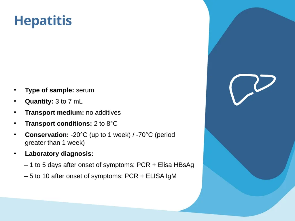
slide20. Hepatitis Type of sample: serum
Quantity: 3 to 7 mL
Transport medium: no additives
Transport conditions: 2 to 8°C
Conservation: -20°C (up to 1 week) / -70°C (period greater than 1 week)
Laboratory diagnosis:
– 1 to 5 days after onset of symptoms: PCR + Elisa HBsAg
– 5 to 10 after onset of symptoms: PCR + ELISA IgM<br>
slide21. Influenza and other respiratory viruses Type of sample: nasopharyngeal swab
Quantity: 2 nylon swabs
Transport medium: viral transport medium or saline solution (3 mL)
Transport conditions: 2 to 8°C
Conservation: 4°C until aliquots are prepared; aliquots to a -20°C (up to 48 hours) and -70°C (period greater than 48 hours)
Laboratory diagnosis:
– PCR or IF (only typification) followed by PCR<br>
slide22. Influenza and other respiratory viruses Type of sample: nasopharyngeal aspiration; nasopharyngeal wash
Material: suction device
Transport medium: saline solution
Transport conditions: 2 to 8°C
Conservation: 4°C until aliquots are prepared; aliquots to a -20°C (up to 48 hours) and -70°C (period greater than 48 hours)
Laboratory diagnosis:
– PCR or IF (only typification) followed by PCR<br>
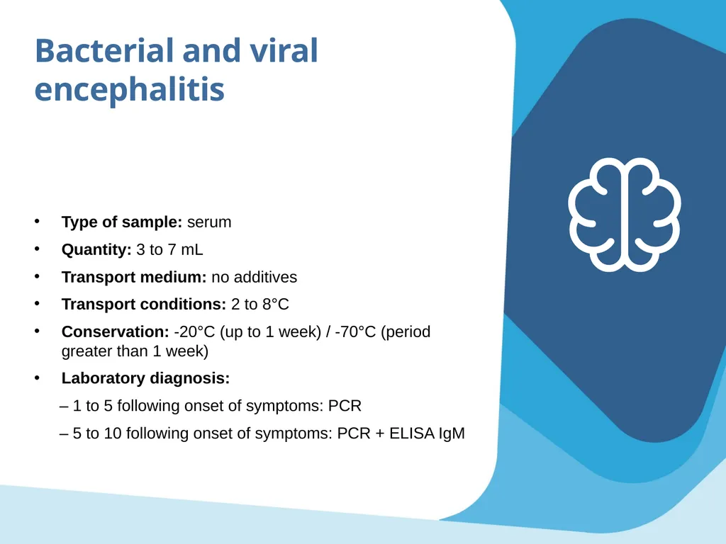
slide23. Bacterial and viral encephalitis Type of sample: serum
Quantity: 3 to 7 mL
Transport medium: no additives
Transport conditions: 2 to 8°C
Conservation: -20°C (up to 1 week) / -70°C (period greater than 1 week)
Laboratory diagnosis:
– 1 to 5 following onset of symptoms: PCR
– 5 to 10 following onset of symptoms: PCR + ELISA IgM<br>
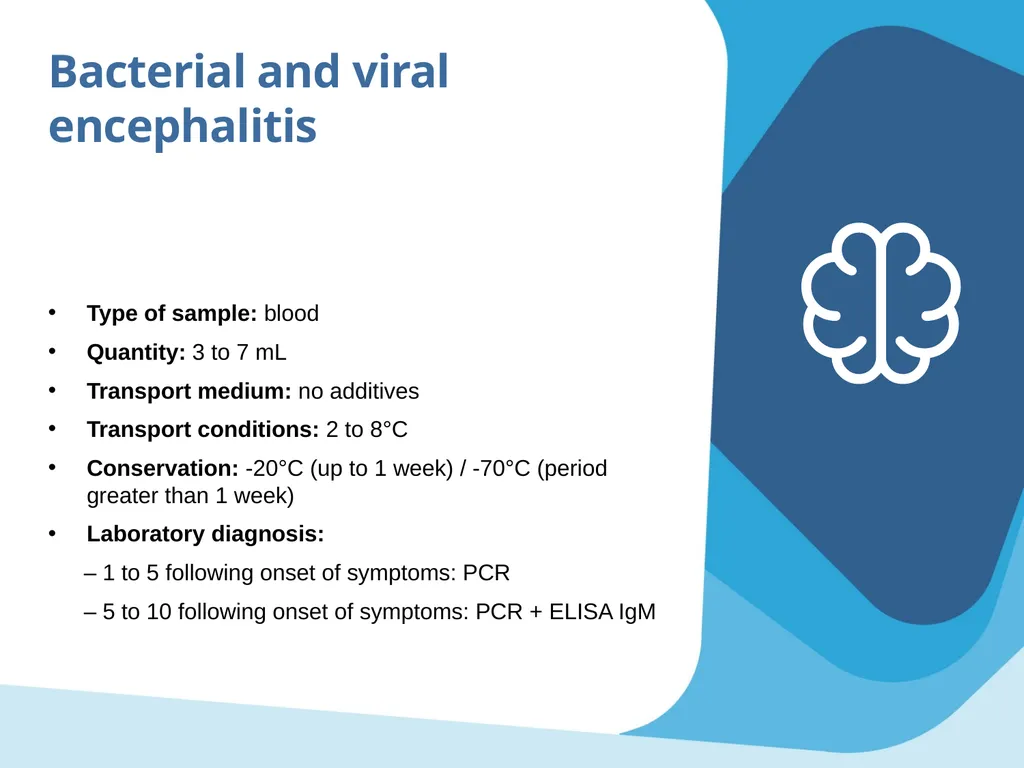
slide24. Bacterial and viral encephalitis Type of sample: blood
Quantity: 3 to 7 mL
Transport medium: no additives
Transport conditions: 2 to 8°C
Conservation: -20°C (up to 1 week) / -70°C (period greater than 1 week)
Laboratory diagnosis:
– 1 to 5 following onset of symptoms: PCR
– 5 to 10 following onset of symptoms: PCR + ELISA IgM<br>
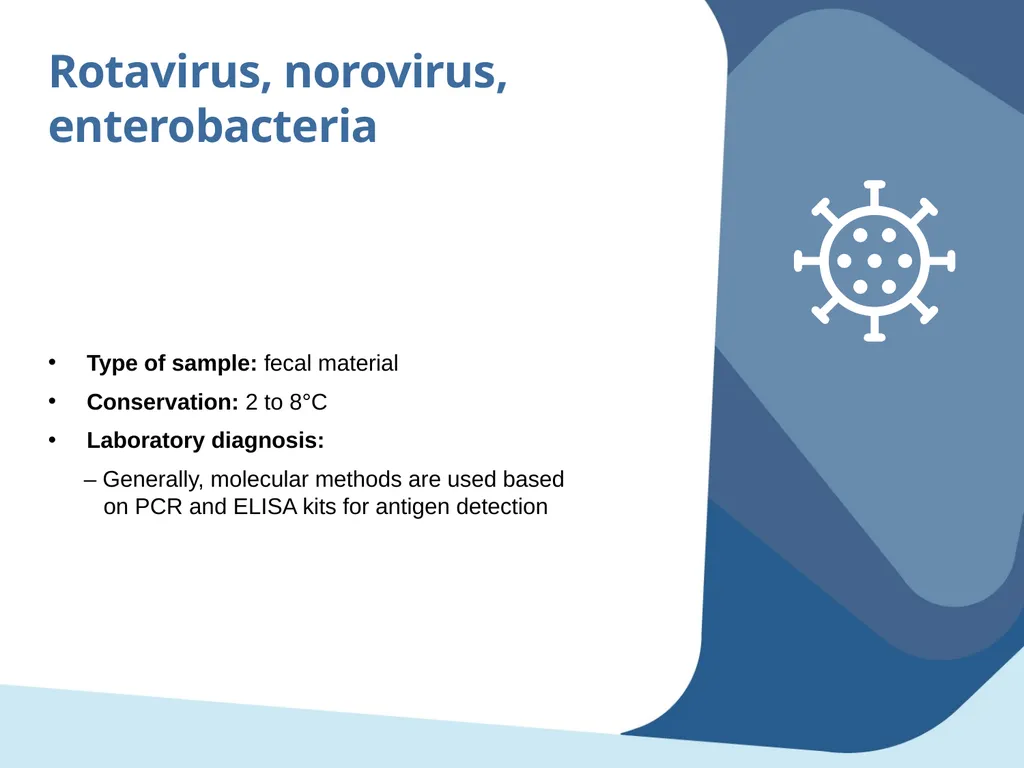
slide25. Rotavirus, norovirus, enterobacteria Type of sample: fecal material
Conservation: 2 to 8°C
Laboratory diagnosis:
– Generally, molecular methods are used based on PCR and ELISA kits for antigen detection<br>
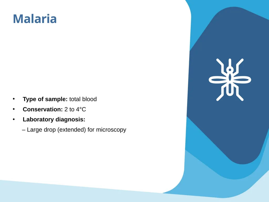
slide26. Malaria Type of sample: total blood
Conservation: 2 to 4°C
Laboratory diagnosis:
– Large drop (extended) for microscopy<br>
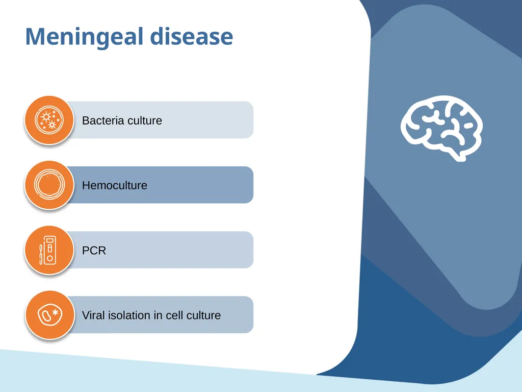
slide27. Meningeal disease Bacteria culture Hemoculture PCR Viral isolation in cell culture<br>
slide28. © Pan American Health Organization, 2023
Property of the Pan American Health Organization. Reproduced with permission pursuant to the Creative Commons Attribution-NonCommercial-NoDerivs 3.0 IGO license (CC BY-NC-ND 3.0 IGO).
In any use of this work, there should be no suggestion that the Pan American Health Organization (PAHO) endorses any specific organization, product, or service. Use of the PAHO logo, in a way that is not already incorporated in the work or consistent with the Creative Commons license CC BY-NC-ND 3.0 IGO, is strictly prohibited. Any use of this work that is inconsistent with or not permitted under the Creative Commons license BY-NC-ND 3.0 IGO requires the express written consent of PAHO.
Further, all reasonable precautions have been taken by PAHO and the GS/OAS to verify the information contained in this publication. However, the published material is being distributed without warranty of any kind, either expressed or implied. The responsibility for the interpretation and use of the material lies with the reader. In no event shall PAHO and/or the OAS or GS/OAS be liable for damages arising from its use<br>
slide2. Objectives Identify the main causes of outbreak in the work zone Know the different types of samples and diagnostic tests that can be conducted, based on the infection dynamics Review the role of the laboratory coordinated with surveillance to characterize outbreaks<br>
slide3. Content Most frequent etiologies in the Americas Most commonly used tests for diagnosing etiologic agents
– Serology
– Molecular
– Microscopic Specimen type, collection, and conservation
– Examples of type of sample and appropriate conservation<br>
slide4. Most common etiologies
in the Americas Febrile syndrome:
– Dengue, Chikungunya, Zika, yellow fever, influenza, leptospirosis, malaria
Icteric febrile syndrome:
– Yellow fever, leptospirosis, malaria, hepatitis
Hemorrhagic icteric febrile syndrome:
– Yellow fever, leptospirosis
Hemorrhagic febrile syndrome:
– Dengue, yellow fever, leptospirosis, hantavirus
Neurological syndromes:
– Bacterial, viral (Zika and viral encephalitis), polio, botulism
Respiratory syndromes:
– Influenza, hantavirus, diphtheria and other respiratory viruses
Acute diarrheic disease:
– Cholera, rotavirus, norovirus, enterobacteria
Exanthematous disease:
– Zika, measles, rubella<br>
slide5. Most used tests for diagnosing etiologic agents Serological:
– ELISA (IgM and IgG)
– Direct and indirect immunofluorescence Molecular:
– Polymerase chain reaction (PCR)
– Real time PCR
– Sequencing Microscopic:
– Optical microscopy<br>
slide6. Serological tests (Serology) Two types: direct and indirect tests
– Direct tests: based on the antigen antibody reaction to investigate the presence of antigen. That is, these tests investigate the presence of the etiologic agent or one of its components. Example: direct immunofluorescence.
– Indirect tests: based on detecting specific antibodies against the agent or one of its components, which indirectly allow us to assume that the pathogen being investigated was present in the host at some point Detect the presence of antibodies Cheaper than molecular tests Based on the antigen antibody reaction and constitute a valuable tool in diagnosing infectious disease<br>
slide7. Serological tests (Serology) Important concepts for interpreting serological results: Positive IgM is related to a recent infection
Increased antibody titer in paired samples indicates a recent infection
– First sample: negative
– Second sample: antibodies present
– Antibody titer in the second sample > 4x the antibody titer in the first sample
Positive IgG indicates (generally) a past infection<br>
slide8. Types of samples and appropriate collection Primary infection Secondary infection<br>
slide9. Serological tests (Serology) Immunofluorescence
– Immunofluorescence techniques may be applied as techniques to investigate the presence of viral, parasitic, bacterial, or mycotic antigens in clinical samples or antibodies produced against them in the patient’s serum.
ELISA (enzyme-linked immunosorbent assay)
– Immunoenzymatic techniques are tests based on antibody antigen reaction, making it possible to detect the presence of antibodies produced against viral, parasitic, bacterial, or mycotic antigens in clinical specimens in the patient’s serum.
– The ELISA technique may be used to detect IgM or IgG.<br>
slide10. Molecular tests Are based on detecting the etiologic agent’s genome.
Make it possible to detect the etiologic agent’s RNA or DNA with high sensitivity and specificity.
Are more costly than serology.
Make it possible to confirm the presence of the etiologic agent in clinical samples.
Make it possible to detect RNA or DNA even in very small sample volumes.<br>
slide11. Molecular tests Polymerase chain reaction (end point PCR)
– The end point PCR is based on amplifying and visualizing the complementary DNA or ADNA (cDNA). This technique consists of amplifying and visualizing a specific DNA fragment from the etiologic agent.
Real time PCR
– Also known as quantitative PCR, this test is a variant of the polymerase chain reaction (PCR).
– Real time PCR makes it possible to amplify and at the same time absolutely quantify specific DNA or cDNA molecules<br>
slide12. Microscopic Consists of using special instruments such as magnifying glasses and microscopes to magnify the size of structures that are invisible to the naked human eye, such as viruses, bacteria, and parasites.
Microscopic observation is necessary in the field of parasitology.
Modes of observation vary based on strategies employed and range from micrometric to nanometric observation.
Therefore, various types of microscopes are required, such as photonic and electronic (scanning, transmission, and atomic energy).<br>
slide13. Microscopic Optical microscopy technique allows us to visualize bacteria and parasites simply by examining the sample with a microscope.
For bacteria, a Gram stain (a purple stain) is often performed first. Bacteria is classified as follows:
– Gram positive (appear blue because they retain the Gram stain)
– Gram negative (appear red because they do not capture the stain)
A blood swab is performed to detect parasites found in the blood, such as filariasis, malaria, or babesiosis. For this test, a drop of blood is placed on a microscope slide, stained, and examined under the microscope to visualize the parasite. Optical microscopy<br>
slide14. Sample type, collection, and conservation Successful laboratory diagnosis depends on two factors prior to the laboratory phase: Selecting the type of sample:
It is important to select the correct type of sample, based on clinical material and days since the onset of symptoms, whenever it is possible to obtain a positive result (serologically or molecularly) Sample quality:
There are three factors that have a direct influence on the quality of the sample to be received by the laboratory:
– Correct collection
– Conservation of the cold chain during transport
– Conservation at the appropriate temperature based on the processing time<br>
slide15. Examples of sample type and appropriate conservation Dengue, Chikungunya, yellow fever, hantavirus, leptospirosis Zika, measles, and rubella Hepatitis Influenza and other respiratory viruses Bacterial and viral encephalitis Rotavirus, norovirus, enterobacteria Malaria Meningeal disease<br>
slide16. Dengue, Chikungunya, yellow fever, hantavirus, leptospirosis Type of sample: serum
Quantity: 3 to 7 mL
Transport medium: no additives
Transport conditions: 2 to 8°C
Conservation: -20°C (up to 1 week) / -70°C (period greater than 1 week)
Laboratory diagnosis:
– 1 to 5 days following onset of symptoms: PCR
– 5 to 10 days following the onset of symptoms: PCR + ELISA IgM<br>
slide17. Zika, measles, and rubella Type of sample: serum
Quantity: 3 to 7 mL
Transport medium: no additives
Transport conditions: 2 to 8°C
Conservation: -20°C (up to 1 week) / -70°C (period greater than one week)
Laboratory diagnosis:
– 1 to 5 following the onset of symptoms: PCR
– 5 to 10 days following the onset of symptoms: PCR + ELISA IgM<br>
slide18. Zika, measles, and rubella Type of sample: urine
Quantity: 3 to 7 mL
Transport medium: no additives
Transport conditions: 2 to 8°C
Conservation: -20°C (up to 1 week) / -70°C (period greater than 1 week)
Laboratory diagnosis:
– 1 to 15 days following the onset of symptoms: PCR<br>
slide19. Zika – congenital syndrome or fatal cases * Ambient temperature
** Under medical indication for suspected neurological syndrome *** Fatal cases: brain, liver, kidney, others<br>
slide20. Hepatitis Type of sample: serum
Quantity: 3 to 7 mL
Transport medium: no additives
Transport conditions: 2 to 8°C
Conservation: -20°C (up to 1 week) / -70°C (period greater than 1 week)
Laboratory diagnosis:
– 1 to 5 days after onset of symptoms: PCR + Elisa HBsAg
– 5 to 10 after onset of symptoms: PCR + ELISA IgM<br>
slide21. Influenza and other respiratory viruses Type of sample: nasopharyngeal swab
Quantity: 2 nylon swabs
Transport medium: viral transport medium or saline solution (3 mL)
Transport conditions: 2 to 8°C
Conservation: 4°C until aliquots are prepared; aliquots to a -20°C (up to 48 hours) and -70°C (period greater than 48 hours)
Laboratory diagnosis:
– PCR or IF (only typification) followed by PCR<br>
slide22. Influenza and other respiratory viruses Type of sample: nasopharyngeal aspiration; nasopharyngeal wash
Material: suction device
Transport medium: saline solution
Transport conditions: 2 to 8°C
Conservation: 4°C until aliquots are prepared; aliquots to a -20°C (up to 48 hours) and -70°C (period greater than 48 hours)
Laboratory diagnosis:
– PCR or IF (only typification) followed by PCR<br>
slide23. Bacterial and viral encephalitis Type of sample: serum
Quantity: 3 to 7 mL
Transport medium: no additives
Transport conditions: 2 to 8°C
Conservation: -20°C (up to 1 week) / -70°C (period greater than 1 week)
Laboratory diagnosis:
– 1 to 5 following onset of symptoms: PCR
– 5 to 10 following onset of symptoms: PCR + ELISA IgM<br>
slide24. Bacterial and viral encephalitis Type of sample: blood
Quantity: 3 to 7 mL
Transport medium: no additives
Transport conditions: 2 to 8°C
Conservation: -20°C (up to 1 week) / -70°C (period greater than 1 week)
Laboratory diagnosis:
– 1 to 5 following onset of symptoms: PCR
– 5 to 10 following onset of symptoms: PCR + ELISA IgM<br>
slide25. Rotavirus, norovirus, enterobacteria Type of sample: fecal material
Conservation: 2 to 8°C
Laboratory diagnosis:
– Generally, molecular methods are used based on PCR and ELISA kits for antigen detection<br>
slide26. Malaria Type of sample: total blood
Conservation: 2 to 4°C
Laboratory diagnosis:
– Large drop (extended) for microscopy<br>
slide27. Meningeal disease Bacteria culture Hemoculture PCR Viral isolation in cell culture<br>
slide28. © Pan American Health Organization, 2023
Property of the Pan American Health Organization. Reproduced with permission pursuant to the Creative Commons Attribution-NonCommercial-NoDerivs 3.0 IGO license (CC BY-NC-ND 3.0 IGO).
In any use of this work, there should be no suggestion that the Pan American Health Organization (PAHO) endorses any specific organization, product, or service. Use of the PAHO logo, in a way that is not already incorporated in the work or consistent with the Creative Commons license CC BY-NC-ND 3.0 IGO, is strictly prohibited. Any use of this work that is inconsistent with or not permitted under the Creative Commons license BY-NC-ND 3.0 IGO requires the express written consent of PAHO.
Further, all reasonable precautions have been taken by PAHO and the GS/OAS to verify the information contained in this publication. However, the published material is being distributed without warranty of any kind, either expressed or implied. The responsibility for the interpretation and use of the material lies with the reader. In no event shall PAHO and/or the OAS or GS/OAS be liable for damages arising from its use<br>