
Neurological Assessment Overview By: Aliyah
TA
Published · 49 slides · 0 views

1 / 1

Description
Neurological Assessment Overview By: Aliyah Mangum, BSN, RN, PCCN,CNIII CTICU Expectations of Care Neuro Anatomy Function Review Frontal Lobe: voluntary movement, memory, concentration, thoughts, personality, emotions, motor control of
Related Topics
Share
Embed code
Download this presentation From Below
"Neurological Assessment Overview By: Aliyah" is the property of its rightful owner. Permission is granted to download and print the materials on this website for personal, non-commercial use only, and to display it on your personal computer provided you do not modify the materials and that you retain all copyright notices contained in the materials. By downloading content from our website, you accept the terms of this agreement.
Presentation Transcript
01
Neurological Assessment
Overview By: Aliyah Mangum, BSN, RN, PCCN,CNIII<br>
02
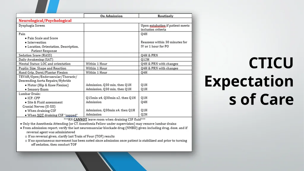
CTICU Expectations of Care<br>
03
Neuro Anatomy & Function Review<br>
04
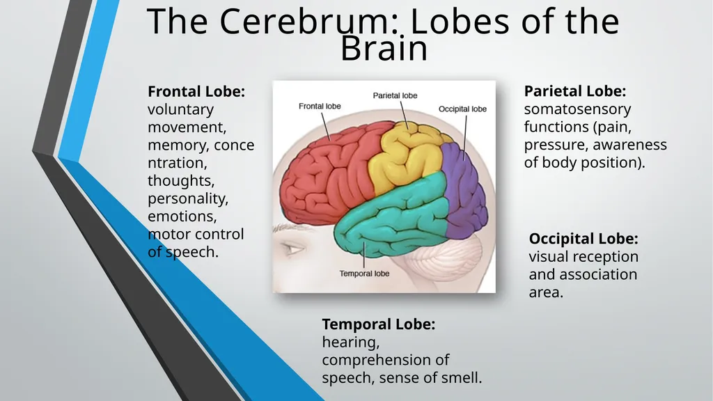
Frontal Lobe: voluntary movement, memory, concentration, thoughts, personality, emotions, motor control of speech. Temporal Lobe: hearing, comprehension of speech, sense of smell. Occipital Lobe: visual reception and association area. Parietal Lobe: somatosensory functions (pain, pressure, awareness of body position). The Cerebrum: Lobes of the Brain<br>
05
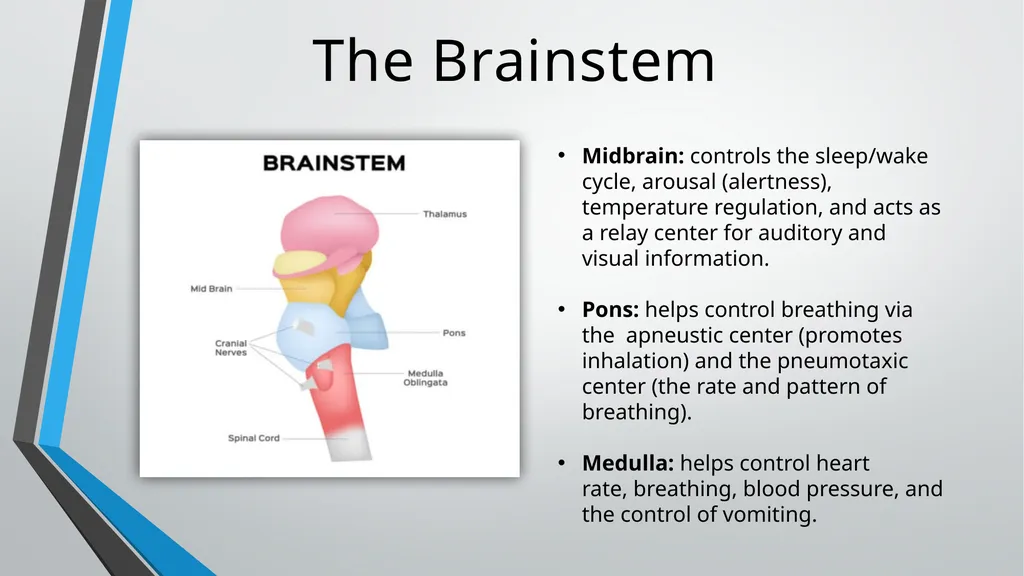
The Brainstem Midbrain: controls the sleep/wake cycle, arousal (alertness), temperature regulation, and acts as a relay center for auditory and visual information.
Pons: helps control breathing via the apneustic center (promotes inhalation) and the pneumotaxic center (the rate and pattern of breathing).
Medulla: helps control heart rate, breathing, blood pressure, and the control of vomiting.<br>
Pons: helps control breathing via the apneustic center (promotes inhalation) and the pneumotaxic center (the rate and pattern of breathing).
Medulla: helps control heart rate, breathing, blood pressure, and the control of vomiting.<br>
06
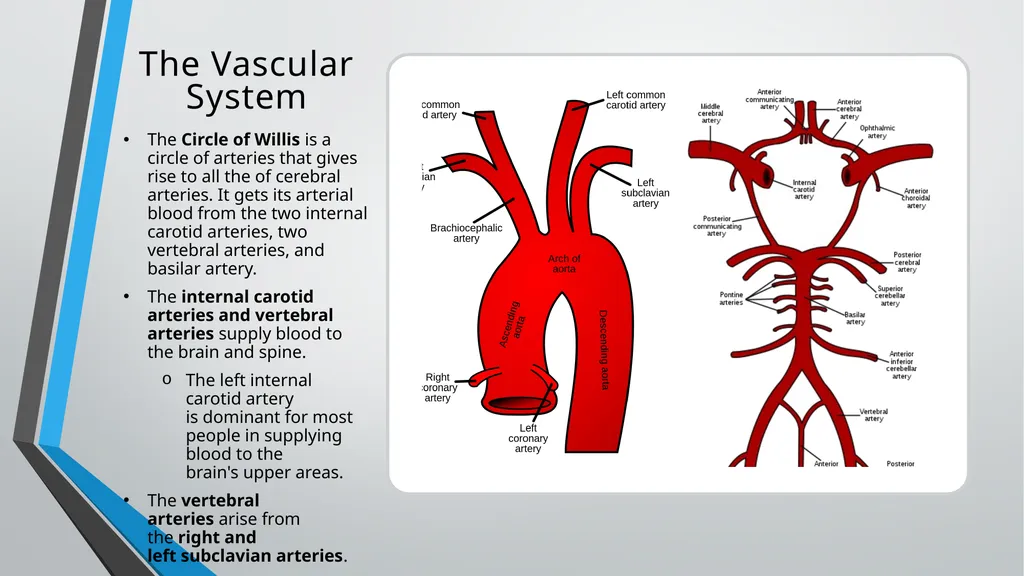
The Vascular System The Circle of Willis is a circle of arteries that gives rise to all the of cerebral arteries. It gets its arterial blood from the two internal carotid arteries, two vertebral arteries, and basilar artery.
The internal carotid arteries and vertebral arteries supply blood to the brain and spine.
The left internal carotid artery is dominant for most people in supplying blood to the brain's upper areas.
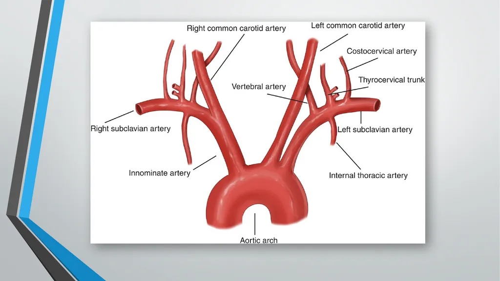
The vertebral arteries arise from the right and left subclavian arteries.<br>
The internal carotid arteries and vertebral arteries supply blood to the brain and spine.
The left internal carotid artery is dominant for most people in supplying blood to the brain's upper areas.
The vertebral arteries arise from the right and left subclavian arteries.<br>
07
The Neurological Assessment “Evaluation of neurological status and level of consciousness is an essential part of nursing at the critical care level. Regardless of diagnosis, patients can experience neurological deficits and changes in mental status based on their current condition, medication regime or acute changes in their overall condition. The recognition of a change in mental status can make a significant impact on a patient's prognosis. Early identification of neurological deterioration is vital to preventing secondary brain injury!”
-Paula Nekic, CNE<br>
-Paula Nekic, CNE<br>
08
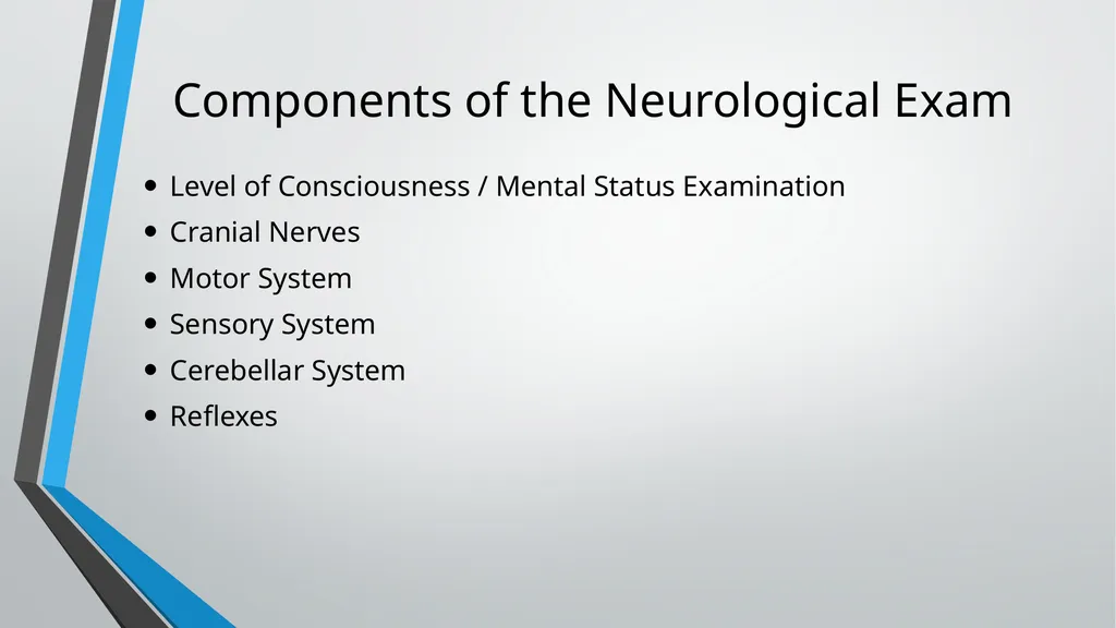
Components of the Neurological Exam Level of Consciousness / Mental Status Examination
Cranial Nerves
Motor System
Sensory System
Cerebellar System
Reflexes<br>
Cranial Nerves
Motor System
Sensory System
Cerebellar System
Reflexes<br>
09
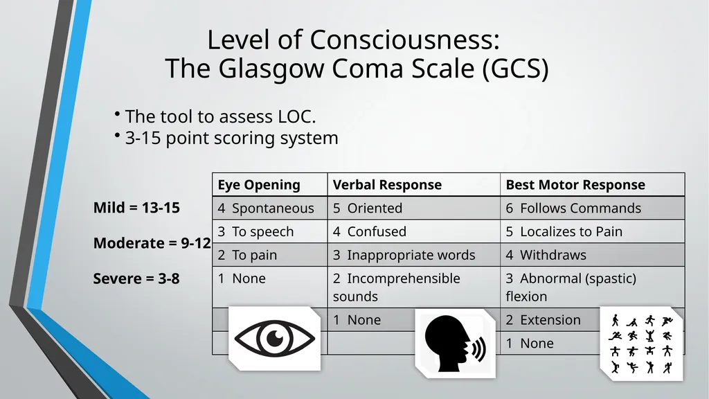
Level of Consciousness:
The Glasgow Coma Scale (GCS) Mild = 13-15
Moderate = 9-12
Severe = 3-8 The tool to assess LOC.
3-15 point scoring system<br>
Moderate = 9-12
Severe = 3-8 The tool to assess LOC.
3-15 point scoring system<br>
10
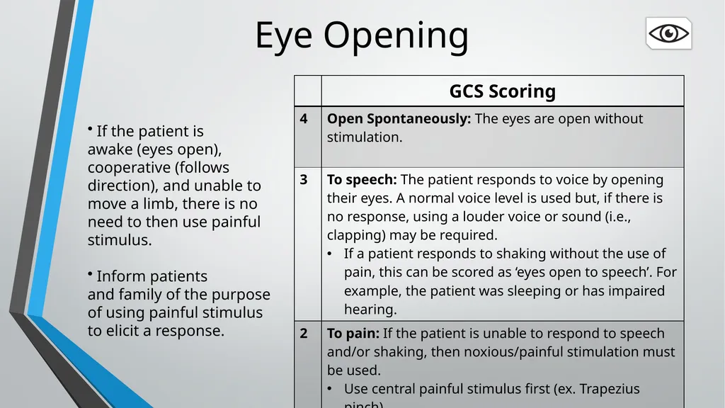
Eye Opening If the patient is awake (eyes open), cooperative (follows direction), and unable to move a limb, there is no need to then use painful stimulus.
Inform patients and family of the purpose of using painful stimulus to elicit a response.<br>
Inform patients and family of the purpose of using painful stimulus to elicit a response.<br>
11
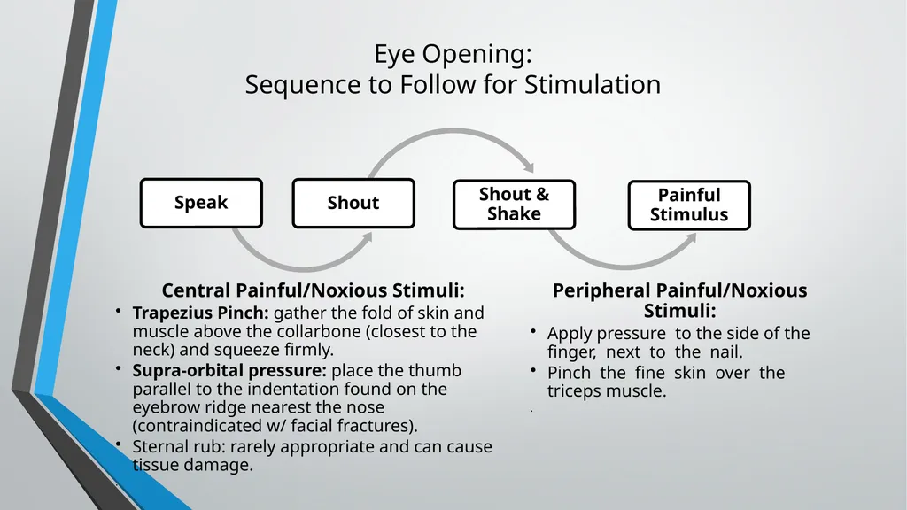
Eye Opening:
Sequence to Follow for Stimulation Central Painful/Noxious Stimuli:
Trapezius Pinch: gather the fold of skin and muscle above the collarbone (closest to the neck) and squeeze firmly.
Supra-orbital pressure: place the thumb parallel to the indentation found on the eyebrow ridge nearest the nose (contraindicated w/ facial fractures).
Sternal rub: rarely appropriate and can cause tissue damage.
. Peripheral Painful/Noxious Stimuli:
Apply pressure to the side of the finger, next to the nail.
Pinch the fine skin over the triceps muscle.
.<br>
Trapezius Pinch: gather the fold of skin and muscle above the collarbone (closest to the neck) and squeeze firmly.
Supra-orbital pressure: place the thumb parallel to the indentation found on the eyebrow ridge nearest the nose (contraindicated w/ facial fractures).
Sternal rub: rarely appropriate and can cause tissue damage.
. Peripheral Painful/Noxious Stimuli:
Apply pressure to the side of the finger, next to the nail.
Pinch the fine skin over the triceps muscle.
.<br>
12
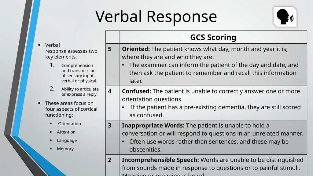
Verbal Response Verbal response assesses two key elements:
Comprehension and transmission of sensory input; verbal or physical.
Ability to articulate or express a reply.
These areas focus on four aspects of cortical functioning:
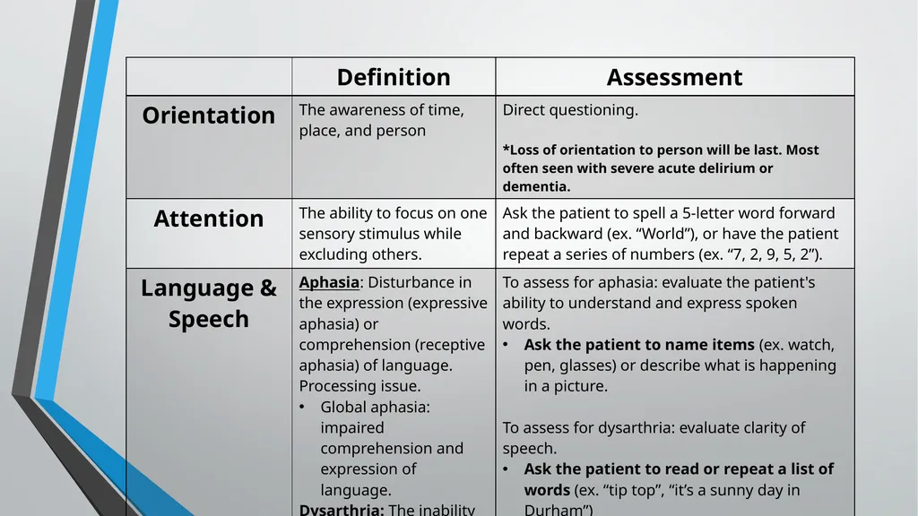
Orientation
Attention
Language
Memory<br>
Comprehension and transmission of sensory input; verbal or physical.
Ability to articulate or express a reply.
These areas focus on four aspects of cortical functioning:
Orientation
Attention
Language
Memory<br>
13
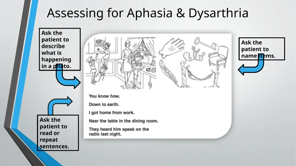
Assessing for Aphasia & Dysarthria Ask the patient to name items. Ask the patient to read or repeat sentences. Ask the patient to describe what is happening in a photo.<br>
14
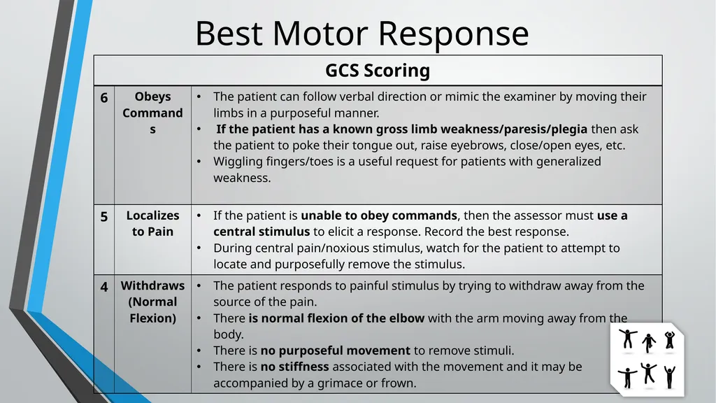
Best Motor Response<br>
15
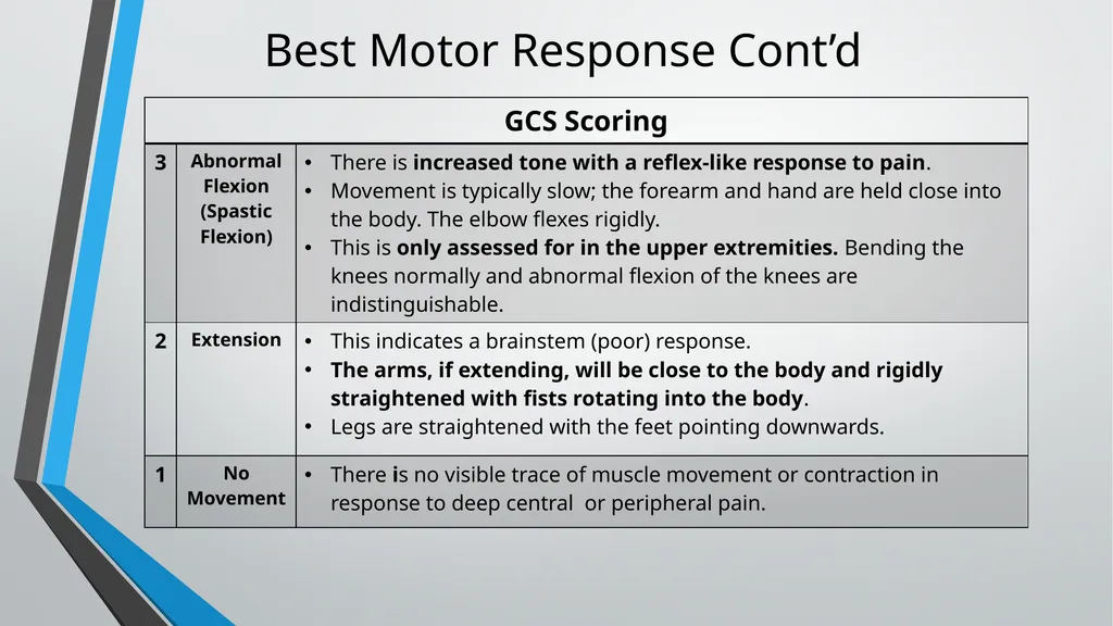
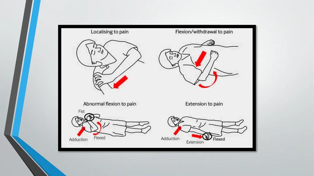
Best Motor Response Cont’d<br>
16
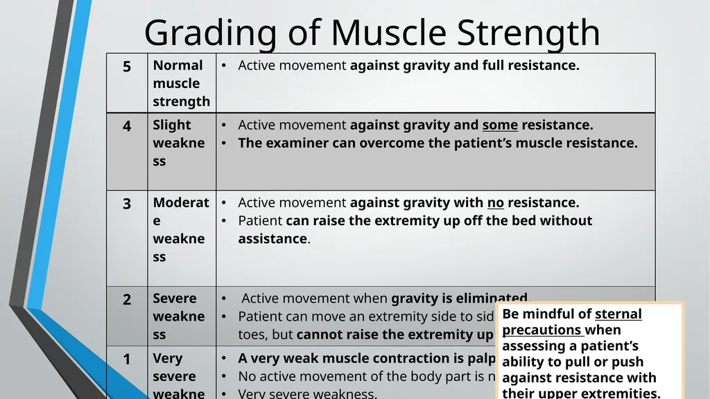
Grading of Muscle Strength Be mindful of sternal precautions when assessing a patient’s ability to pull or push against resistance with their upper extremities.<br>
17
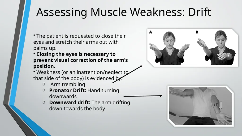
Assessing Muscle Weakness: Drift The patient is requested to close their eyes and stretch their arms out with palms up.
Closing the eyes is necessary to prevent visual correction of the arm's position.
Weakness (or an inattention/neglect to that side of the body) is evidenced by:
Arm trembling
Pronator Drift: Hand turning downwards
Downward drift: The arm drifting down towards the body<br>
Closing the eyes is necessary to prevent visual correction of the arm's position.
Weakness (or an inattention/neglect to that side of the body) is evidenced by:
Arm trembling
Pronator Drift: Hand turning downwards
Downward drift: The arm drifting down towards the body<br>
18
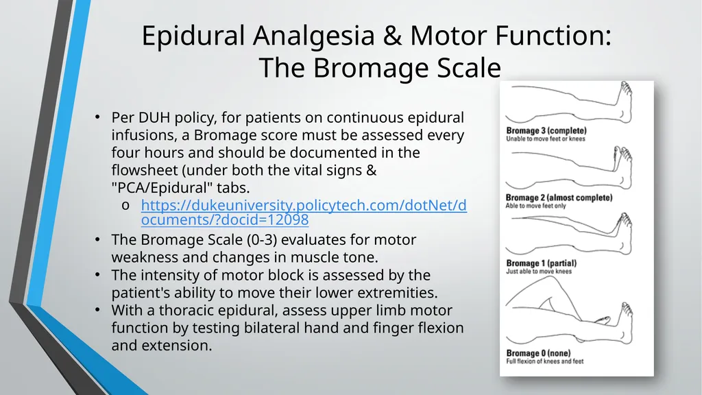
Epidural Analgesia & Motor Function:
The Bromage Scale Per DUH policy, for patients on continuous epidural infusions, a Bromage score must be assessed every four hours and should be documented in the flowsheet (under both the vital signs & "PCA/Epidural" tabs.
https://dukeuniversity.policytech.com/dotNet/documents/?docid=12098
The Bromage Scale (0-3) evaluates for motor weakness and changes in muscle tone.
The intensity of motor block is assessed by the patient's ability to move their lower extremities.
With a thoracic epidural, assess upper limb motor function by testing bilateral hand and finger flexion and extension.<br>
https://dukeuniversity.policytech.com/dotNet/documents/?docid=12098
The Bromage Scale (0-3) evaluates for motor weakness and changes in muscle tone.
The intensity of motor block is assessed by the patient's ability to move their lower extremities.
With a thoracic epidural, assess upper limb motor function by testing bilateral hand and finger flexion and extension.<br>
19
Sensory Assessment The examiner determines the patient’s ability to perceive various types of sensations with the eyes closed.
There are three main pathways for sensation, and they should be compared bilaterally:
Light touch
Position sense (proprioception)
Pain and temperature sensation
Commonly evaluated body parts: face, neck, deltoid regions, forearm, hands (top side), chest, abdomen, thighs, lower legs, and feet (top side).
Rating scale:
Normal
Present, but diminished (abnormal)
Absent
Sensory deficits are generally on the same side as motor deficits.<br>
There are three main pathways for sensation, and they should be compared bilaterally:
Light touch
Position sense (proprioception)
Pain and temperature sensation
Commonly evaluated body parts: face, neck, deltoid regions, forearm, hands (top side), chest, abdomen, thighs, lower legs, and feet (top side).
Rating scale:
Normal
Present, but diminished (abnormal)
Absent
Sensory deficits are generally on the same side as motor deficits.<br>
20
Cranial Nerve II-XII Assessment https://youtu.be/oZGFrwogx14<br>
21
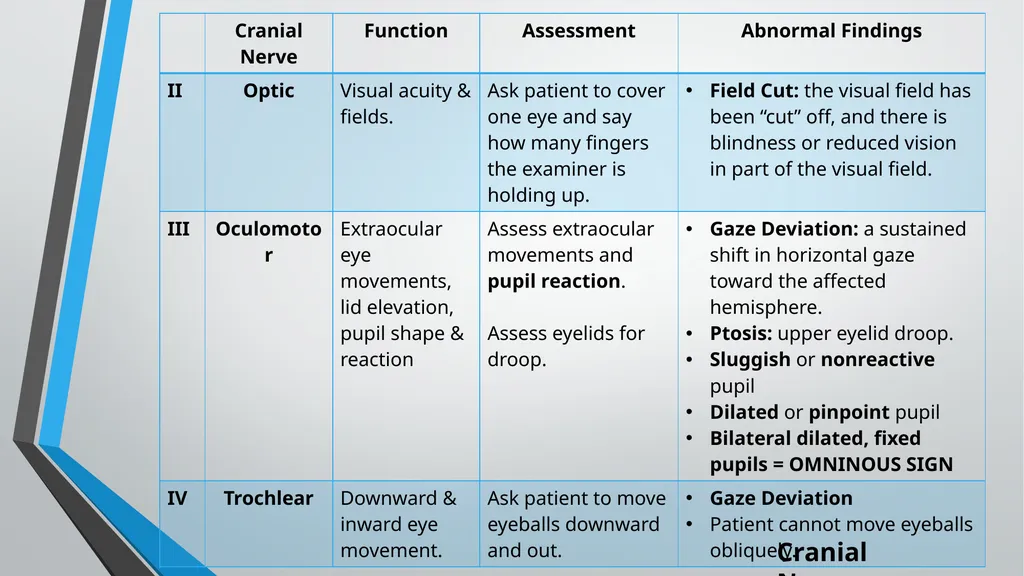
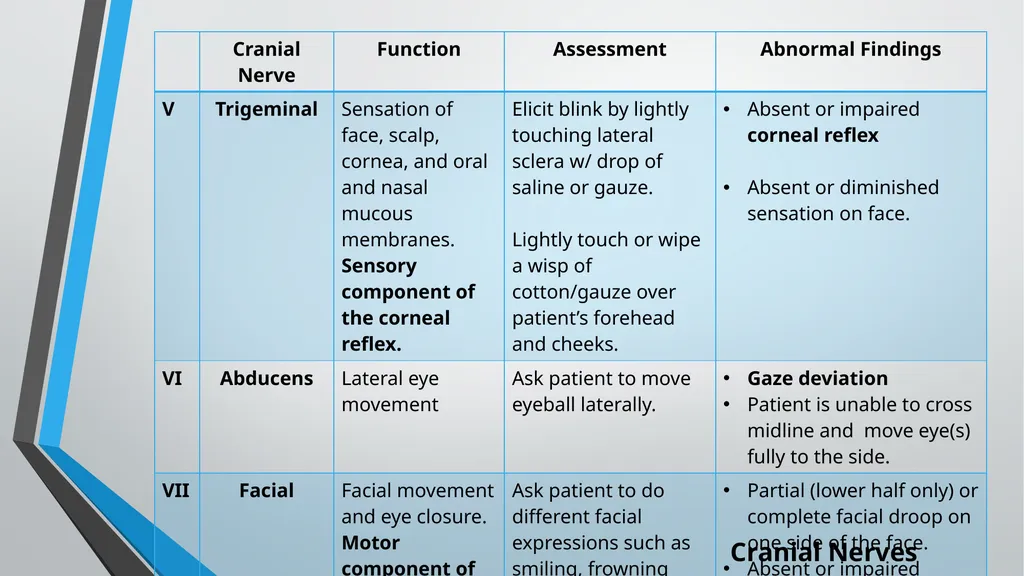
Cranial Nerves<br>
22
Cranial Nerves<br>
23
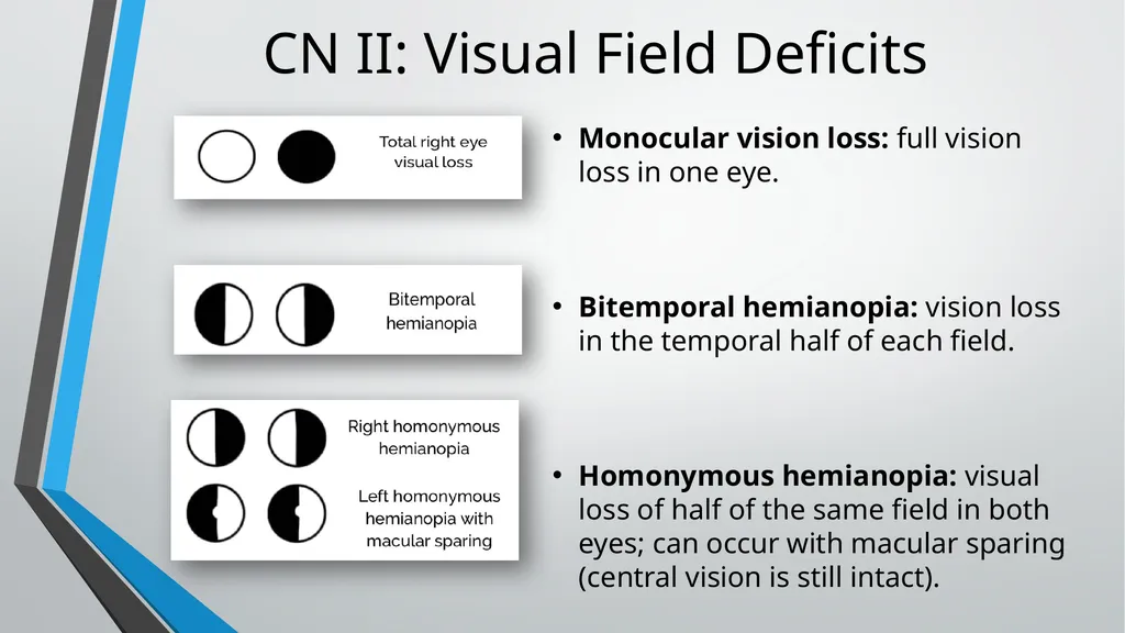
Monocular vision loss: full vision loss in one eye.
Bitemporal hemianopia: vision loss in the temporal half of each field.
Homonymous hemianopia: visual loss of half of the same field in both eyes; can occur with macular sparing (central vision is still intact). CN II: Visual Field Deficits<br>
Bitemporal hemianopia: vision loss in the temporal half of each field.
Homonymous hemianopia: visual loss of half of the same field in both eyes; can occur with macular sparing (central vision is still intact). CN II: Visual Field Deficits<br>
24
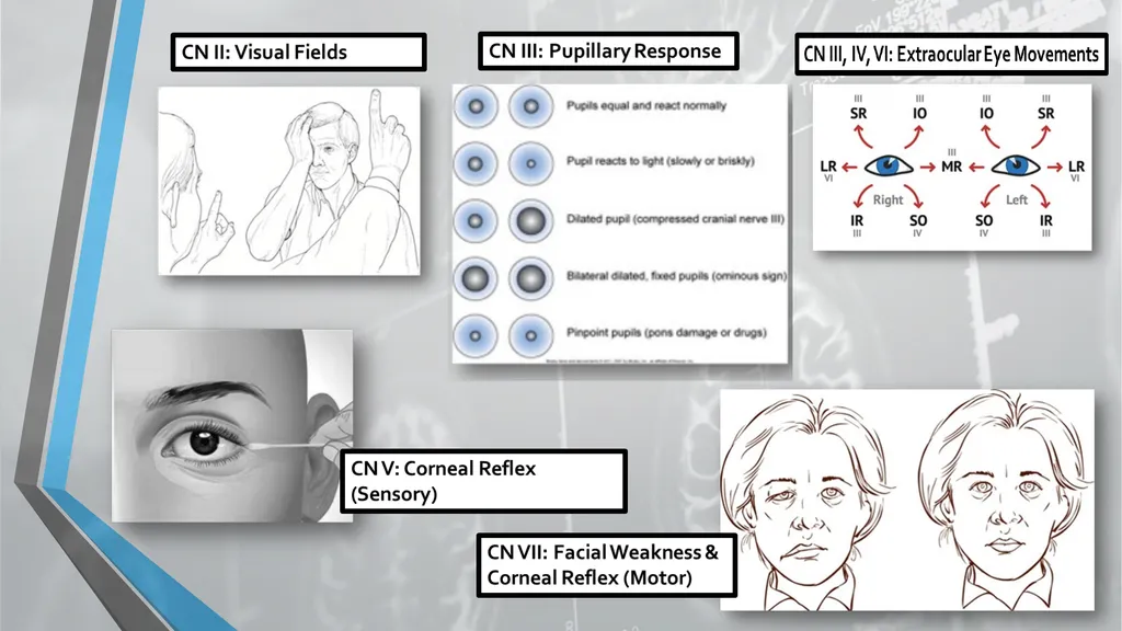
Cranial Nerves<br>
25
Cranial Nerves<br>
26
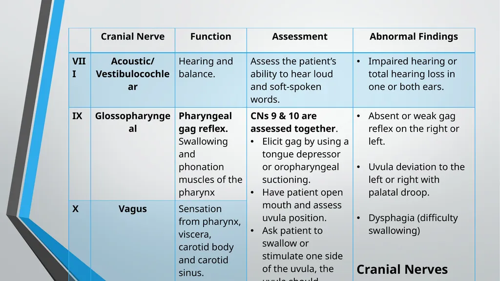
CN VIII: Hearing<br>
27
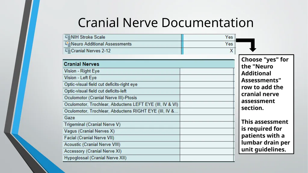
Cranial Nerve Documentation Choose "yes" for the "Neuro Additional Assessments" row to add the cranial nerve assessment section.
This assessment is required for patients with a lumbar drain per unit guidelines.<br>
This assessment is required for patients with a lumbar drain per unit guidelines.<br>
28
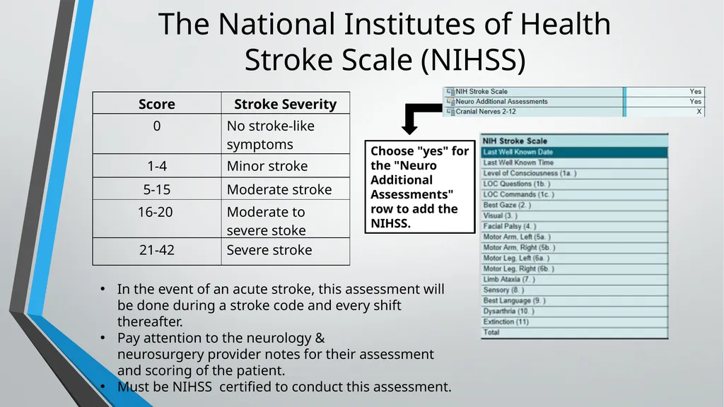
The National Institutes of Health
Stroke Scale (NIHSS) Choose "yes" for the "Neuro Additional Assessments" row to add the NIHSS. In the event of an acute stroke, this assessment will be done during a stroke code and every shift thereafter.
Pay attention to the neurology & neurosurgery provider notes for their assessment and scoring of the patient.
Must be NIHSS certified to conduct this assessment.<br>
Pay attention to the neurology & neurosurgery provider notes for their assessment and scoring of the patient.
Must be NIHSS certified to conduct this assessment.<br>
29
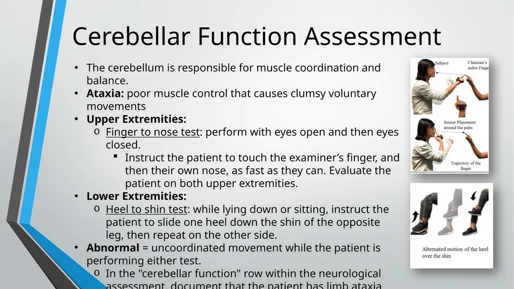
Cerebellar Function Assessment The cerebellum is responsible for muscle coordination and balance.
Ataxia: poor muscle control that causes clumsy voluntary movements
Upper Extremities:
Finger to nose test: perform with eyes open and then eyes closed.
Instruct the patient to touch the examiner’s finger, and then their own nose, as fast as they can. Evaluate the patient on both upper extremities.
Lower Extremities:
Heel to shin test: while lying down or sitting, instruct the patient to slide one heel down the shin of the opposite leg, then repeat on the other side.
Abnormal = uncoordinated movement while the patient is performing either test.
In the "cerebellar function" row within the neurological assessment, document that the patient has limb ataxia and specify which limb(s) are affected.<br>
Ataxia: poor muscle control that causes clumsy voluntary movements
Upper Extremities:
Finger to nose test: perform with eyes open and then eyes closed.
Instruct the patient to touch the examiner’s finger, and then their own nose, as fast as they can. Evaluate the patient on both upper extremities.
Lower Extremities:
Heel to shin test: while lying down or sitting, instruct the patient to slide one heel down the shin of the opposite leg, then repeat on the other side.
Abnormal = uncoordinated movement while the patient is performing either test.
In the "cerebellar function" row within the neurological assessment, document that the patient has limb ataxia and specify which limb(s) are affected.<br>
30
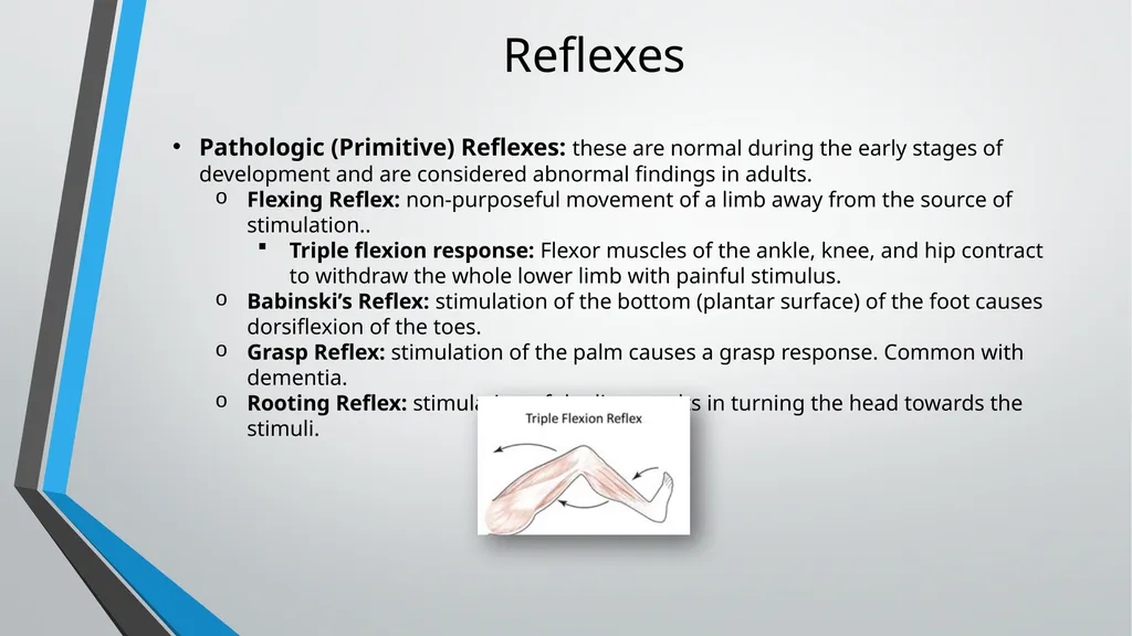
Reflexes Pathologic (Primitive) Reflexes: these are normal during the early stages of development and are considered abnormal findings in adults.
Flexing Reflex: non-purposeful movement of a limb away from the source of stimulation..
Triple flexion response: Flexor muscles of the ankle, knee, and hip contract to withdraw the whole lower limb with painful stimulus.
Babinski’s Reflex: stimulation of the bottom (plantar surface) of the foot causes dorsiflexion of the toes.
Grasp Reflex: stimulation of the palm causes a grasp response. Common with dementia.
Rooting Reflex: stimulation of the lips results in turning the head towards the stimuli.<br>
Flexing Reflex: non-purposeful movement of a limb away from the source of stimulation..
Triple flexion response: Flexor muscles of the ankle, knee, and hip contract to withdraw the whole lower limb with painful stimulus.
Babinski’s Reflex: stimulation of the bottom (plantar surface) of the foot causes dorsiflexion of the toes.
Grasp Reflex: stimulation of the palm causes a grasp response. Common with dementia.
Rooting Reflex: stimulation of the lips results in turning the head towards the stimuli.<br>
31
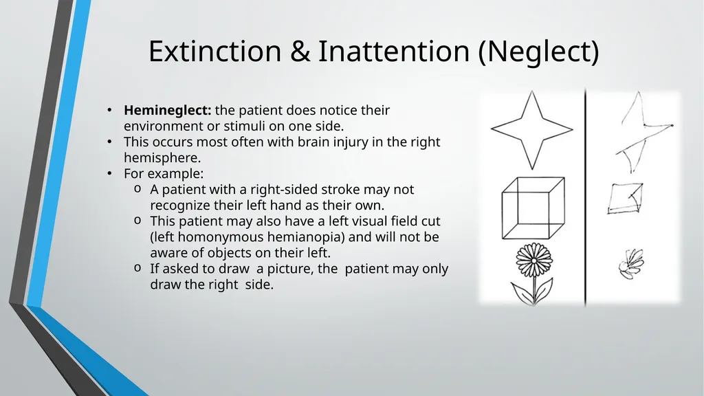
Extinction & Inattention (Neglect) Hemineglect: the patient does notice their environment or stimuli on one side.
This occurs most often with brain injury in the right hemisphere.
For example:
A patient with a right-sided stroke may not recognize their left hand as their own.
This patient may also have a left visual field cut (left homonymous hemianopia) and will not be aware of objects on their left.
If asked to draw a picture, the patient may only draw the right side.<br>
This occurs most often with brain injury in the right hemisphere.
For example:
A patient with a right-sided stroke may not recognize their left hand as their own.
This patient may also have a left visual field cut (left homonymous hemianopia) and will not be aware of objects on their left.
If asked to draw a picture, the patient may only draw the right side.<br>
32
Neurological Symptoms of
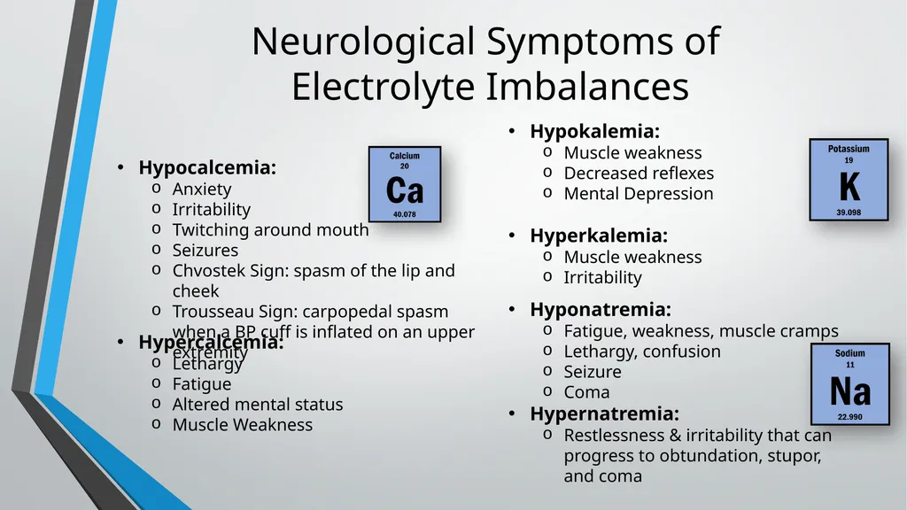
Electrolyte Imbalances Hypocalcemia:
Anxiety
Irritability
Twitching around mouth
Seizures
Chvostek Sign: spasm of the lip and cheek
Trousseau Sign: carpopedal spasm when a BP cuff is inflated on an upper extremity Hypercalcemia:
Lethargy
Fatigue
Altered mental status
Muscle Weakness Hypokalemia:
Muscle weakness
Decreased reflexes
Mental Depression
Hyperkalemia:
Muscle weakness
Irritability Hyponatremia:
Fatigue, weakness, muscle cramps
Lethargy, confusion
Seizure
Coma
Hypernatremia:
Restlessness & irritability that can progress to obtundation, stupor, and coma<br>
Anxiety
Irritability
Twitching around mouth
Seizures
Chvostek Sign: spasm of the lip and cheek
Trousseau Sign: carpopedal spasm when a BP cuff is inflated on an upper extremity Hypercalcemia:
Lethargy
Fatigue
Altered mental status
Muscle Weakness Hypokalemia:
Muscle weakness
Decreased reflexes
Mental Depression
Hyperkalemia:
Muscle weakness
Irritability Hyponatremia:
Fatigue, weakness, muscle cramps
Lethargy, confusion
Seizure
Coma
Hypernatremia:
Restlessness & irritability that can progress to obtundation, stupor, and coma<br>
33
Neurological Symptoms of
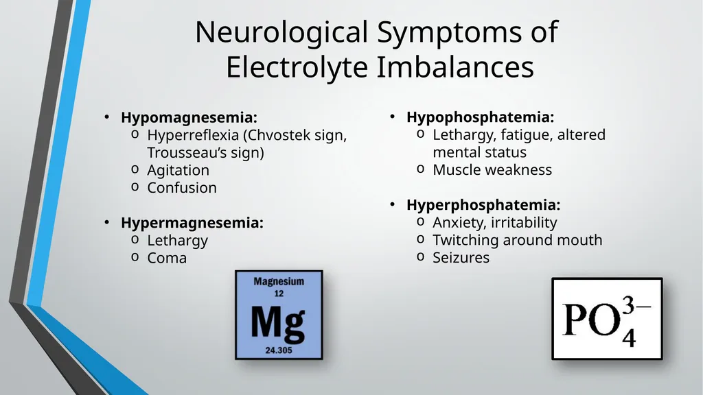
Electrolyte Imbalances Hypomagnesemia:
Hyperreflexia (Chvostek sign, Trousseau’s sign)
Agitation
Confusion
Hypermagnesemia:
Lethargy
Coma Hypophosphatemia:
Lethargy, fatigue, altered mental status
Muscle weakness
Hyperphosphatemia:
Anxiety, irritability
Twitching around mouth
Seizures<br>
Hyperreflexia (Chvostek sign, Trousseau’s sign)
Agitation
Confusion
Hypermagnesemia:
Lethargy
Coma Hypophosphatemia:
Lethargy, fatigue, altered mental status
Muscle weakness
Hyperphosphatemia:
Anxiety, irritability
Twitching around mouth
Seizures<br>
34
Neurological Exam
of the Comatose
Patient<br>
35
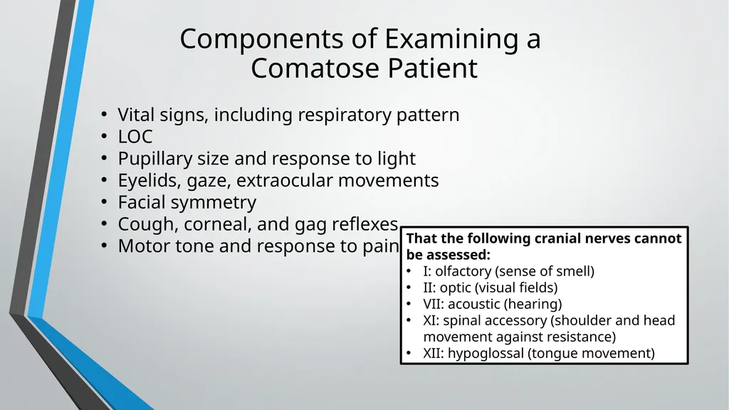
Components of Examining a
Comatose Patient Vital signs, including respiratory pattern
LOC
Pupillary size and response to light
Eyelids, gaze, extraocular movements
Facial symmetry
Cough, corneal, and gag reflexes
Motor tone and response to pain That the following cranial nerves cannot be assessed:
I: olfactory (sense of smell)
II: optic (visual fields)
VII: acoustic (hearing)
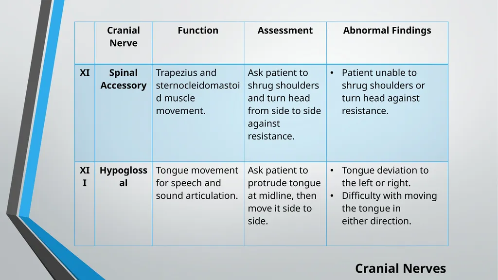
XI: spinal accessory (shoulder and head movement against resistance)
XII: hypoglossal (tongue movement)<br>
LOC
Pupillary size and response to light
Eyelids, gaze, extraocular movements
Facial symmetry
Cough, corneal, and gag reflexes
Motor tone and response to pain That the following cranial nerves cannot be assessed:
I: olfactory (sense of smell)
II: optic (visual fields)
VII: acoustic (hearing)
XI: spinal accessory (shoulder and head movement against resistance)
XII: hypoglossal (tongue movement)<br>
36
Vital Sign Changes with Neurological Injury Vital sign changes are a late sign with brain injury, lesion, and hemorrhage.
Cushing's Triad: signs of brain herniation, which results from cerebral edema.
Increased systolic BP with widening pulse pressure
Bradycardia
Bradypnea
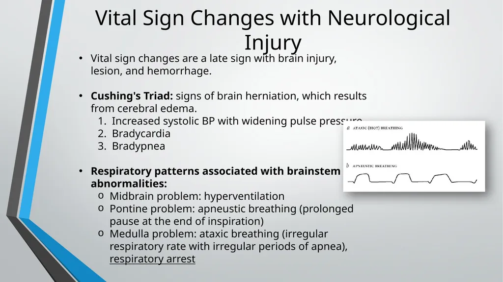
Respiratory patterns associated with brainstem abnormalities:
Midbrain problem: hyperventilation
Pontine problem: apneustic breathing (prolonged pause at the end of inspiration)
Medulla problem: ataxic breathing (irregular respiratory rate with irregular periods of apnea), respiratory arrest<br>
Cushing's Triad: signs of brain herniation, which results from cerebral edema.
Increased systolic BP with widening pulse pressure
Bradycardia
Bradypnea
Respiratory patterns associated with brainstem abnormalities:
Midbrain problem: hyperventilation
Pontine problem: apneustic breathing (prolonged pause at the end of inspiration)
Medulla problem: ataxic breathing (irregular respiratory rate with irregular periods of apnea), respiratory arrest<br>
37
Level of Consciousness Determine the extent of coma.
1st assess for a response to verbal stimulus or loud noise (clapping).
2nd if there is no response to verbal stimulus, use noxious stimuli.
If the patient’s family is in the room, do not assess for a response to pain without explaining the purpose of the assessment to them.<br>
1st assess for a response to verbal stimulus or loud noise (clapping).
2nd if there is no response to verbal stimulus, use noxious stimuli.
If the patient’s family is in the room, do not assess for a response to pain without explaining the purpose of the assessment to them.<br>
38
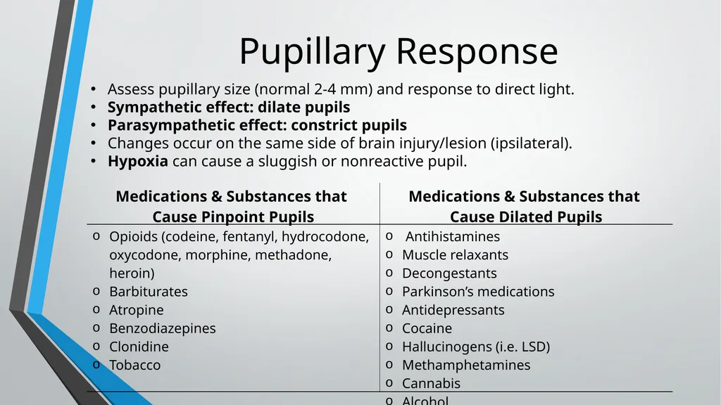
Pupillary Response Assess pupillary size (normal 2-4 mm) and response to direct light.
Sympathetic effect: dilate pupils
Parasympathetic effect: constrict pupils
Changes occur on the same side of brain injury/lesion (ipsilateral).
Hypoxia can cause a sluggish or nonreactive pupil.<br>
Sympathetic effect: dilate pupils
Parasympathetic effect: constrict pupils
Changes occur on the same side of brain injury/lesion (ipsilateral).
Hypoxia can cause a sluggish or nonreactive pupil.<br>
39
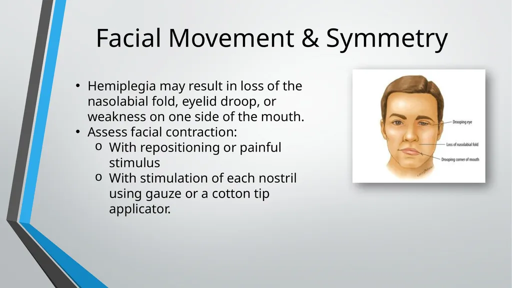
Facial Movement & Symmetry Hemiplegia may result in loss of the nasolabial fold, eyelid droop, or weakness on one side of the mouth.
Assess facial contraction:
With repositioning or painful stimulus
With stimulation of each nostril using gauze or a cotton tip applicator.<br>
Assess facial contraction:
With repositioning or painful stimulus
With stimulation of each nostril using gauze or a cotton tip applicator.<br>
40
Eyelids, Gaze, & Ocular Movement Gently raise the eyelids to inspect the position and movement of the eyes. In a comatose patient, the eyelids will close slowly when released.
Roving Eye Movement: spontaneous movement of the eyes. This is seen in comatose patients with intact oculomotor (CN III) function.
Gaze Preference:
Stoke: Eyes deviate toward the side of the lesion; usually lasts for minutes to hours.
Seizure: Eyes deviate away from the discharging focus; can last several days.
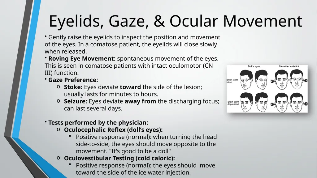
Tests performed by the physician:
Oculocephalic Reflex (doll’s eyes):
Positive response (normal): when turning the head side-to-side, the eyes should move opposite to the movement. "It's good to be a doll"
Oculovestibular Testing (cold caloric):
Positive response (normal): the eyes should move toward the side of the ice water injection.<br>
Roving Eye Movement: spontaneous movement of the eyes. This is seen in comatose patients with intact oculomotor (CN III) function.
Gaze Preference:
Stoke: Eyes deviate toward the side of the lesion; usually lasts for minutes to hours.
Seizure: Eyes deviate away from the discharging focus; can last several days.
Tests performed by the physician:
Oculocephalic Reflex (doll’s eyes):
Positive response (normal): when turning the head side-to-side, the eyes should move opposite to the movement. "It's good to be a doll"
Oculovestibular Testing (cold caloric):
Positive response (normal): the eyes should move toward the side of the ice water injection.<br>
41
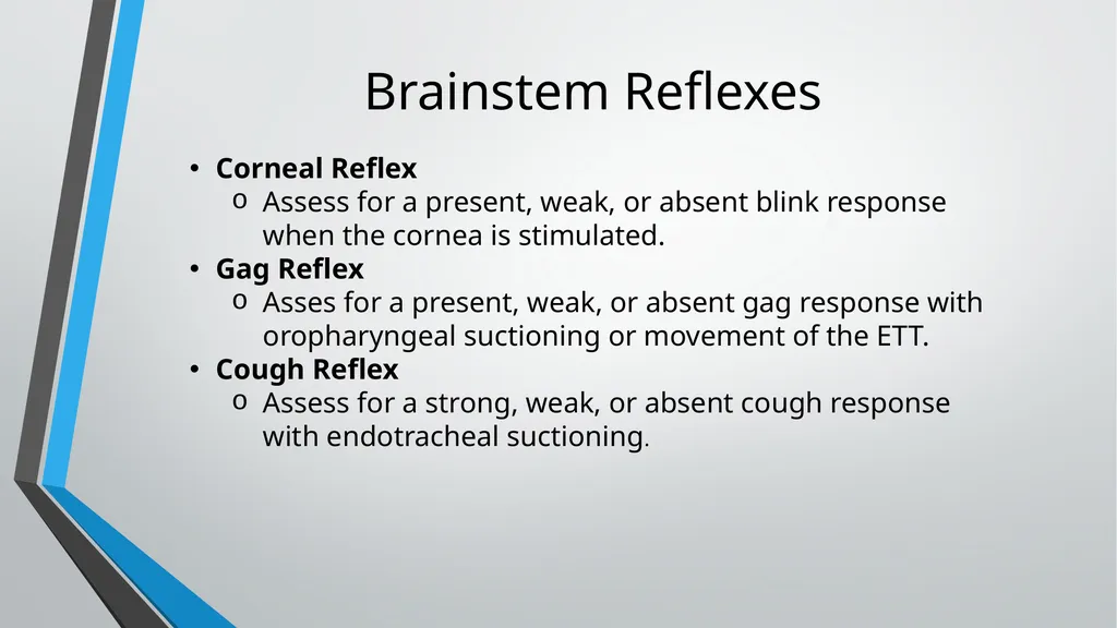
Brainstem Reflexes Corneal Reflex
Assess for a present, weak, or absent blink response when the cornea is stimulated.
Gag Reflex
Asses for a present, weak, or absent gag response with oropharyngeal suctioning or movement of the ETT.
Cough Reflex
Assess for a strong, weak, or absent cough response with endotracheal suctioning.<br>
Assess for a present, weak, or absent blink response when the cornea is stimulated.
Gag Reflex
Asses for a present, weak, or absent gag response with oropharyngeal suctioning or movement of the ETT.
Cough Reflex
Assess for a strong, weak, or absent cough response with endotracheal suctioning.<br>
42
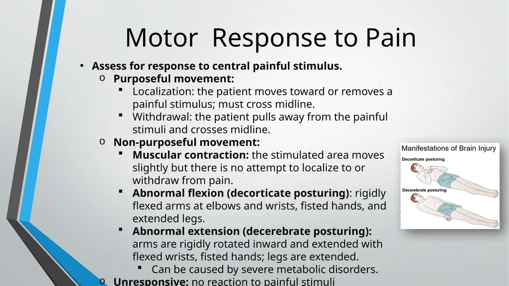
Motor Response to Pain Assess for response to central painful stimulus.
Purposeful movement:
Localization: the patient moves toward or removes a painful stimulus; must cross midline.
Withdrawal: the patient pulls away from the painful stimuli and crosses midline.
Non-purposeful movement:
Muscular contraction: the stimulated area moves slightly but there is no attempt to localize to or withdraw from pain.
Abnormal flexion (decorticate posturing): rigidly flexed arms at elbows and wrists, fisted hands, and extended legs.
Abnormal extension (decerebrate posturing): arms are rigidly rotated inward and extended with flexed wrists, fisted hands; legs are extended.
Can be caused by severe metabolic disorders.
Unresponsive: no reaction to painful stimuli<br>
Purposeful movement:
Localization: the patient moves toward or removes a painful stimulus; must cross midline.
Withdrawal: the patient pulls away from the painful stimuli and crosses midline.
Non-purposeful movement:
Muscular contraction: the stimulated area moves slightly but there is no attempt to localize to or withdraw from pain.
Abnormal flexion (decorticate posturing): rigidly flexed arms at elbows and wrists, fisted hands, and extended legs.
Abnormal extension (decerebrate posturing): arms are rigidly rotated inward and extended with flexed wrists, fisted hands; legs are extended.
Can be caused by severe metabolic disorders.
Unresponsive: no reaction to painful stimuli<br>
43
Motor Size & Tone Observe muscles for size.
Palpate muscles at rest and during passive movement to assess tone.
Abnormalities in muscle tone:
Spasticity: increased resistance to passive movement.
Rigidity: a state of increased resistance.
Flaccidity (hypotonia): decreased muscle tone; the muscle is weak, soft, and fatigues easily.<br>
Palpate muscles at rest and during passive movement to assess tone.
Abnormalities in muscle tone:
Spasticity: increased resistance to passive movement.
Rigidity: a state of increased resistance.
Flaccidity (hypotonia): decreased muscle tone; the muscle is weak, soft, and fatigues easily.<br>
44
Helpful Neuro Assessment Tips A change in LOC is always the first sign of a neurological problem (with an exception of epidural hematomas).
Eyes (gaze) will deviate towards the problem with acute stroke.
Eyes (gaze) will deviate away from the problem with seizures.
Pupil changes are ipsilateral to the problem (same side).
Motor, sensory, and visual changes are contralateral to the problem (opposite side).
Extinction/Inattention (neglect) is contralateral to the problem.<br>
Eyes (gaze) will deviate towards the problem with acute stroke.
Eyes (gaze) will deviate away from the problem with seizures.
Pupil changes are ipsilateral to the problem (same side).
Motor, sensory, and visual changes are contralateral to the problem (opposite side).
Extinction/Inattention (neglect) is contralateral to the problem.<br>
45
Sources Hickey, Joanne V. The Clinical Practice of Neurological and Neurosurgical Nursing. Edited by Andrea L. Strayer, 8th ed., Wolters Kluwer, 2020.
Juarez, Pat. Barron's CCRN Exam. Barron's, 2015.
Nekic, Paula. “Neurological Learning Package.” 2014. https://www.studocu.com/en-us/document/texas-womans-university/med-surg/neurological-learning-package/7764848<br>
Juarez, Pat. Barron's CCRN Exam. Barron's, 2015.
Nekic, Paula. “Neurological Learning Package.” 2014. https://www.studocu.com/en-us/document/texas-womans-university/med-surg/neurological-learning-package/7764848<br>