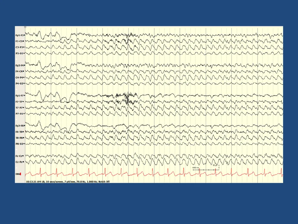
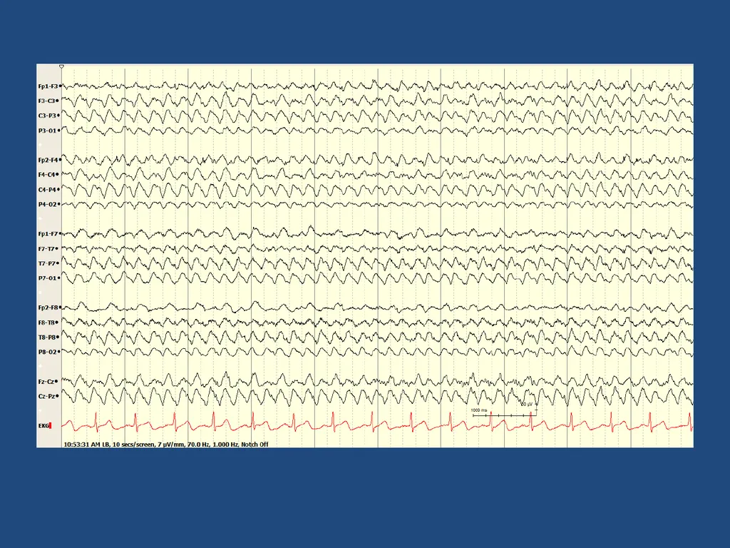
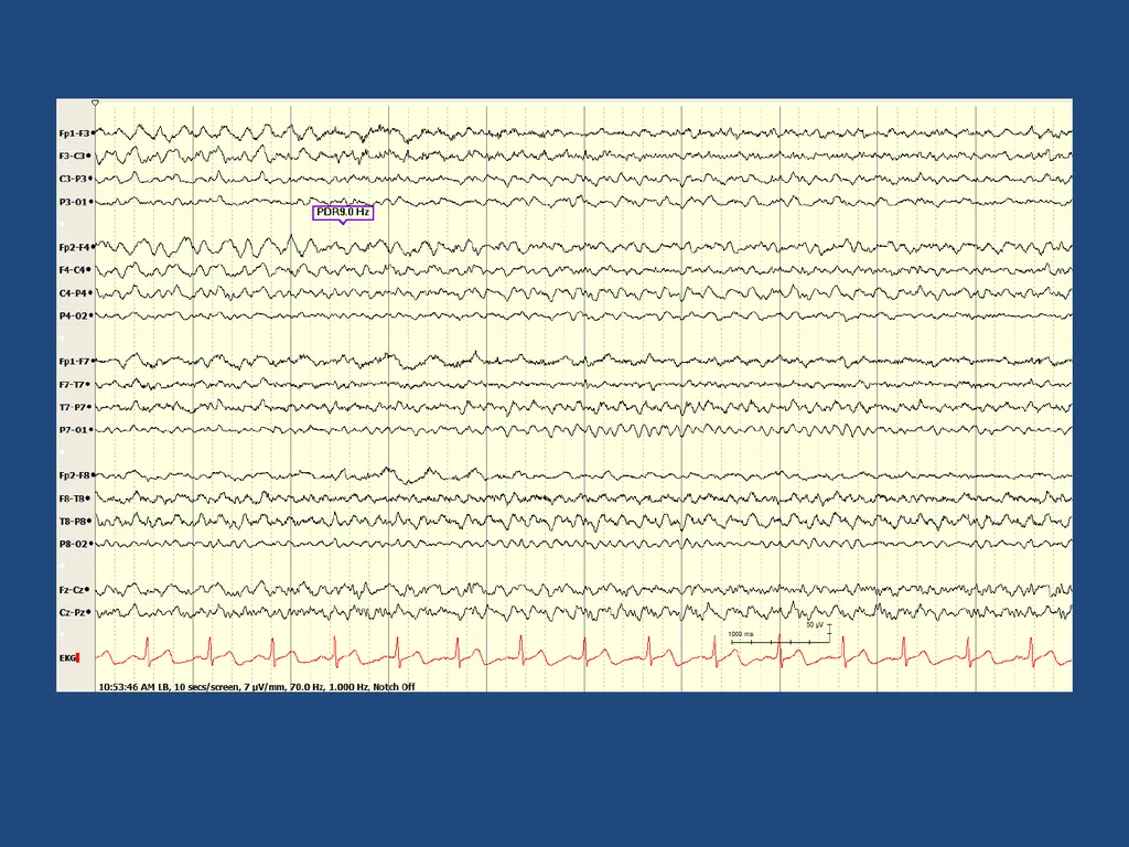
04
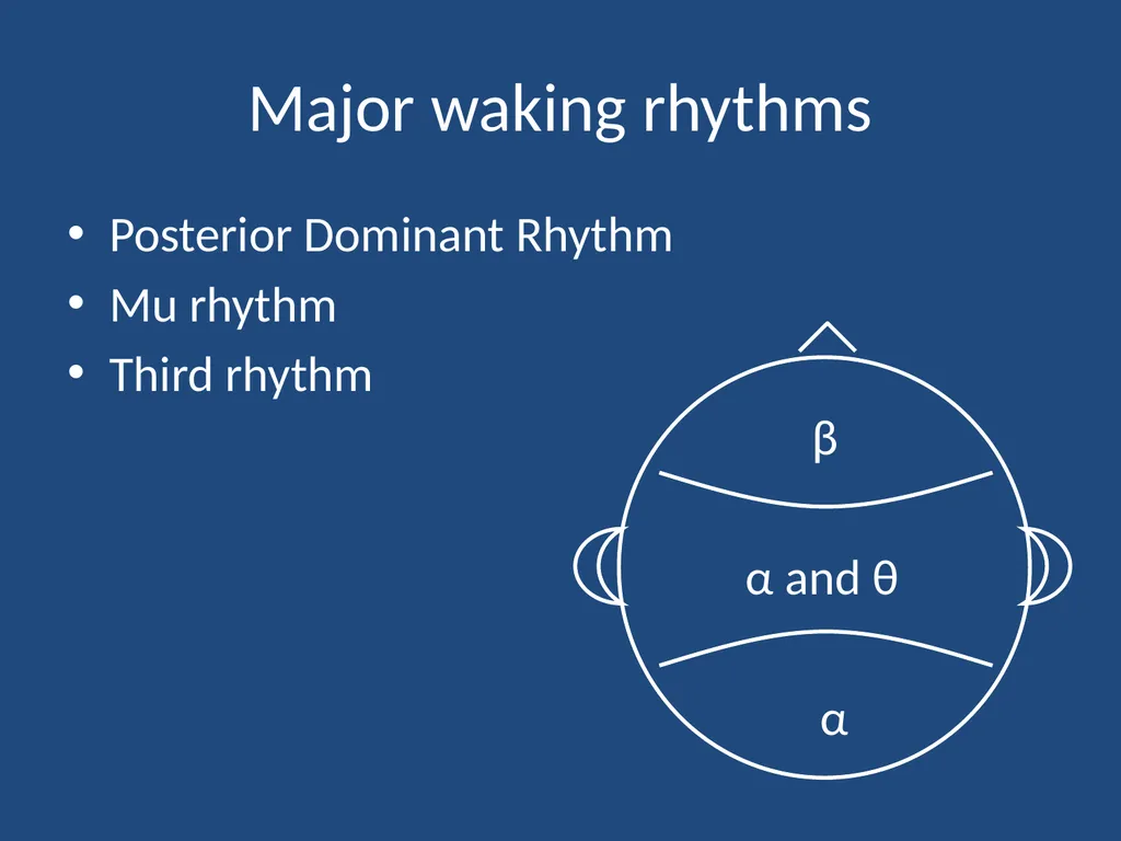
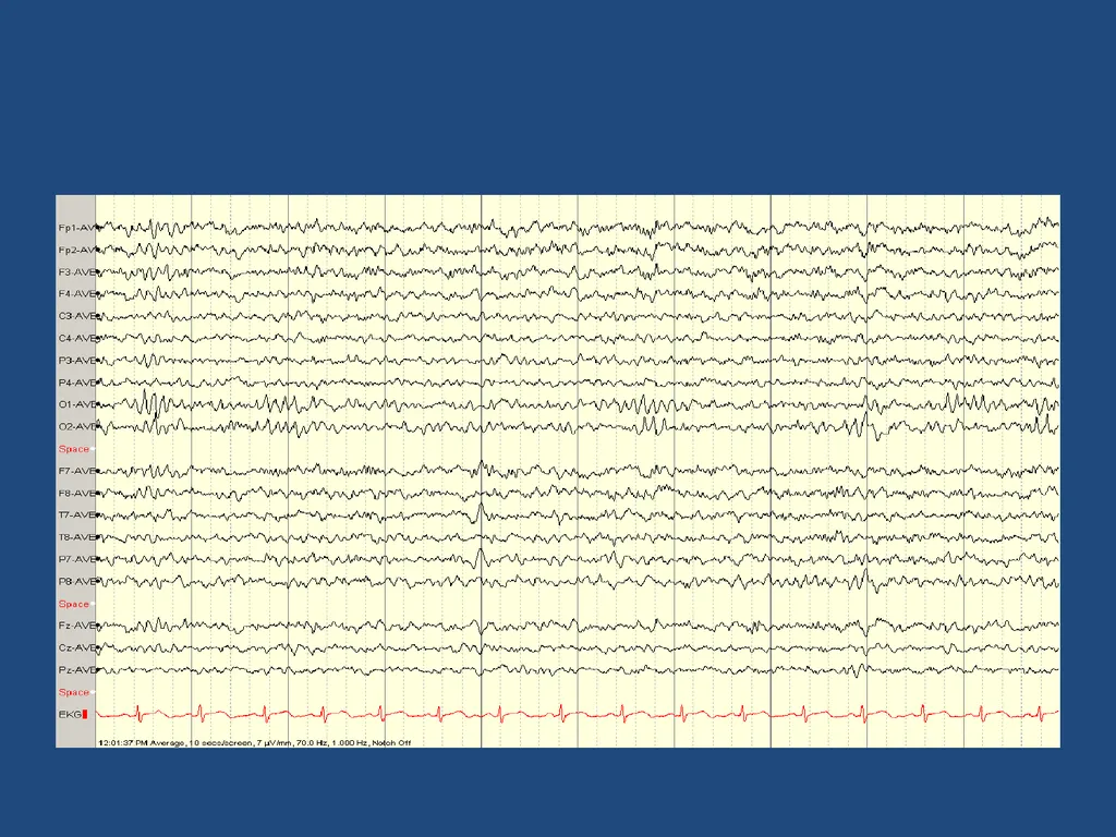
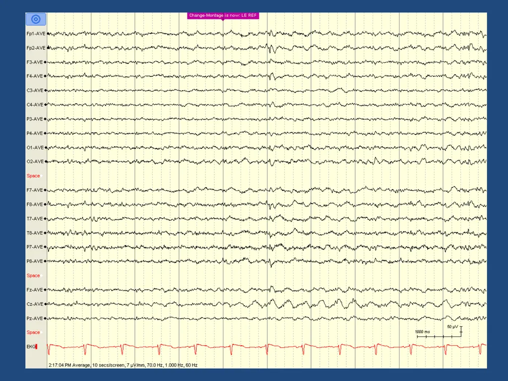
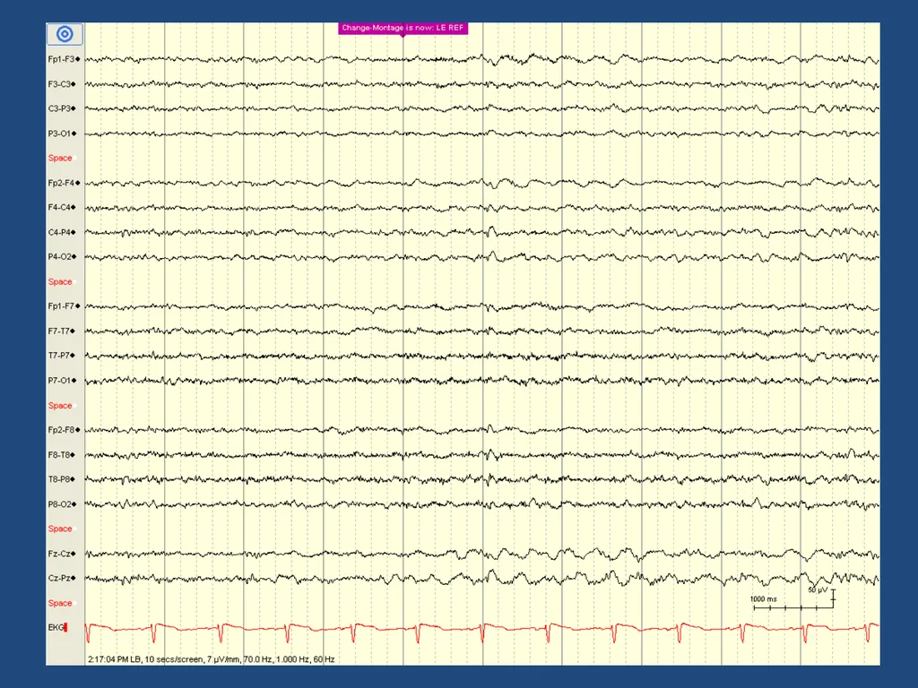
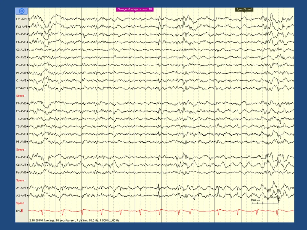
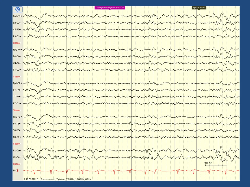
Major waking rhythms Posterior Dominant Rhythm
Mu rhythm
Third rhythm β α α and θ<br>
05
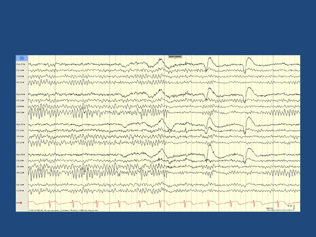
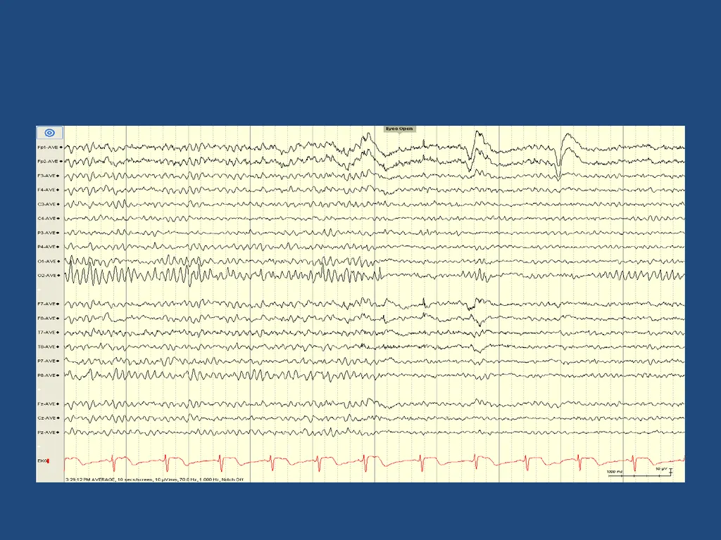
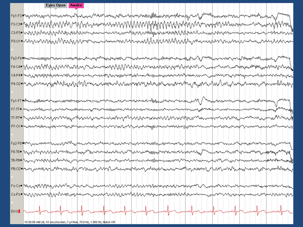
Posterior Dominant Rhythm “the Alpha rhythm” (resting rhythm of occipital cortex)
Variants
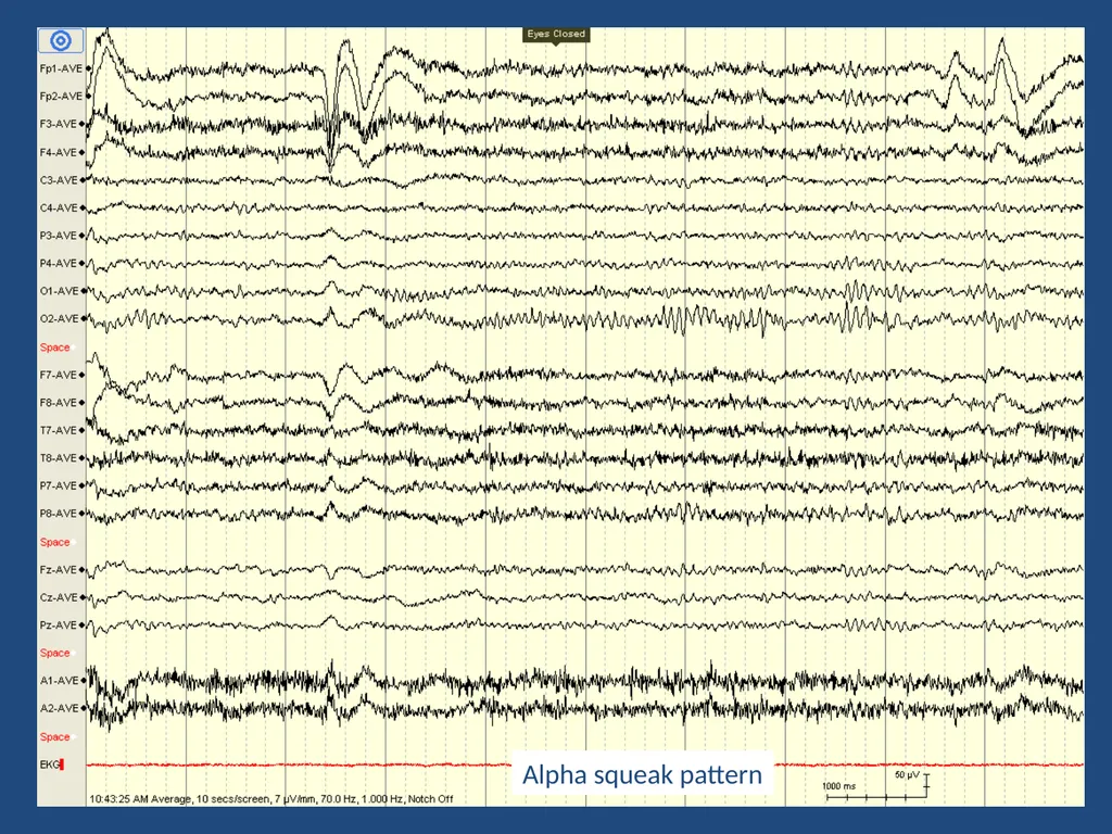
alpha squeak
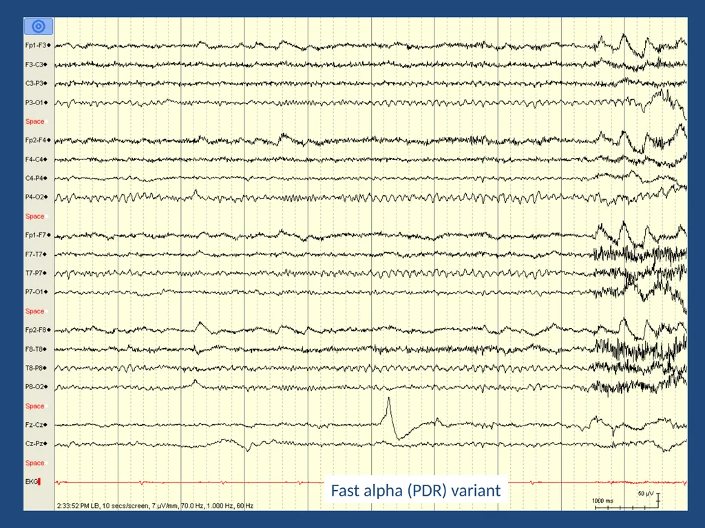
slow and fast variants
paradoxical alpha – increases with alertness
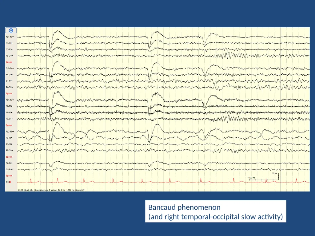
Bancaud’s phenomenon (abnormal) – failure to attenuate with eye closure (ipsilateral pathway lesion)
Should be symmetric…?<br>
06
What is the allowable, normal asymmetry regarding the posterior dominant rhythm (alpha rhythm)?
Up to 35% higher amplitude on the right, and up to 35% higher amplitude on the left.
Up to 50% higher amplitude on the right, and up to 35% higher amplitude on the left.
Up to 35% higher amplitude on the right, and up to 50% higher amplitude on the left.
Up to 50% higher amplitude on the right, and up to 50% higher amplitude on the left.
Any asymmetry is considered abnormal<br>
07
Alpha squeak pattern<br>
08
Fast alpha (PDR) variant<br>
09
Bancaud phenomenon
(and right temporal-occipital slow activity)<br>
10
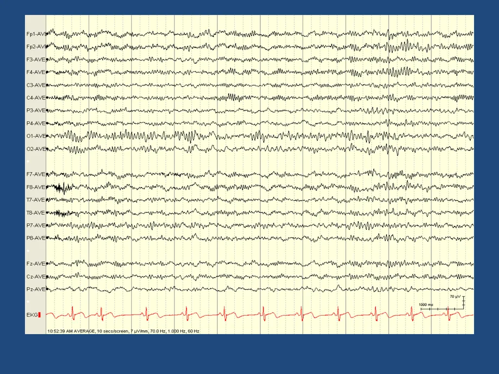
Mu rhythm resting rhythm of central (premotor) cortex
Alpha or theta (7-11 Hz) spiky rhythm
Looks like the letter “µ”
Attenuates with contralateral limb movement (or even thinking about movement)
Often enhanced in presence of breach rhythm<br>
11
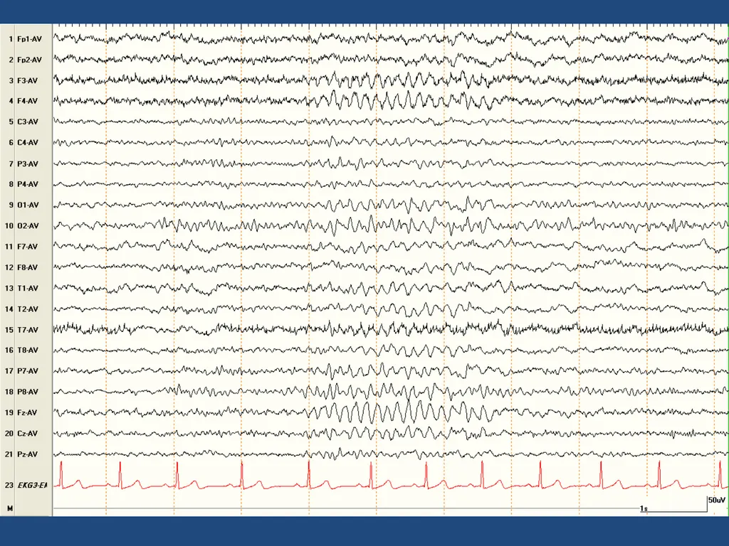
Midline theta a.k.a. Ciganek rhythm
5-7 Hz sinusoidal activity maximal at Cz or Fz
may be spiky or arciform (mu-like)
Present in awake and drowsy states
Unrelated to eye opening, alerting, limb mvmt
May enhance with concentration (midline frontal theta)<br>
12
Which of the following is NOT a characteristic of Stage N1 Sleep?
Slow lateral eye movements
Attenuation of posterior dominant rhythm
Emergence of sleep spindles
Emergence of vertex waves
Emergence of theta activity<br>
13
Drowsy EEG Changes in PDR
Attenuation without eye opening
May slow by up to 1 Hz
May become anteriorly projected
Slow lateral eye movements
Emergence of theta activity (often bursts)
Emergence of frontal beta activity<br>
14
Frontal Beta Activity Resting rhythm of the frontal lobes
Best seen in drowsiness
Abnormal*
Generalized
High voltage (>30 µV)
Persistent in sleep or awake states *think benzodiazepines, barbiturates, and propofol<br>
15
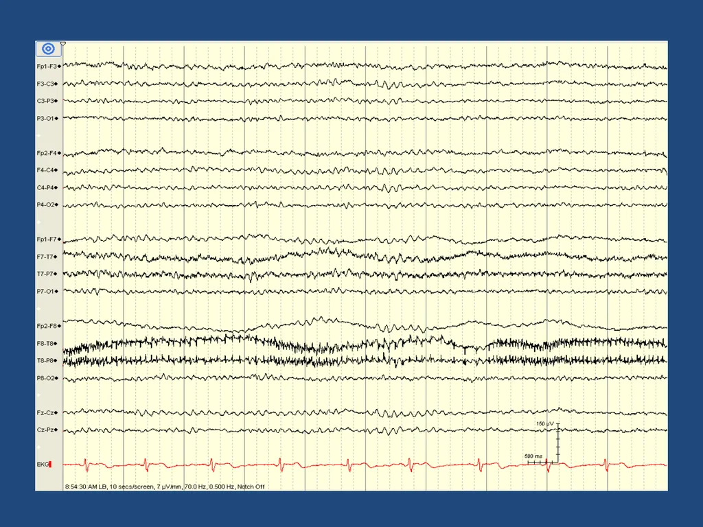
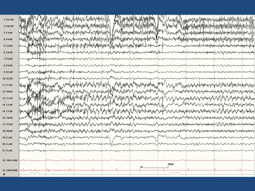
Third rhythm Resting rhythm of temporal cortex
Alpha or theta (7-11 Hz) sinusoidal rhythm
May be asynchronous or unilateral
Can be seen in waking or drowsiness
If not seen, EEG may still be normal<br>
16
Which of the following features is especially useful in differentiating normal / benign variants from true epileptiform discharges?
Benign variants attenuate during deeper sleep
Benign variants disrupt the background rhythms
Benign variants have associated slow activity
Benign variants are blunt (not sharp or spike)
Benign variants are low voltage<br>
17
Benign Variants May be rhythmic or occur in isolation
May be high or low voltage (typically low)
May be quite “sharp” or “spiky”
Usually in drowsiness (not in deeper sleep)
Should not disrupt the background
Have a “smooth” rhythm
No associated slow / delta activity<br>
18
Which of the following normal / benign variants occurs in waking (and not drowsiness)?
Wicket waves
14- and 6-Hz positive bursts
Small sharp spikes
Lambda waves
Psychomotor variant<br>
19
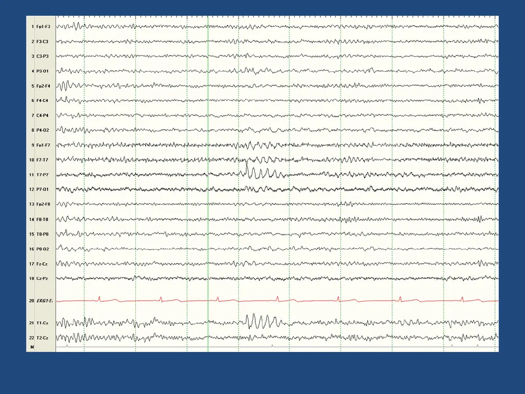
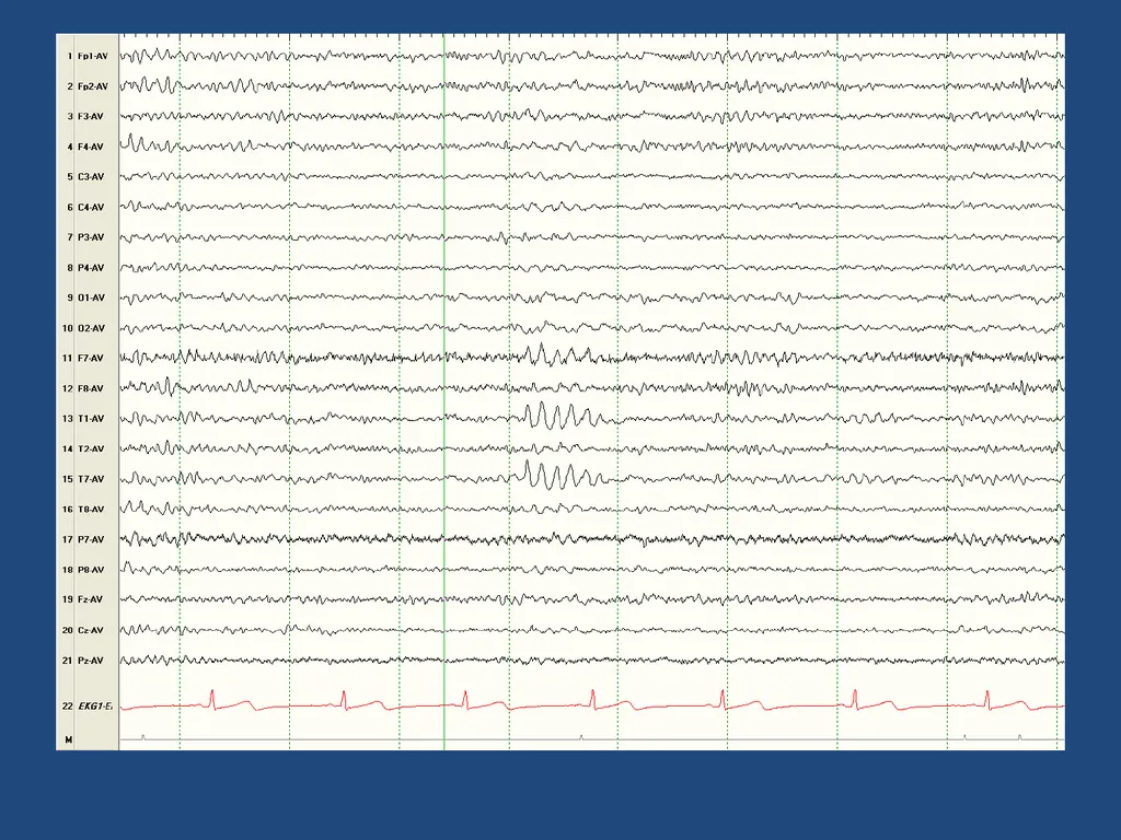
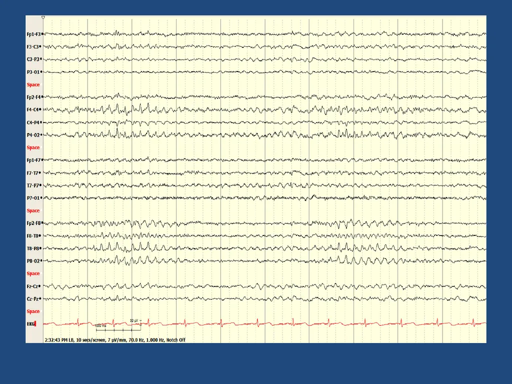
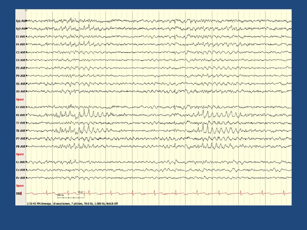
Wickets Rhythmic bursts of monophasic 6-11 Hz activity
Seen bitemporally in drowsiness (not in deep sleep)
Typically occur in trains or runs
don’t disrupt background
tend to be “isosceles” (no aftergoing slow wave)
when seen as single waves – may be overinterpreted
surrounded by similar waves (may be lower amplitude)
On a spectrum with third rhythm<br>
20
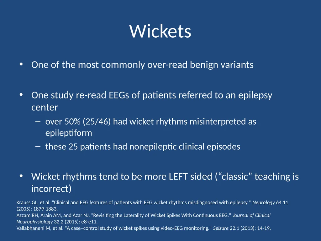
Wickets One of the most commonly over-read benign variants
One study re-read EEGs of patients referred to an epilepsy center
over 50% (25/46) had wicket rhythms misinterpreted as epileptiform
these 25 patients had nonepileptic clinical episodes
Wicket rhythms tend to be more LEFT sided (“classic” teaching is incorrect) Krauss GL, et al. "Clinical and EEG features of patients with EEG wicket rhythms misdiagnosed with epilepsy." Neurology 64.11 (2005): 1879-1883.
Azzam RH, Arain AM, and Azar NJ. "Revisiting the Laterality of Wicket Spikes With Continuous EEG." Journal of Clinical Neurophysiology 32.2 (2015): e8-e11.
Vallabhaneni M, et al. "A case–control study of wicket spikes using video-EEG monitoring." Seizure 22.1 (2013): 14-19.<br>
21
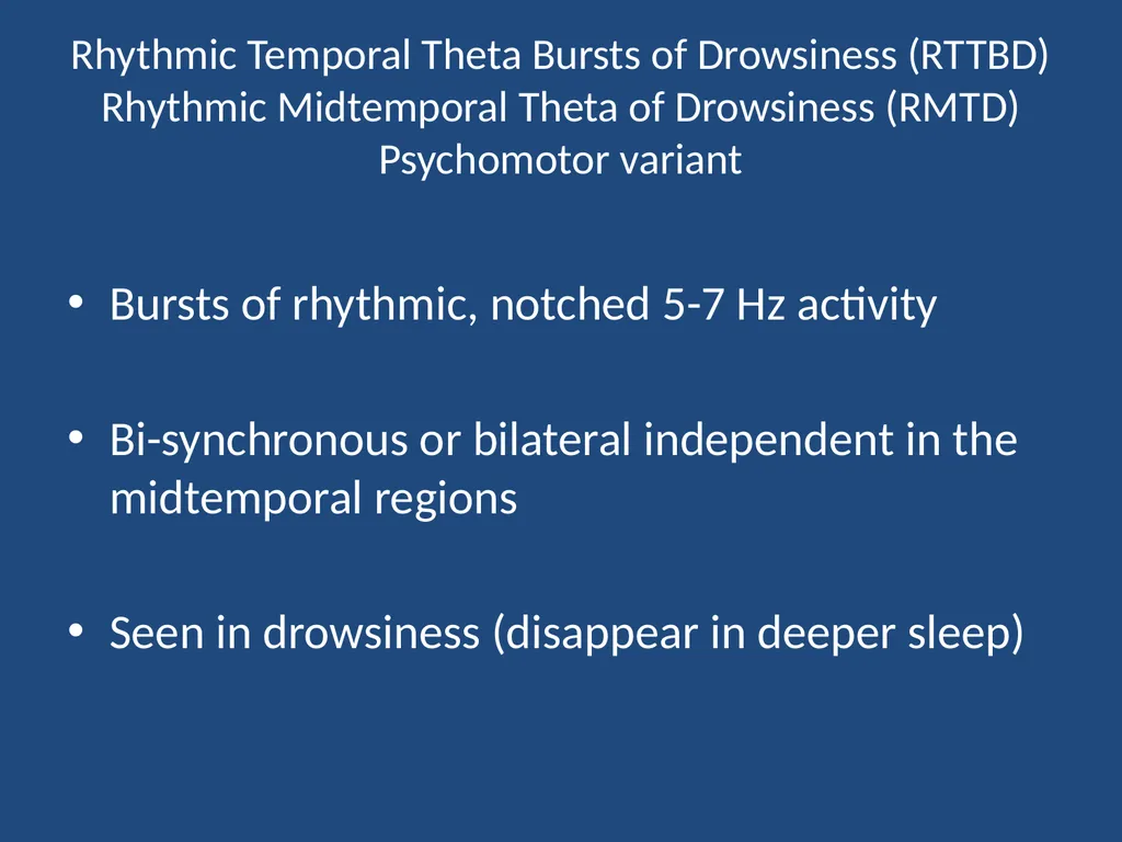
Rhythmic Temporal Theta Bursts of Drowsiness (RTTBD)�Rhythmic Midtemporal Theta of Drowsiness (RMTD)�Psychomotor variant Bursts of rhythmic, notched 5-7 Hz activity
Bi-synchronous or bilateral independent in the midtemporal regions
Seen in drowsiness (disappear in deeper sleep)<br>
22
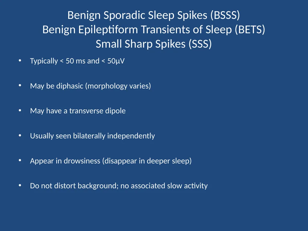
Benign Sporadic Sleep Spikes (BSSS)�Benign Epileptiform Transients of Sleep (BETS)�Small Sharp Spikes (SSS) Typically < 50 ms and < 50μV
May be diphasic (morphology varies)
May have a transverse dipole
Usually seen bilaterally independently
Appear in drowsiness (disappear in deeper sleep)
Do not distort background; no associated slow activity<br>
23
14- and 6-Hz positive bursts (ctenoids) Intermixed 14 Hz and 6-7 Hz activity
Wide field positive polarity (posterior temporal predominance)
Best confirmed on contralateral ear montage (long distance referential)
Occur in N1 or N2 sleep
Seen in normal adolescents but also in hepatic disease (Reye syndrome, hepatic encephalopathy)<br>
24
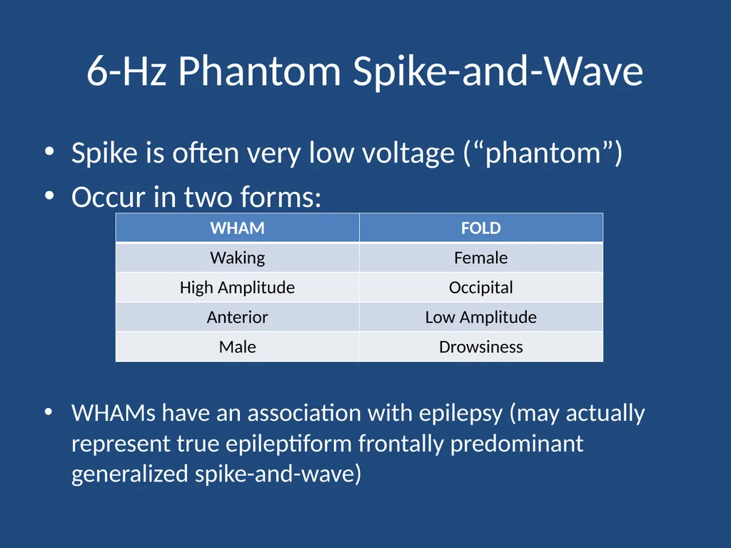
6-Hz Phantom Spike-and-Wave Spike is often very low voltage (“phantom”)
Occur in two forms:
WHAMs have an association with epilepsy (may actually represent true epileptiform frontally predominant generalized spike-and-wave)<br>
25
Which of the following features is necessary to diagnosis SREDA (subclinical rhythmic electrographic discharges of adults)?
Age > 65 years
Lack of clinical signs during discharge
Normal EEG outside of SREDA
Occurs during N2 sleep
Occurs during N3 sleep<br>
26
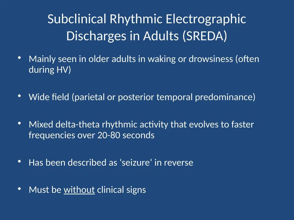
Subclinical Rhythmic Electrographic Discharges in Adults (SREDA) Mainly seen in older adults in waking or drowsiness (often during HV)
Wide field (parietal or posterior temporal predominance)
Mixed delta-theta rhythmic activity that evolves to faster frequencies over 20-80 seconds
Has been described as ‘seizure’ in reverse
Must be without clinical signs<br>
27
Sleep EEG Stage N1 - drowsiness
Stage N2 - specific architecture
Stage N3 - slow wave sleep
REM - rapid eye movement<br>
28
Patterns in Stage N2 Sleep* Sleep spindles
K complexes
Vertex waves (can also be in stage 1)
POSTS
* all are normal, though some appear “sharp”<br>
29
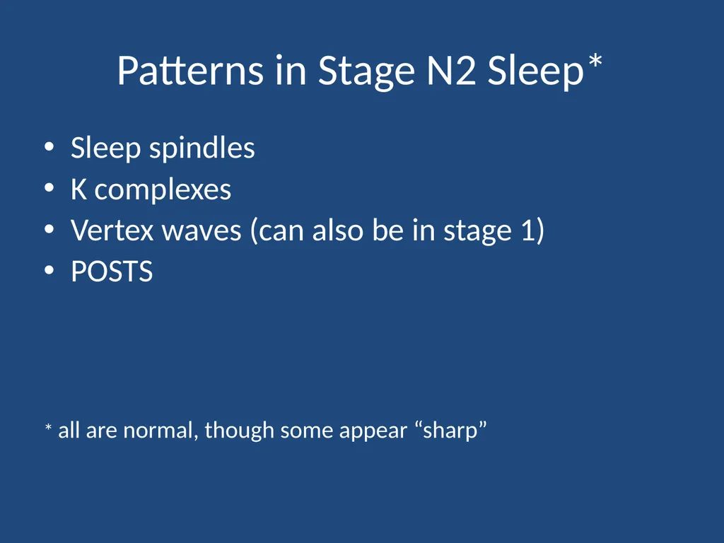
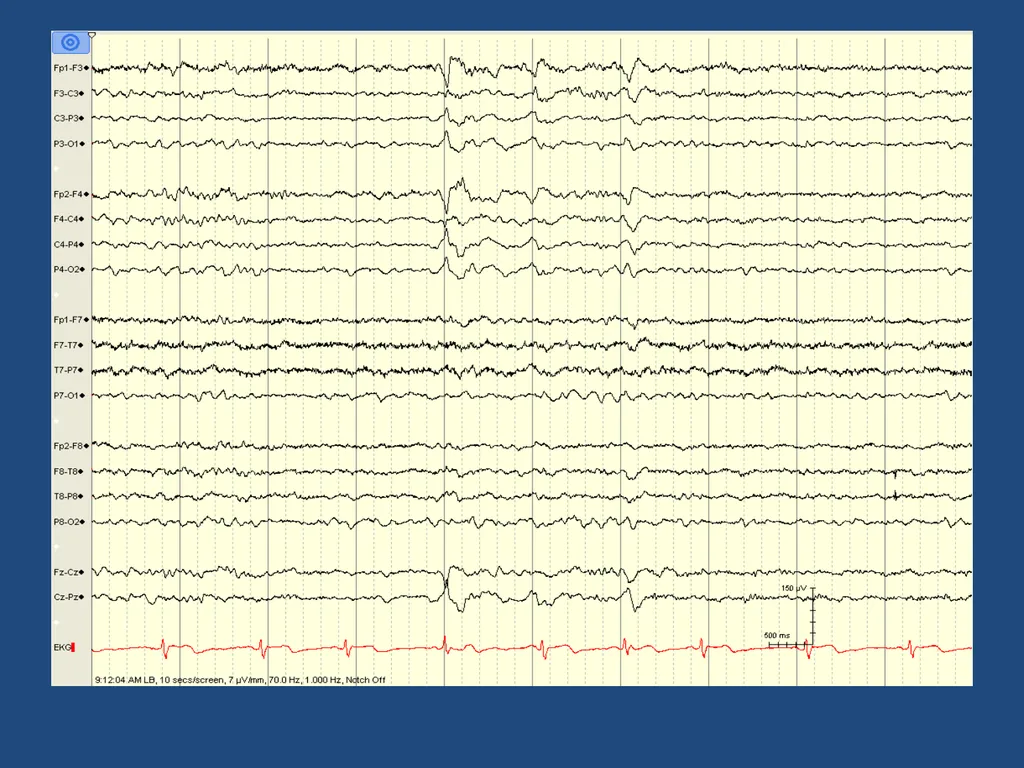
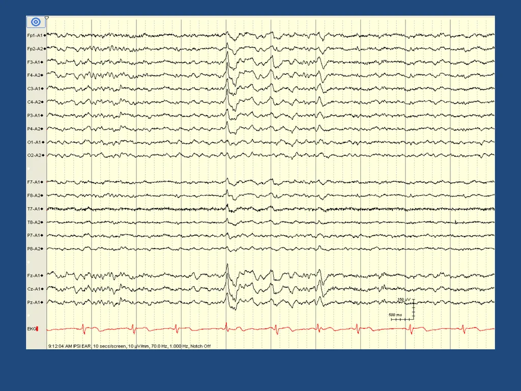
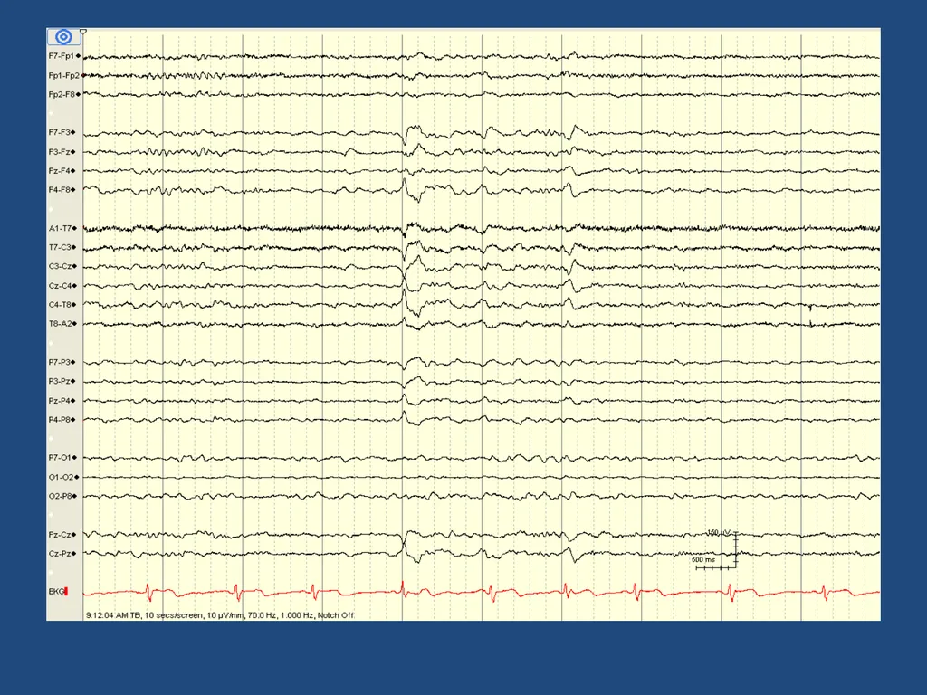
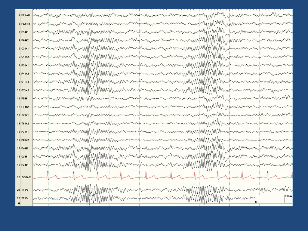
Vertex Waves High voltage, sharp looking, surface negative waves
Originate at Cz (the vertex)
Thought to be generated by the thalamus<br>
30
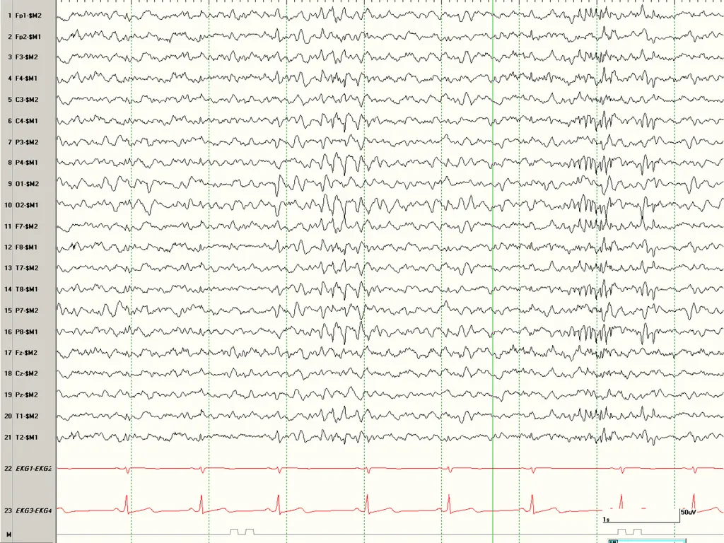
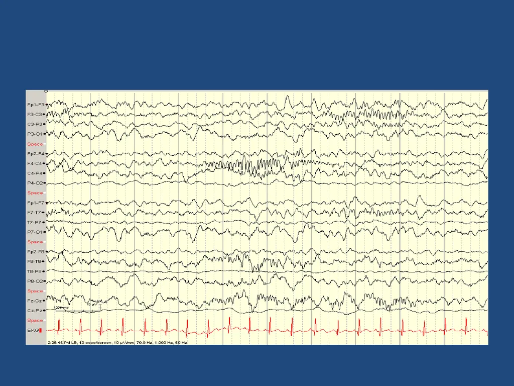
Sleep spindles Bursts of beta activity (12-16 Hz)
symmetric and synchronous (age > 2 yrs)
fronto-central head region
last 1-1.5 seconds
Thought to be generated by the thalamus<br>
31
By what age should sleep spindles become synchronous?
2 months
6 months
12 months
2 years
6 years<br>
32
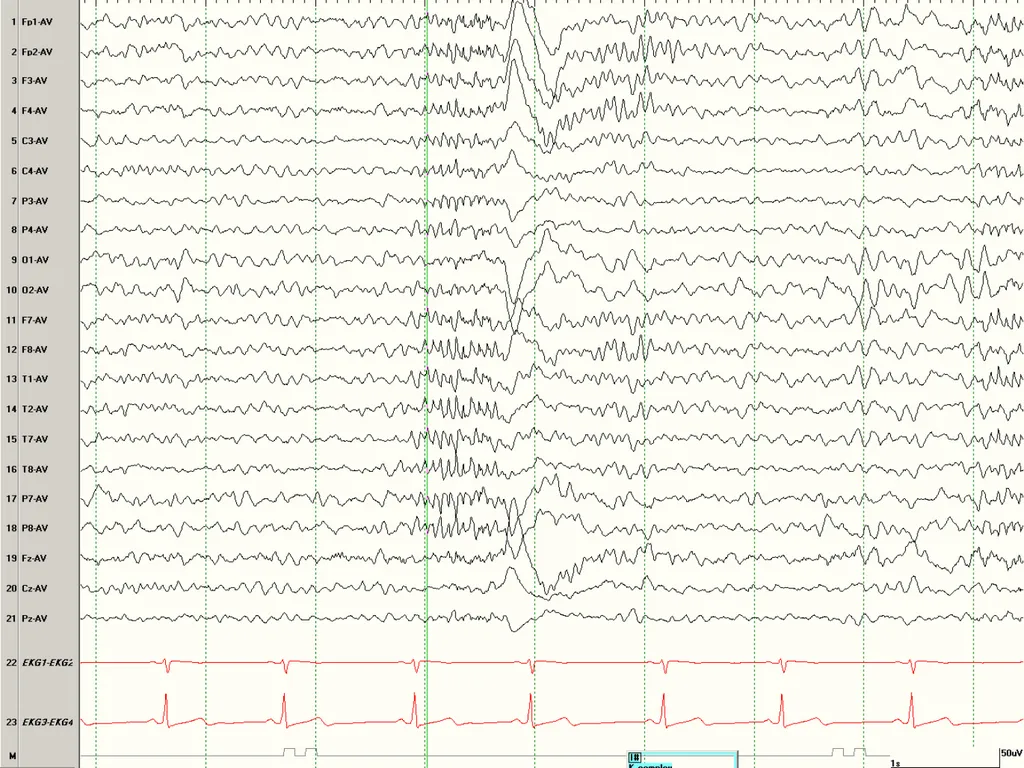
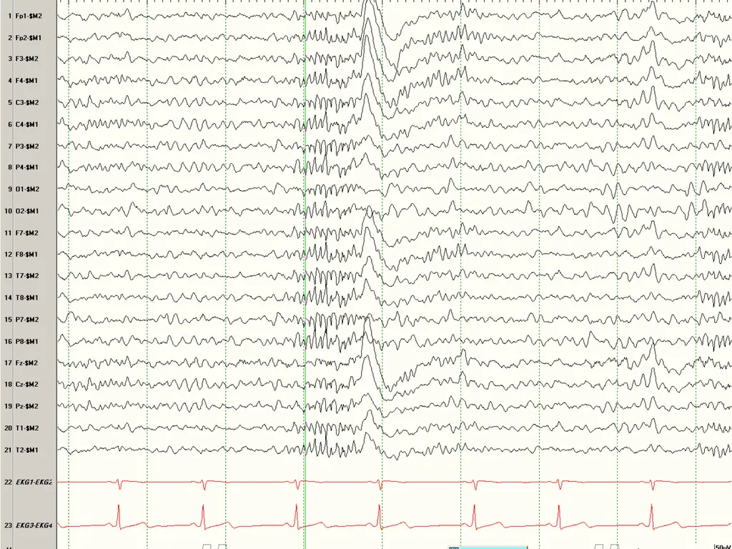
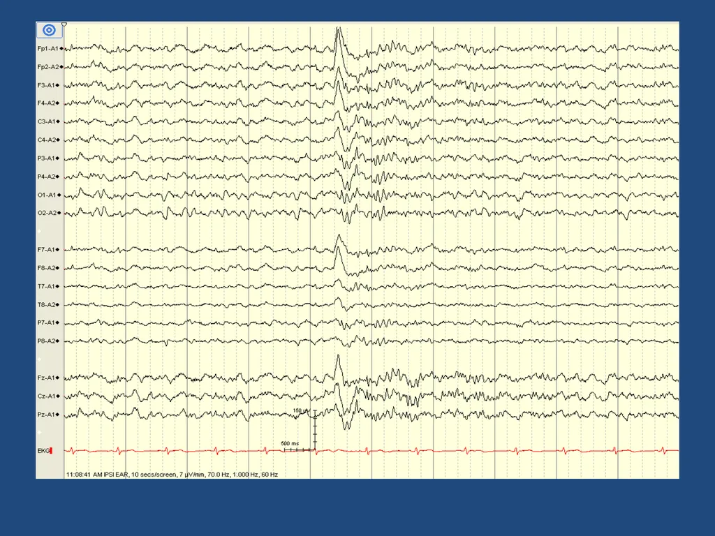
K complexes Broad 0.5-2 second long wave
Fronto-central predominance
Might be followed by a sleep spindle
related to arousal (noise, clapping, knock)<br>
33
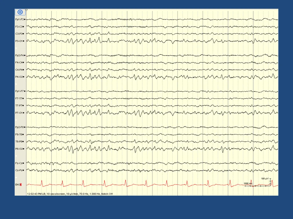
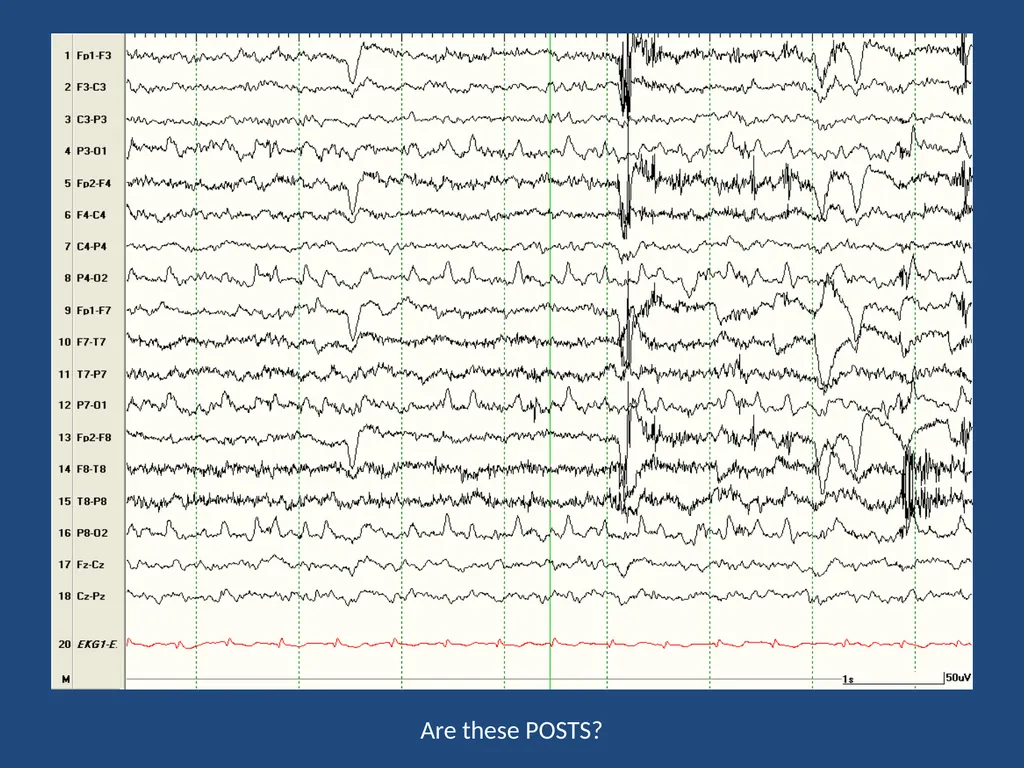
POSTS Positive Occipital Sharp Transients of Sleep
That’s exactly what they are.
Should be symmetric and synchronous.<br>
35
Lambda Waves A sharp looking wave that has a positive polarity in the occipital regions
looks like the letter “λ”
synchronous and symmetric
Occurs when scanning lines or looking at an picture (visual activity)<br>
36
Summary Awake EEG includes PDR, Mu, and third rhythm
Benign variants do not disrupt the background, do not persist into deep sleep, and must not be over-interpreted
Normal N2 sleep structures include K complexes, vertex waves, sleep spindles, and POSTS<br>