NUCLEAR MAGNETIC RESONANCE spectroscopy NMR
Description: NUCLEAR MAGNETIC RESONANCE spectroscopy NMR spectroscopy Alkhair Adam Khalil, B. Pharm., M. Pharm. Department of Pharmaceutical Chemistry College of Pharmacy Karary University Weve seen up to this point that IR spectroscopy provides
Related Topics
Download Presentation
"NUCLEAR MAGNETIC RESONANCE spectroscopy NMR" is the property of its rightful owner. Permission is granted to download and print the materials on this website for personal, non-commercial use only, and to display it on your personal computer provided you do not modify the materials and that you retain all copyright notices contained in the materials. By downloading content from our website, you accept the terms of this agreement.
Presentation Transcript
slide1. NUCLEAR MAGNETIC RESONANCE spectroscopyNMR spectroscopy Alkhair Adam Khalil, B. Pharm., M. Pharm.
Department of Pharmaceutical Chemistry
College of Pharmacy – Karary University<br>
slide2. We’ve seen up to this point that IR spectroscopy provides information about a molecule’s functional groups and that UV spectroscopy provides information about a molecule’s conjugated electron system.
Nuclear magnetic resonance(NMR) spectroscopy complements these techniques by providing a “map” of the carbon–hydrogen framework in an organic molecule.
Taken together, IR, UV, and NMR spectroscopies often make it possible to find the structures of even very complex molecules.<br>
slide3. In this lecture we are going to know about a new spectroscopic technique:
NMR Spectroscopy
The physics behind NMR:
Nuclear spin
Charges and magnetic field
Effect of Radio waves on nuclear spin
Effect of electron’s local magnetic field
“shielded” and “deshielded” nuclei<br>
slide4. Two common types of NMR spectroscopy are used to characterize organic structure:
• 1H NMR (proton NMR) is used to determine the number and type of hydrogen atoms in a molecule; and
• 13C NMR (carbon NMR) is used to determine the type of carbon atoms in a molecule.<br>
slide5. Before you can learn how to use NMR spectroscopy to determine the structure of a compound,
you need to understand a bit about the physics behind it. Keep in mind, though, that NMR stems from the same basic principle as all other forms of spectroscopy.
Energy interacts with a molecule, and absorptions occur only when the incident energy matches the energy difference between two states.<br>
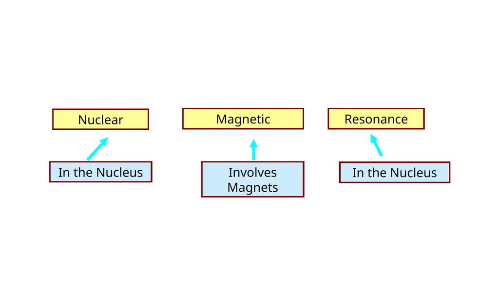
slide6. Magnetic Nuclear Resonance<br>
slide7. Basic principles of NMR-Spectroscopy<br>
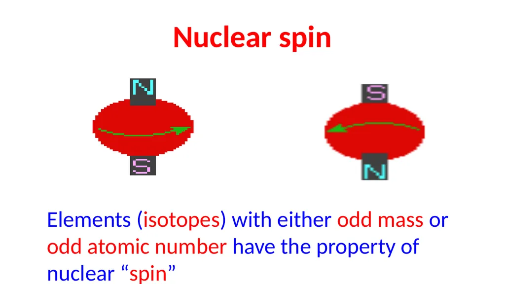
slide9. Nuclear spin Elements (isotopes) with either odd mass or odd atomic number have the property of nuclear “spin”<br>
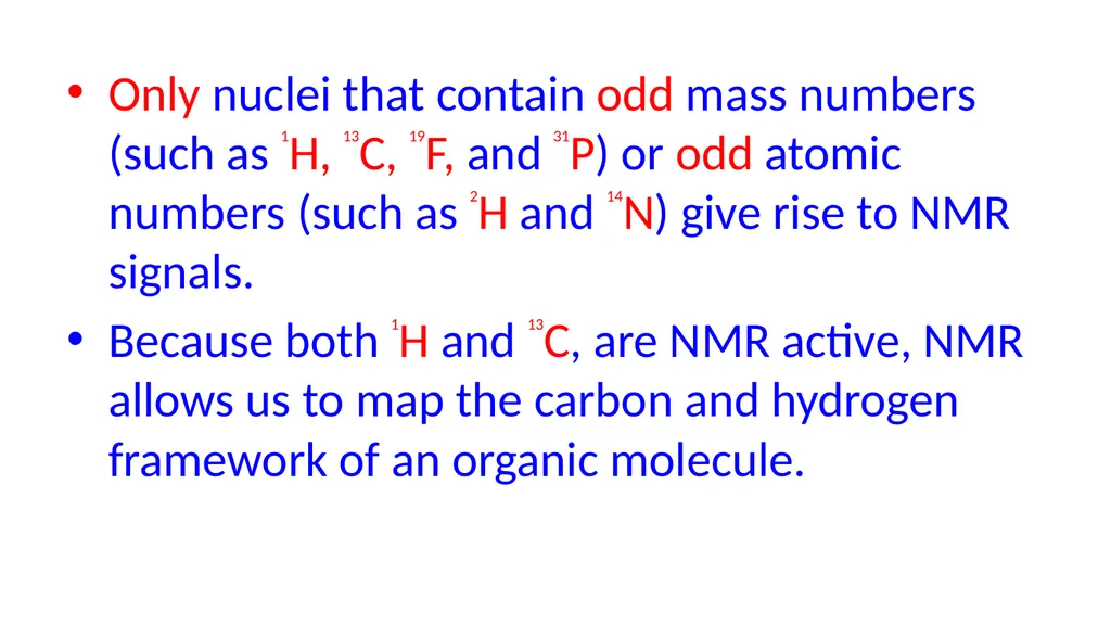
slide10. Only nuclei that contain odd mass numbers (such as 1H, 13C, 19F, and 31P) or odd atomic numbers (such as 2H and 14N) give rise to NMR signals.
Because both 1H and 13C, are NMR active, NMR allows us to map the carbon and hydrogen framework of an organic molecule.<br>
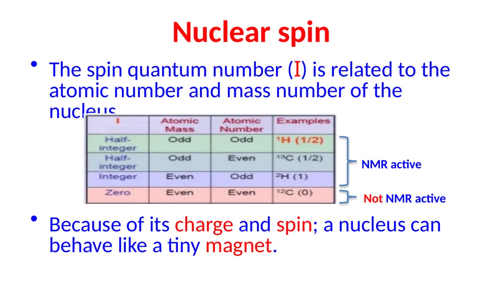
slide11. Nuclear spin The spin quantum number (I) is related to the atomic number and mass number of the nucleus.
Because of its charge and spin; a nucleus can behave like a tiny magnet. NMR active Not NMR active<br>
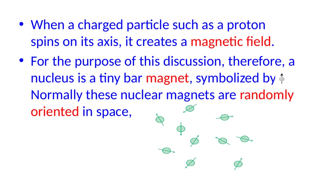
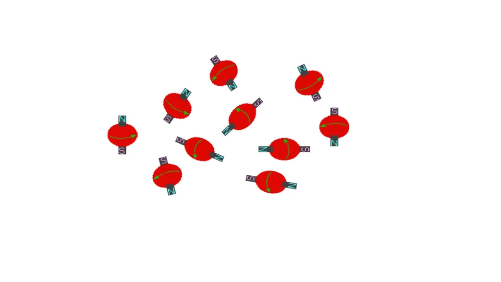
slide12. When a charged particle such as a proton spins on its axis, it creates a magnetic field.
For the purpose of this discussion, therefore, a nucleus is a tiny bar magnet, symbolized by Normally these nuclear magnets are randomly oriented in space,<br>
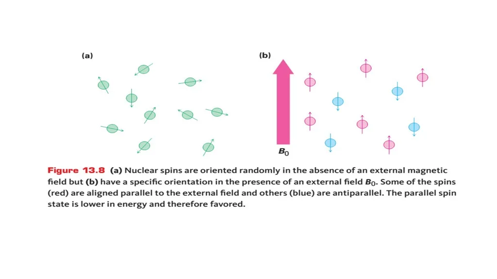
slide14. But in the presence of an external magnetic field, (symbolized by B0), they are oriented with or against this applied field.
More nuclei are oriented with the applied field because this arrangement is lower in energy, but the energy difference between these two states is very small (< 0.4 J/mole)<br>
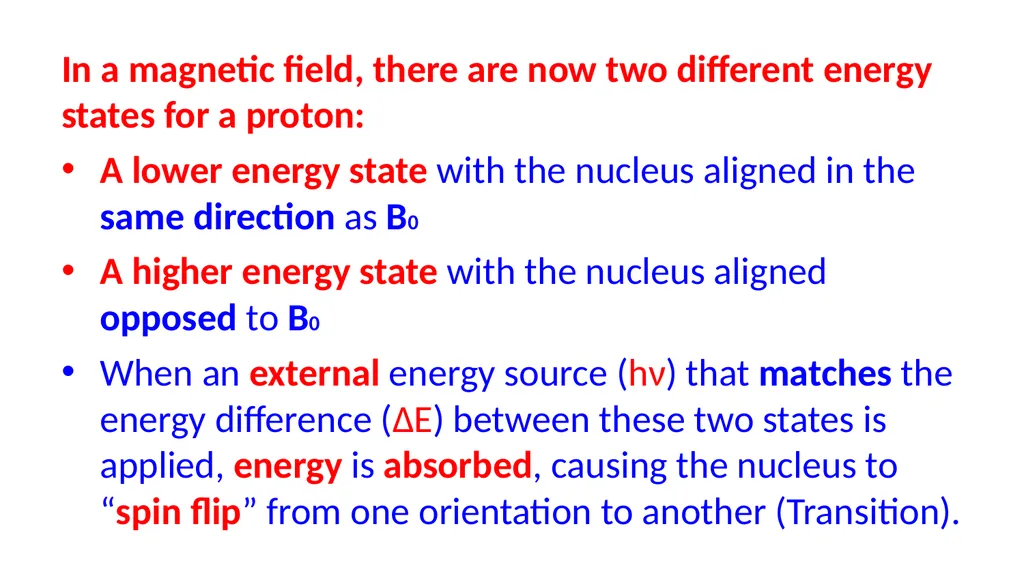
slide16. In a magnetic field, there are now two different energy states for a proton:
A lower energy state with the nucleus aligned in the same direction as B0
A higher energy state with the nucleus aligned opposed to B0
When an external energy source (hν) that matches the energy difference (∆E) between these two states is applied, energy is absorbed, causing the nucleus to “spin flip” from one orientation to another (Transition).<br>
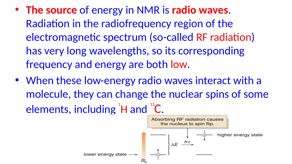
slide17. The source of energy in NMR is radio waves. Radiation in the radiofrequency region of the electromagnetic spectrum (so-called RF radiation) has very long wavelengths, so its corresponding frequency and energy are both low.
When these low-energy radio waves interact with a molecule, they can change the nuclear spins of some elements, including 1H and 13C.<br>
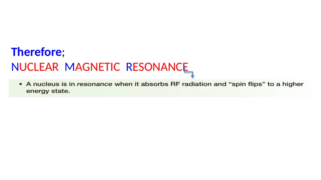
slide18. Therefore;
NUCLEAR MAGNETIC RESONANCE<br>
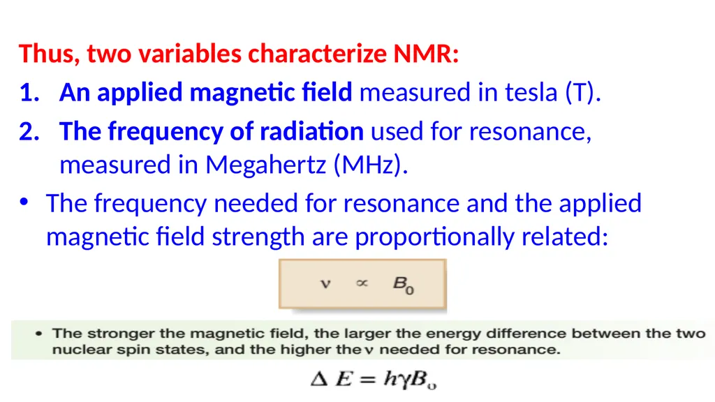
slide19. Thus, two variables characterize NMR:
An applied magnetic field measured in tesla (T).
The frequency of radiation used for resonance, measured in Megahertz (MHz).
The frequency needed for resonance and the applied magnetic field strength are proportionally related:<br>
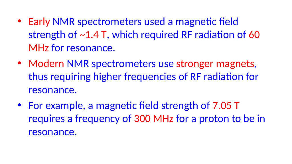
slide20. Early NMR spectrometers used a magnetic field strength of ~1.4 T, which required RF radiation of 60 MHz for resonance.
Modern NMR spectrometers use stronger magnets, thus requiring higher frequencies of RF radiation for resonance.
For example, a magnetic field strength of 7.05 T requires a frequency of 300 MHz for a proton to be in resonance.<br>
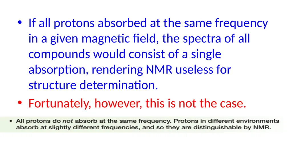
slide21. If all protons absorbed at the same frequency in a given magnetic field, the spectra of all compounds would consist of a single absorption, rendering NMR useless for structure determination.
Fortunately, however, this is not the case.<br>
slide22. Local field The frequency at which a particular proton absorbs is determined by its electronic environment.
Because electrons are moving charged particles, they create a magnetic field (local field) opposed to the applied field B0, and the size of the local magnetic field generated by the electrons around a nucleus determines where it absorbs.<br>
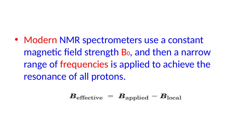
slide23. Modern NMR spectrometers use a constant magnetic field strength B0, and then a narrow range of frequencies is applied to achieve the resonance of all protons.<br>
slide24. Effect of local field Nuclei that are not in identical structural situations do not experience the external magnetic field to the same extent.
The nuclei are “shielded” or “deshielded” due to small local fields generated by circulating sigma, pi and lone pair electrons.<br>
slide25. Effect of local field When the electrons circulate, they generate a small magnetic field that happens to point in the opposite direction to the external field Therefore, the nucleus experiences a reduced external magnetic field and resonate at lower frequency.
This is typically known as “shielding”, e.g. Hydrogens like those in methane are said to be “shielded”.<br>
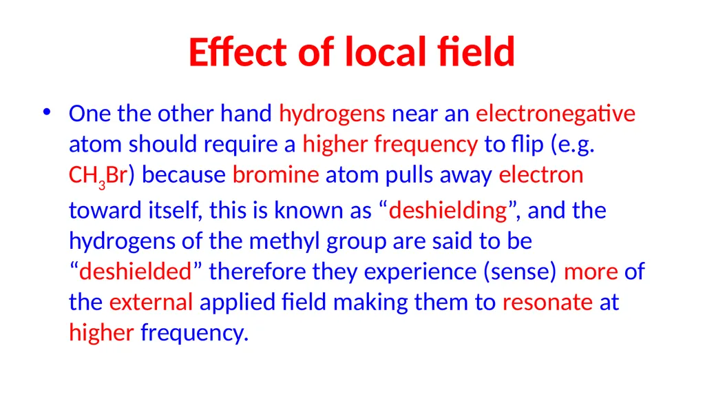
slide26. Effect of local field One the other hand hydrogens near an electronegative atom should require a higher frequency to flip (e.g. CH3Br) because bromine atom pulls away electron toward itself, this is known as “deshielding”, and the hydrogens of the methyl group are said to be “deshielded” therefore they experience (sense) more of the external applied field making them to resonate at higher frequency.<br>
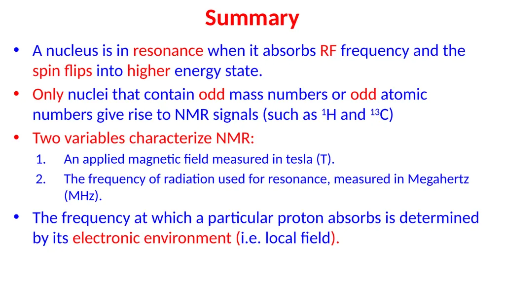
slide27. Summary A nucleus is in resonance when it absorbs RF frequency and the spin flips into higher energy state.
Only nuclei that contain odd mass numbers or odd atomic numbers give rise to NMR signals (such as 1H and 13C)
Two variables characterize NMR:
An applied magnetic field measured in tesla (T).
The frequency of radiation used for resonance, measured in Megahertz (MHz).
The frequency at which a particular proton absorbs is determined by its electronic environment (i.e. local field).<br>
slide28. Questions !<br>
slide29. 1H NMR Spectrum To be continued next lecture.<br>

Department of Pharmaceutical Chemistry
College of Pharmacy – Karary University<br>
slide2. We’ve seen up to this point that IR spectroscopy provides information about a molecule’s functional groups and that UV spectroscopy provides information about a molecule’s conjugated electron system.
Nuclear magnetic resonance(NMR) spectroscopy complements these techniques by providing a “map” of the carbon–hydrogen framework in an organic molecule.
Taken together, IR, UV, and NMR spectroscopies often make it possible to find the structures of even very complex molecules.<br>
slide3. In this lecture we are going to know about a new spectroscopic technique:
NMR Spectroscopy
The physics behind NMR:
Nuclear spin
Charges and magnetic field
Effect of Radio waves on nuclear spin
Effect of electron’s local magnetic field
“shielded” and “deshielded” nuclei<br>
slide4. Two common types of NMR spectroscopy are used to characterize organic structure:
• 1H NMR (proton NMR) is used to determine the number and type of hydrogen atoms in a molecule; and
• 13C NMR (carbon NMR) is used to determine the type of carbon atoms in a molecule.<br>
slide5. Before you can learn how to use NMR spectroscopy to determine the structure of a compound,
you need to understand a bit about the physics behind it. Keep in mind, though, that NMR stems from the same basic principle as all other forms of spectroscopy.
Energy interacts with a molecule, and absorptions occur only when the incident energy matches the energy difference between two states.<br>
slide6. Magnetic Nuclear Resonance<br>
slide7. Basic principles of NMR-Spectroscopy<br>
slide9. Nuclear spin Elements (isotopes) with either odd mass or odd atomic number have the property of nuclear “spin”<br>
slide10. Only nuclei that contain odd mass numbers (such as 1H, 13C, 19F, and 31P) or odd atomic numbers (such as 2H and 14N) give rise to NMR signals.
Because both 1H and 13C, are NMR active, NMR allows us to map the carbon and hydrogen framework of an organic molecule.<br>
slide11. Nuclear spin The spin quantum number (I) is related to the atomic number and mass number of the nucleus.
Because of its charge and spin; a nucleus can behave like a tiny magnet. NMR active Not NMR active<br>
slide12. When a charged particle such as a proton spins on its axis, it creates a magnetic field.
For the purpose of this discussion, therefore, a nucleus is a tiny bar magnet, symbolized by Normally these nuclear magnets are randomly oriented in space,<br>
slide14. But in the presence of an external magnetic field, (symbolized by B0), they are oriented with or against this applied field.
More nuclei are oriented with the applied field because this arrangement is lower in energy, but the energy difference between these two states is very small (< 0.4 J/mole)<br>
slide16. In a magnetic field, there are now two different energy states for a proton:
A lower energy state with the nucleus aligned in the same direction as B0
A higher energy state with the nucleus aligned opposed to B0
When an external energy source (hν) that matches the energy difference (∆E) between these two states is applied, energy is absorbed, causing the nucleus to “spin flip” from one orientation to another (Transition).<br>
slide17. The source of energy in NMR is radio waves. Radiation in the radiofrequency region of the electromagnetic spectrum (so-called RF radiation) has very long wavelengths, so its corresponding frequency and energy are both low.
When these low-energy radio waves interact with a molecule, they can change the nuclear spins of some elements, including 1H and 13C.<br>
slide18. Therefore;
NUCLEAR MAGNETIC RESONANCE<br>
slide19. Thus, two variables characterize NMR:
An applied magnetic field measured in tesla (T).
The frequency of radiation used for resonance, measured in Megahertz (MHz).
The frequency needed for resonance and the applied magnetic field strength are proportionally related:<br>
slide20. Early NMR spectrometers used a magnetic field strength of ~1.4 T, which required RF radiation of 60 MHz for resonance.
Modern NMR spectrometers use stronger magnets, thus requiring higher frequencies of RF radiation for resonance.
For example, a magnetic field strength of 7.05 T requires a frequency of 300 MHz for a proton to be in resonance.<br>
slide21. If all protons absorbed at the same frequency in a given magnetic field, the spectra of all compounds would consist of a single absorption, rendering NMR useless for structure determination.
Fortunately, however, this is not the case.<br>
slide22. Local field The frequency at which a particular proton absorbs is determined by its electronic environment.
Because electrons are moving charged particles, they create a magnetic field (local field) opposed to the applied field B0, and the size of the local magnetic field generated by the electrons around a nucleus determines where it absorbs.<br>
slide23. Modern NMR spectrometers use a constant magnetic field strength B0, and then a narrow range of frequencies is applied to achieve the resonance of all protons.<br>
slide24. Effect of local field Nuclei that are not in identical structural situations do not experience the external magnetic field to the same extent.
The nuclei are “shielded” or “deshielded” due to small local fields generated by circulating sigma, pi and lone pair electrons.<br>
slide25. Effect of local field When the electrons circulate, they generate a small magnetic field that happens to point in the opposite direction to the external field Therefore, the nucleus experiences a reduced external magnetic field and resonate at lower frequency.
This is typically known as “shielding”, e.g. Hydrogens like those in methane are said to be “shielded”.<br>
slide26. Effect of local field One the other hand hydrogens near an electronegative atom should require a higher frequency to flip (e.g. CH3Br) because bromine atom pulls away electron toward itself, this is known as “deshielding”, and the hydrogens of the methyl group are said to be “deshielded” therefore they experience (sense) more of the external applied field making them to resonate at higher frequency.<br>
slide27. Summary A nucleus is in resonance when it absorbs RF frequency and the spin flips into higher energy state.
Only nuclei that contain odd mass numbers or odd atomic numbers give rise to NMR signals (such as 1H and 13C)
Two variables characterize NMR:
An applied magnetic field measured in tesla (T).
The frequency of radiation used for resonance, measured in Megahertz (MHz).
The frequency at which a particular proton absorbs is determined by its electronic environment (i.e. local field).<br>
slide28. Questions !<br>
slide29. 1H NMR Spectrum To be continued next lecture.<br>