/
Analysis   Infrared Spectroscopy Analysis   Infrared Spectroscopy

Analysis Infrared Spectroscopy - PowerPoint Presentation

danika-pritchard
danika-pritchard . @danika-pritchard
Follow
348 views
Uploaded On 2018-11-05

Analysis Infrared Spectroscopy - PPT Presentation

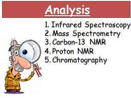
Mass Spectrometry Carbon13 NMR Proton NMR Chromatography 1 Infrared Spectroscopy Absorption of infrared radiation causes bonds to vibrate Different bonds absorb different wavelengths These can be identified using the table on the data sheet ID: 716315

proton peak nmr protons peak proton protons nmr carbon phase chemical spectroscopy peaks chromatography due magnetic shift ion number resonance molecular gas

Share:

Link:

Embed:

Download Presentation from below link

Download Presentation The PPT/PDF document "Analysis Infrared Spectroscopy" is the property of its rightful owner. Permission is granted to download and print the materials on this web site for personal, non-commercial use only, and to display it on your personal computer provided you do not modify the materials and that you retain all copyright notices contained in the materials. By downloading content from our website, you accept the terms of this agreement.


Presentation Transcript

Slide1

Analysis

Infrared Spectroscopy Mass Spectrometry Carbon-13 NMR Proton NMR Chromatography Slide2

1. Infrared Spectroscopy

Absorption of infrared radiation causes bonds to vibrate. Different bonds absorb different wavelengths. These can be identified using the table on the data sheet.

-O-H

carboxylic acid

C=O

Modern breathalysers measure ethanol in the breath by analysis using IR spectroscopy. Slide3

2. Mass Spectrometry

When an organic compound is placed in a mass spectrometer, some molecules lose an electron and become ionised. The resulting positive ion is called the molecular ion, M+. The molecular ion produces the peak with the highest m/z value in the mass spectrum. The M+ has a molecular mass equal to the Mr

of the compound

.

Ethanol

Excess energy from the ionisation process can be transferred to the molecular ion, causing bonds to weaken, and the molecular ion can split into pieces by

fragmentation. The positive molecular ion will split into 2 pieces, one of which will be a positive fragment ion.

Let’s take ethanol , C

2

H

5

OH as an example. When it loses an electron, it will form a molecular ion C

2

H

5

OH

+

. This produces a peak at m/z 46, which means that the Mr of the compound is 46.

Other fragment ions that could be produced are:

CH

2

OH

+

= 31

CH

3

+

= 15

C

2

H

5

+

= 29Slide4

3. Carbon-13 Nuclear

Magnetic Resonance Spectroscopy

Absorption of

low energy radiofrequency electromagnetic radiation

in the presence of a strong magnetic field can cause the energy state of carbon-13 nuclei to change.

Tetramethylsilane (TMS)

is used as the standard for chemical shift measurements because it is

chemically unreactive

and it is

volatile

, so it can easily be recovered from a sample at the end of an experiment. TMS produces an NMR signal at a chemical shift of 0

ppm

.

Chemical shift,

δ

,

is a scale that compares the frequency of an NMR absorption with the frequency of the reference peak of TMS at

δ

=0

ppm

. Slide5

3. Carbon-13 Nuclear

Magnetic Resonance Spectroscopy

Analysis usually occurs in solution. Most organic solvents would produce an NMR signal because they contain carbon-13 or hydrogen-1. Instead, we use a

deuterated solvent,

which has had its hydrogen nuclei replaced by deuterium. An example is

CDCl

3

.

Carbon-13 NMR spectra provide 2 pieces of information about the structure of a molecule:

Number of peaks present in the spectrum = how many different carbon environments there are in the molecule

Chemical shift of each peak tells us about the nature of the carbon environment responsible for that peak.

This is the C-13 NMR spectrum of

but-3-en-2-one

4 peaks due to 4 carbon environments

Peak at 26ppm due to C-C bond of methyl group

Peaks at 137 and 129 are due to C=C double bond

Peak at 198 due to C=O double bond Slide6

4. Proton N

uclear Magnetic Resonance Spectroscopy

It is the spin of the protons in the nucleus that is detected as peaks of chemical shift when exposed to radio waves.

If the number of protons is even, the spin is cancelled out and there is no detection of peaks as chemical shift.

If the number of protons is

odd

, there is an

uneven distribution of spin following exposure to the radio waves

and the resonance is detected as a peak of

chemical shift.

Low energy radio frequency electromagnetic radiation

Tetramethylsilane (TMS) used as reference standardDeuterated solvents such as CDCl

3 used

Low resolution proton NMR gives 3 pieces of information about a compound:

Number of peaks = number of proton environments

Chemical shift values = type of proton environment

Integration trace value = relative number of hydrogen atoms in each chemical environment

A proton NMR of methyl ethanoate would have 2 peaks because there are 2 proton environments. The ratio would be 1:1 as there are 3 protons in each environment. The first proton environment is HC-C=O (

δ

2-3

ppm

) and the second is HC-O (

δ

3-4

ppm

)Slide7

4. Proton N

uclear Magnetic Resonance Spectroscopy High resolution proton-NMR provides one additional piece of information about the molecule. This results from the interaction of protons with adjacent protons – this is known as

spin-spin coupling resulting in a splitting pattern

. The splitting of a peak can be predicted using the

n+1 rule.

“For n protons on an adjacent carbon atom, the splitting pattern in n+1”

Peak with Splitting patter

n (n+1)

Number

of H on adjacent carbon (n)

Singlet

0

Doublet

1Triplet2

Quartet

3

Quintet

4

Sextet

5

Heptet

6

Multiplet

>6 or aromatic

H Slide8

4. Proton N

uclear Magnetic Resonance Spectroscopy

This peak is due to the

HC-O proton.

The peak is split into a

quartet

(4 parts). This means that the neighbouring carbon must contain

3 carbons

. This is correct – there are 3 hydrogens on the -CH

3

group.

This peak is due to the –O-H proton. The peak is a singlet, because the hydrogen atom is attached to an oxygen atom (which has no other hydrogens attached).

This peak is due to the

R-CH

protons in the CH

3

group. This peak is split into a

triplet

which indicates that the adjacent carbon must have 2 carbons. Slide9

4. Proton N

uclear Magnetic Resonance Spectroscopy It can be difficult to identify compounds containing –OH and –NH protons because peaks can appear over a wide range of chemical shift values, signals can be broad and they tend not to show a splitting pattern.

A proton NMR spectrum is run on the sample.

The D

2

O is added to the same compound under investigation.

Second NMR spectrum is run. Any peak due to –OH or –NH protons disappears .

It is now easy to identify which peaks were due to –OH or –NH protons by comparing the 2 spectra.

It is possible to remove the –OH or –NH

peaks using

deuterium oxide.

Deuterium will exchange with the –OH or –NH protons when added to samples under test. The peak will therefore disappear.

MRI in medicine Slide10

4. Proton N

uclear Magnetic

R

esonance

Spectroscopy

NMR in medicine – used in the diagnosis, treatment and follow up of cancer and identifying the extent of soft tissue injuries such as tears in ligaments and muscles.

Patient placed in a large, cylindrical electromagnet

Radio waves are sent through the body. Protons in the body resonate in response to pulses of radiofrequency radiation.

The image is a 3D NMR spectrum of protons in water and other hydrogen containing compounds in the body.

Many diseases change the water content of tissues and organs so the scanner is able to detect these differences.

Advantages

Harmless as it uses low energy radiofrequency electromagnetic waves

Non invasive

Patient does not feel anything

Good for soft tissue scans

Disadvantages

E

lectromagnet is so strong that anything magnetic flies across the room which is hazardous

Patients cannot have an MRI scan if they have a pacemaker or metal implant (such as a pin in the leg) Slide11

5

. ChromatographyChromatography is an analytical technique that separates components in a mixture between a mobile phase and a stationary phase.

Thin Layer Chromatography

The

mobile phase

is the phase

which moves and may be a liquid or a gas

.

The

stationary phase

is the

phase that does not move and may be a solid

(as in thin-layer chromatography)

or either a solid or liquid on a solid support

(as in gas chromatography)

Stationary Phase:

Solid

s

ilica or alumina

Mobile phase:

solvent

Separation onto stationary phase:

Adsorption onto sold

Data collection

:

R

f

values

Stationary Phase:

Solid silicone polymer or liquid long chain

alkane

Mobile phase:

gas

Separation onto stationary phase:

Adsorption onto solid or solubility into liquid

Data collection

: retention times

Gas Chromatography Slide12

5

. ChromatographySlide13

5

. ChromatographyDistance moved by component from base line

Distance moved by solvent front

R

f

value =

Retention time:

time taken form injection of sample for component to leave the column.

Greater solubility – longer retention time

Greater adsorption = longer retention time

Disadvantages of gas chromatography:

Compounds with similar structures such as stereoisomers or same functional groups have similar retention times

Unknown compounds have no reference times for comparison Slide14

Uses of GC-MS:Forensic science – identifying compounds found at the scene of a crimeEnvironmental analysis

– monitoring and analysing pollutants and the content of water Airport security – detecting explosive substances Space probes – collect and analyse material form other planets.

Combining Gas Chromatography and Mass Spectrometry: GC-MS

These two techniques have

different strengths

: gas chromatography can separate component s but cannot identify them conclusively; mass spectrometry can provide detailed structural information on components but cannot separate them.