
Overview Last Updated: March 7, 2025 Retinal
MJ
Published · 53 slides · 0 views

1 / 1





Description
Overview Last Updated: March 7, 2025 Retinal Degenerations Rare: less common than 1:3,500 worldwide Inherited: families may be concentrated in areas with limited access to specialist care Extreme genetic heterogeneity: caused by genetic
Related Topics
Share
Embed code
Download this presentation From Below
"Overview Last Updated: March 7, 2025 Retinal" is the property of its rightful owner. Permission is granted to download and print the materials on this website for personal, non-commercial use only, and to display it on your personal computer provided you do not modify the materials and that you retain all copyright notices contained in the materials. By downloading content from our website, you accept the terms of this agreement.
Presentation Transcript
01
Overview Last Updated: March 7, 2025<br>
02
Retinal Degenerations Rare: less common than 1:3,500 worldwide
Inherited: families may be concentrated in areas with limited access to specialist care
Extreme genetic heterogeneity: caused by genetic changes in over 300 genes, with variable and widely ranging clinical manifestations associated with each gene
Relentless: genetic changes cause progressive dysfunction and death of rod and cone photoreceptors
No effective treatments for most<br>
Inherited: families may be concentrated in areas with limited access to specialist care
Extreme genetic heterogeneity: caused by genetic changes in over 300 genes, with variable and widely ranging clinical manifestations associated with each gene
Relentless: genetic changes cause progressive dysfunction and death of rod and cone photoreceptors
No effective treatments for most<br>
03
Challenges to Developing Treatments Uncommon: few patients available for study, limited information exists
Wide range of phenotypes requires evaluation using specialty testing and expert clinical evaluation
Limited standardized prospective natural history data on IRDs
Unclear what outcome measures will be most likely to demonstrate change over time, or safety and efficacy of treatments<br>
Wide range of phenotypes requires evaluation using specialty testing and expert clinical evaluation
Limited standardized prospective natural history data on IRDs
Unclear what outcome measures will be most likely to demonstrate change over time, or safety and efficacy of treatments<br>
04
Mission Statement The Foundation seeks to build a Consortium for the purpose of conducting clinical studies in patients with rare inherited retinal disorders (IRDs). The goal is to accelerate the development of treatments for IRDs. To accomplish this, the Foundation plans to build centers of excellence to participate in sponsored clinical studies. Investigators from participating clinical centers will collaborate on ideas for hypotheses, study designs, analysis plans, and publications. Data from the completed trials will be archived in an open central repository to stimulate further hypothesis generation and innovation.<br>
05
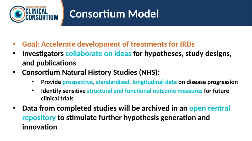
Consortium Model Goal: Accelerate development of treatments for IRDs
Investigators collaborate on ideas for hypotheses, study designs, and publications
Consortium Natural History Studies (NHS):
Provide prospective, standardized, longitudinal data on disease progression
Identify sensitive structural and functional outcome measures for future clinical trials
Data from completed studies will be archived in an open central repository to stimulate further hypothesis generation and innovation<br>
Investigators collaborate on ideas for hypotheses, study designs, and publications
Consortium Natural History Studies (NHS):
Provide prospective, standardized, longitudinal data on disease progression
Identify sensitive structural and functional outcome measures for future clinical trials
Data from completed studies will be archived in an open central repository to stimulate further hypothesis generation and innovation<br>
06
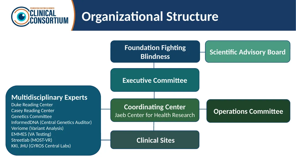
Organizational Structure Clinical Sites Executive Committee Operations Committee Foundation Fighting Blindness Scientific Advisory Board Coordinating Center
Jaeb Center for Health Research Multidisciplinary Experts
Duke Reading Center
Casey Reading Center
Genetics Committee
InformedDNA (Central Genetics Auditor)
Veriome (Variant Analysis)
EMMES (VA Testing)
Streetlab (MOST-VR)
KKI, JHU (GYROS Central Labs)<br>
Jaeb Center for Health Research Multidisciplinary Experts
Duke Reading Center
Casey Reading Center
Genetics Committee
InformedDNA (Central Genetics Auditor)
Veriome (Variant Analysis)
EMMES (VA Testing)
Streetlab (MOST-VR)
KKI, JHU (GYROS Central Labs)<br>
07
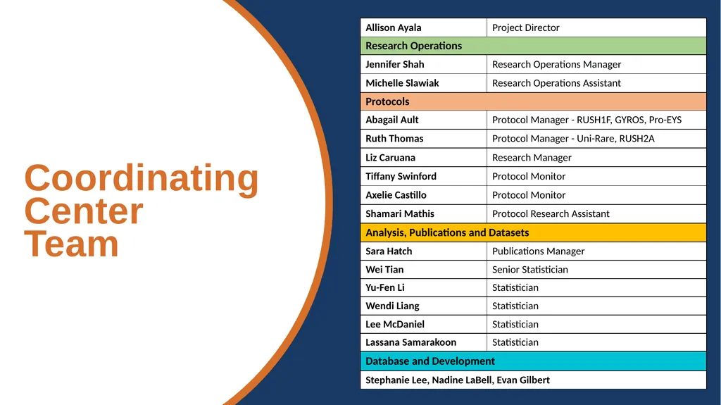
Coordinating Center
Team<br>
08
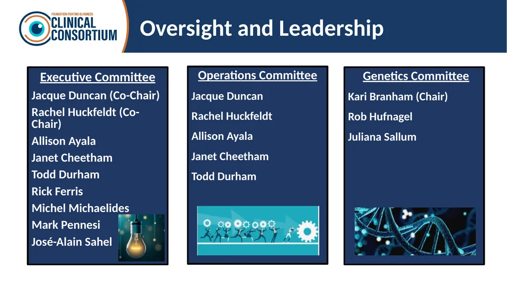
Oversight and Leadership Executive Committee
Jacque Duncan (Co-Chair)
Rachel Huckfeldt (Co-Chair)
Allison Ayala
Janet Cheetham
Todd Durham
Rick Ferris
Michel Michaelides
Mark Pennesi
José-Alain Sahel Operations Committee
Jacque Duncan
Rachel Huckfeldt
Allison Ayala
Janet Cheetham
Todd Durham Genetics Committee
Kari Branham (Chair)
Rob Hufnagel
Juliana Sallum<br>
Jacque Duncan (Co-Chair)
Rachel Huckfeldt (Co-Chair)
Allison Ayala
Janet Cheetham
Todd Durham
Rick Ferris
Michel Michaelides
Mark Pennesi
José-Alain Sahel Operations Committee
Jacque Duncan
Rachel Huckfeldt
Allison Ayala
Janet Cheetham
Todd Durham Genetics Committee
Kari Branham (Chair)
Rob Hufnagel
Juliana Sallum<br>
09
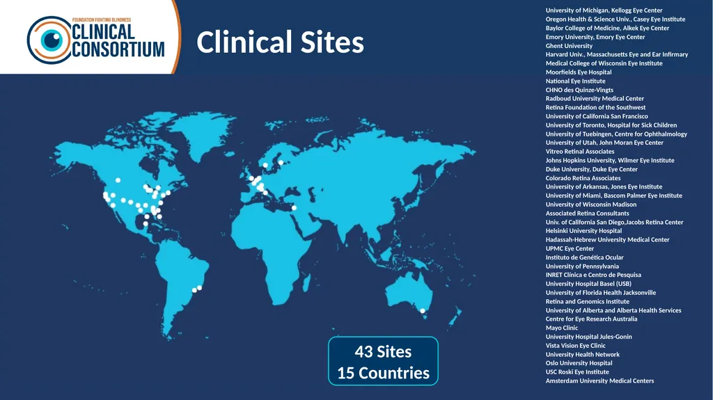
Clinical Sites 43 Sites
15 Countries<br>
15 Countries<br>
10
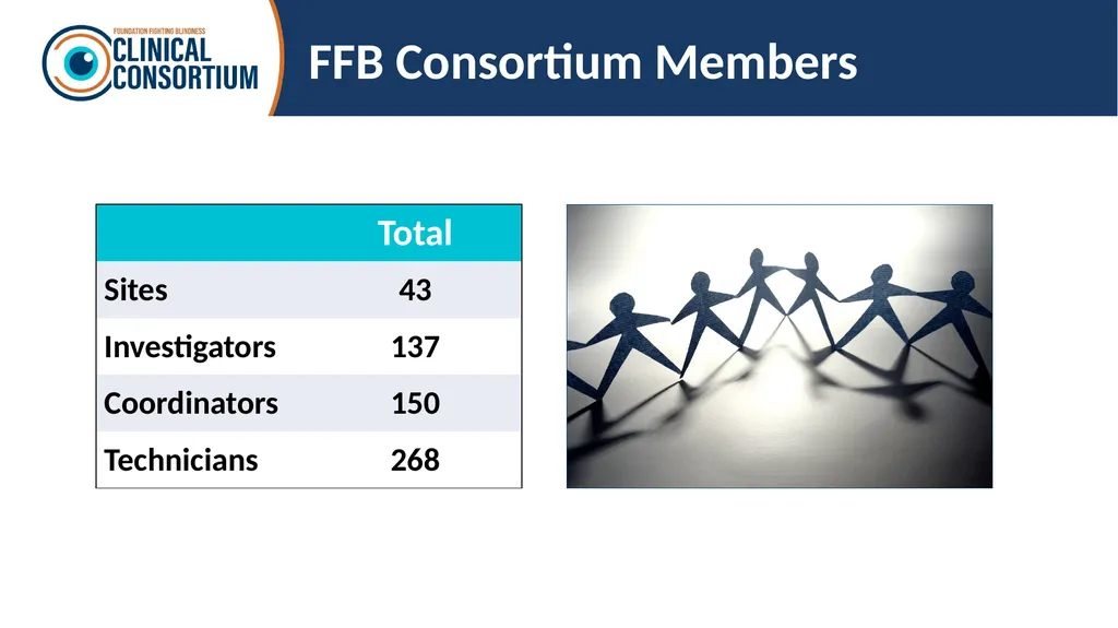
FFB Consortium Members<br>
11
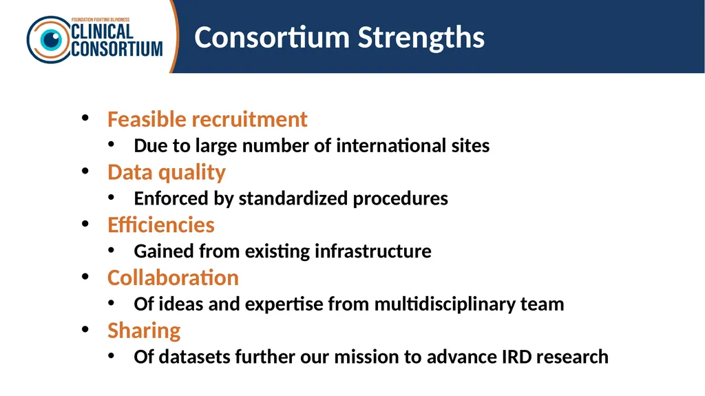
Consortium Strengths Feasible recruitment
Due to large number of international sites
Data quality
Enforced by standardized procedures
Efficiencies
Gained from existing infrastructure
Collaboration
Of ideas and expertise from multidisciplinary team
Sharing
Of datasets further our mission to advance IRD research<br>
Due to large number of international sites
Data quality
Enforced by standardized procedures
Efficiencies
Gained from existing infrastructure
Collaboration
Of ideas and expertise from multidisciplinary team
Sharing
Of datasets further our mission to advance IRD research<br>
12
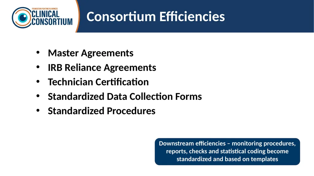
Consortium Efficiencies Master Agreements
IRB Reliance Agreements
Technician Certification
Standardized Data Collection Forms
Standardized Procedures Downstream efficiencies – monitoring procedures, reports, checks and statistical coding become standardized and based on templates<br>
IRB Reliance Agreements
Technician Certification
Standardized Data Collection Forms
Standardized Procedures Downstream efficiencies – monitoring procedures, reports, checks and statistical coding become standardized and based on templates<br>
13
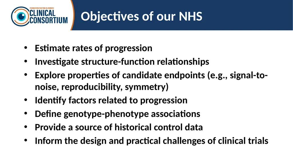
Objectives of our NHS Estimate rates of progression
Investigate structure-function relationships
Explore properties of candidate endpoints (e.g., signal-to-noise, reproducibility, symmetry)
Identify factors related to progression
Define genotype-phenotype associations
Provide a source of historical control data
Inform the design and practical challenges of clinical trials<br>
Investigate structure-function relationships
Explore properties of candidate endpoints (e.g., signal-to-noise, reproducibility, symmetry)
Identify factors related to progression
Define genotype-phenotype associations
Provide a source of historical control data
Inform the design and practical challenges of clinical trials<br>
14
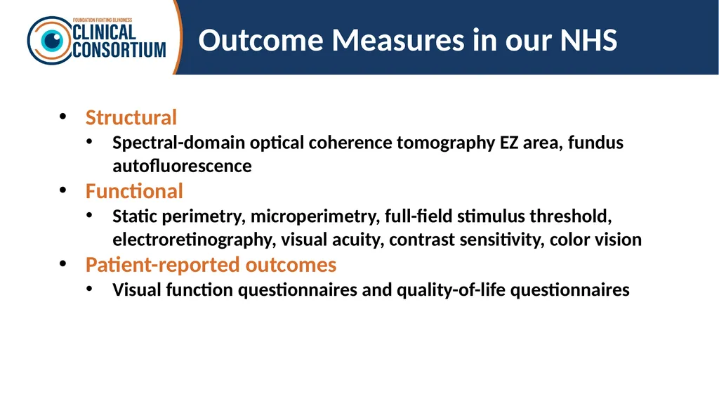
Outcome Measures in our NHS Structural
Spectral-domain optical coherence tomography EZ area, fundus autofluorescence
Functional
Static perimetry, microperimetry, full-field stimulus threshold, electroretinography, visual acuity, contrast sensitivity, color vision
Patient-reported outcomes
Visual function questionnaires and quality-of-life questionnaires<br>
Spectral-domain optical coherence tomography EZ area, fundus autofluorescence
Functional
Static perimetry, microperimetry, full-field stimulus threshold, electroretinography, visual acuity, contrast sensitivity, color vision
Patient-reported outcomes
Visual function questionnaires and quality-of-life questionnaires<br>
15
Studies<br>
16
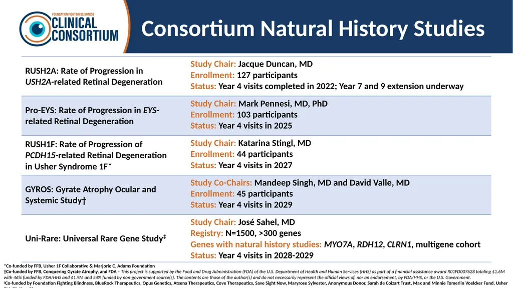
Consortium Natural History Studies *Co-funded by FFB, Usher 1F Collaborative & Marjorie C. Adams Foundation
†Co-funded by FFB, Conquering Gyrate Atrophy, and FDA – This project is supported by the Food and Drug Administration (FDA) of the U.S. Department of Health and Human Services (HHS) as part of a financial assistance award R01FD007628 totaling $1.6M with 46% funded by FDA/HHS and $1.9M and 54% funded by non-government source(s). The contents are those of the author(s) and do not necessarily represent the official views of, nor an endorsement, by FDA/HHS, or the U.S. Government.
‡Co-funded by Foundation Fighting Blindness, BlueRock Therapeutics, Opus Genetics, Atsena Therapeutics, Cove Therapeutics, Save Sight Now, Maryrose Sylvester, Anonymous Donor, Sarah de Coizart Trust, Max and Minnie Tomerlin Voelcker Fund, Usher III Initiative, Akouos.<br>
†Co-funded by FFB, Conquering Gyrate Atrophy, and FDA – This project is supported by the Food and Drug Administration (FDA) of the U.S. Department of Health and Human Services (HHS) as part of a financial assistance award R01FD007628 totaling $1.6M with 46% funded by FDA/HHS and $1.9M and 54% funded by non-government source(s). The contents are those of the author(s) and do not necessarily represent the official views of, nor an endorsement, by FDA/HHS, or the U.S. Government.
‡Co-funded by Foundation Fighting Blindness, BlueRock Therapeutics, Opus Genetics, Atsena Therapeutics, Cove Therapeutics, Save Sight Now, Maryrose Sylvester, Anonymous Donor, Sarah de Coizart Trust, Max and Minnie Tomerlin Voelcker Fund, Usher III Initiative, Akouos.<br>
17
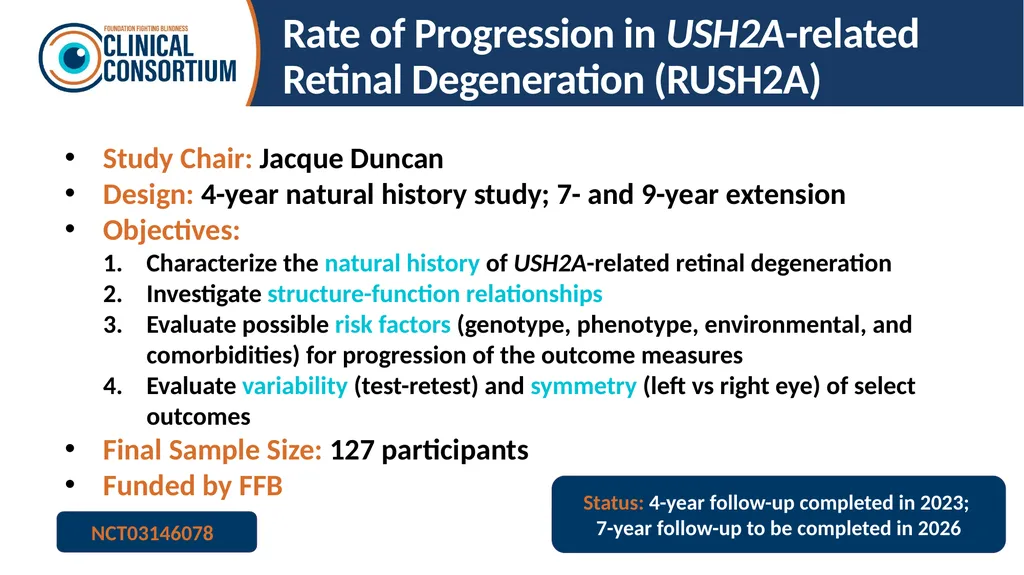
Rate of Progression in USH2A-related Retinal Degeneration (RUSH2A) Study Chair: Jacque Duncan
Design: 4-year natural history study; 7- and 9-year extension
Objectives:
Characterize the natural history of USH2A-related retinal degeneration
Investigate structure-function relationships
Evaluate possible risk factors (genotype, phenotype, environmental, and comorbidities) for progression of the outcome measures
Evaluate variability (test-retest) and symmetry (left vs right eye) of select outcomes
Final Sample Size: 127 participants
Funded by FFB Status: 4-year follow-up completed in 2023;
7-year follow-up to be completed in 2026 NCT03146078<br>
Design: 4-year natural history study; 7- and 9-year extension
Objectives:
Characterize the natural history of USH2A-related retinal degeneration
Investigate structure-function relationships
Evaluate possible risk factors (genotype, phenotype, environmental, and comorbidities) for progression of the outcome measures
Evaluate variability (test-retest) and symmetry (left vs right eye) of select outcomes
Final Sample Size: 127 participants
Funded by FFB Status: 4-year follow-up completed in 2023;
7-year follow-up to be completed in 2026 NCT03146078<br>
18
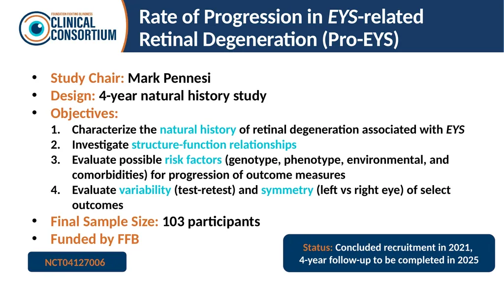
Rate of Progression in EYS-related
Retinal Degeneration (Pro-EYS) Study Chair: Mark Pennesi
Design: 4-year natural history study
Objectives:
Characterize the natural history of retinal degeneration associated with EYS
Investigate structure-function relationships
Evaluate possible risk factors (genotype, phenotype, environmental, and comorbidities) for progression of outcome measures
Evaluate variability (test-retest) and symmetry (left vs right eye) of select outcomes
Final Sample Size: 103 participants
Funded by FFB Status: Concluded recruitment in 2021,
4-year follow-up to be completed in 2025 NCT04127006<br>
Design: 4-year natural history study
Objectives:
Characterize the natural history of retinal degeneration associated with EYS
Investigate structure-function relationships
Evaluate possible risk factors (genotype, phenotype, environmental, and comorbidities) for progression of outcome measures
Evaluate variability (test-retest) and symmetry (left vs right eye) of select outcomes
Final Sample Size: 103 participants
Funded by FFB Status: Concluded recruitment in 2021,
4-year follow-up to be completed in 2025 NCT04127006<br>
19
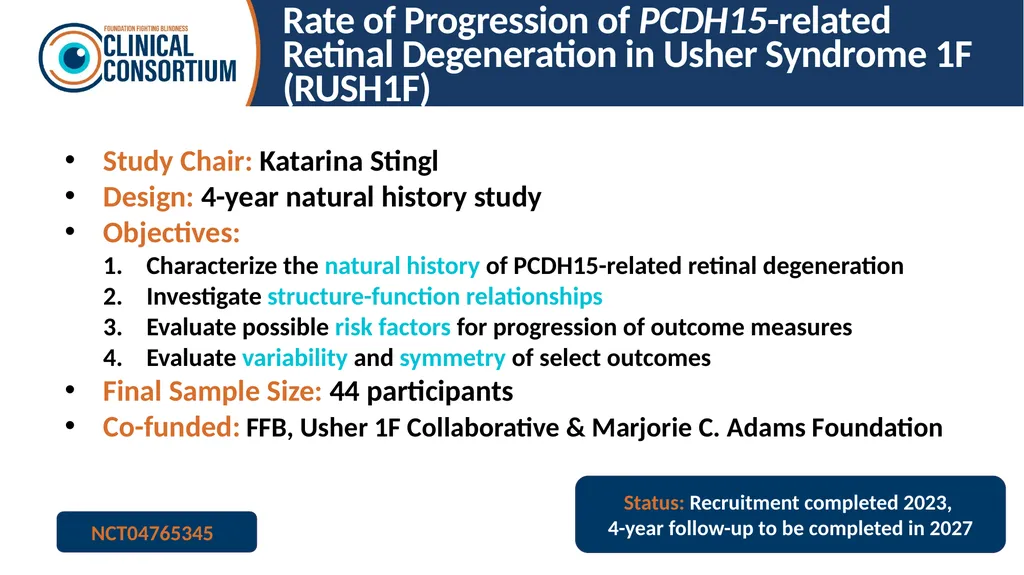
Rate of Progression of PCDH15-related Retinal Degeneration in Usher Syndrome 1F (RUSH1F) Study Chair: Katarina Stingl
Design: 4-year natural history study
Objectives:
Characterize the natural history of PCDH15-related retinal degeneration
Investigate structure-function relationships
Evaluate possible risk factors for progression of outcome measures
Evaluate variability and symmetry of select outcomes
Final Sample Size: 44 participants
Co-funded: FFB, Usher 1F Collaborative & Marjorie C. Adams Foundation Status: Recruitment completed 2023,
4-year follow-up to be completed in 2027 NCT04765345<br>
Design: 4-year natural history study
Objectives:
Characterize the natural history of PCDH15-related retinal degeneration
Investigate structure-function relationships
Evaluate possible risk factors for progression of outcome measures
Evaluate variability and symmetry of select outcomes
Final Sample Size: 44 participants
Co-funded: FFB, Usher 1F Collaborative & Marjorie C. Adams Foundation Status: Recruitment completed 2023,
4-year follow-up to be completed in 2027 NCT04765345<br>
20
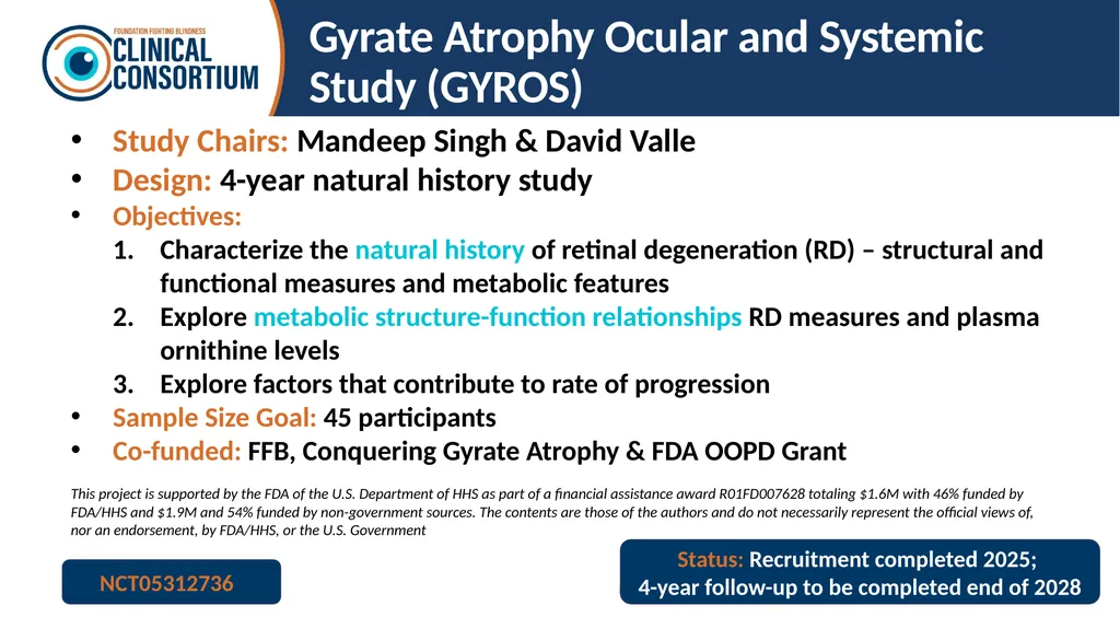
Gyrate Atrophy Ocular and Systemic Study (GYROS) Study Chairs: Mandeep Singh & David Valle
Design: 4-year natural history study
Objectives:
Characterize the natural history of retinal degeneration (RD) – structural and functional measures and metabolic features
Explore metabolic structure-function relationships RD measures and plasma ornithine levels
Explore factors that contribute to rate of progression
Sample Size Goal: 45 participants
Co-funded: FFB, Conquering Gyrate Atrophy & FDA OOPD Grant
This project is supported by the FDA of the U.S. Department of HHS as part of a financial assistance award R01FD007628 totaling $1.6M with 46% funded by FDA/HHS and $1.9M and 54% funded by non-government sources. The contents are those of the authors and do not necessarily represent the official views of, nor an endorsement, by FDA/HHS, or the U.S. Government Status: Recruitment completed 2025;
4-year follow-up to be completed end of 2028 NCT05312736<br>
Design: 4-year natural history study
Objectives:
Characterize the natural history of retinal degeneration (RD) – structural and functional measures and metabolic features
Explore metabolic structure-function relationships RD measures and plasma ornithine levels
Explore factors that contribute to rate of progression
Sample Size Goal: 45 participants
Co-funded: FFB, Conquering Gyrate Atrophy & FDA OOPD Grant
This project is supported by the FDA of the U.S. Department of HHS as part of a financial assistance award R01FD007628 totaling $1.6M with 46% funded by FDA/HHS and $1.9M and 54% funded by non-government sources. The contents are those of the authors and do not necessarily represent the official views of, nor an endorsement, by FDA/HHS, or the U.S. Government Status: Recruitment completed 2025;
4-year follow-up to be completed end of 2028 NCT05312736<br>
21
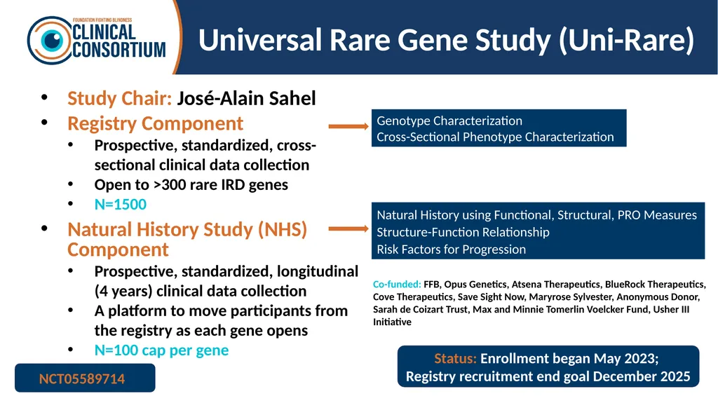
Universal Rare Gene Study (Uni-Rare) Study Chair: José-Alain Sahel
Registry Component
Prospective, standardized, cross-sectional clinical data collection
Open to >300 rare IRD genes
N=1500
Natural History Study (NHS) Component
Prospective, standardized, longitudinal (4 years) clinical data collection
A platform to move participants from the registry as each gene opens
N=100 cap per gene NCT05589714 Genotype Characterization
Cross-Sectional Phenotype Characterization Natural History using Functional, Structural, PRO Measures
Structure-Function Relationship
Risk Factors for Progression Co-funded: FFB, Opus Genetics, Atsena Therapeutics, BlueRock Therapeutics, Cove Therapeutics, Save Sight Now, Maryrose Sylvester, Anonymous Donor, Sarah de Coizart Trust, Max and Minnie Tomerlin Voelcker Fund, Usher III Initiative Status: Enrollment began May 2023;
Registry recruitment end goal December 2025<br>
Registry Component
Prospective, standardized, cross-sectional clinical data collection
Open to >300 rare IRD genes
N=1500
Natural History Study (NHS) Component
Prospective, standardized, longitudinal (4 years) clinical data collection
A platform to move participants from the registry as each gene opens
N=100 cap per gene NCT05589714 Genotype Characterization
Cross-Sectional Phenotype Characterization Natural History using Functional, Structural, PRO Measures
Structure-Function Relationship
Risk Factors for Progression Co-funded: FFB, Opus Genetics, Atsena Therapeutics, BlueRock Therapeutics, Cove Therapeutics, Save Sight Now, Maryrose Sylvester, Anonymous Donor, Sarah de Coizart Trust, Max and Minnie Tomerlin Voelcker Fund, Usher III Initiative Status: Enrollment began May 2023;
Registry recruitment end goal December 2025<br>
22
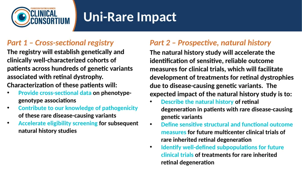
Uni-Rare Impact Part 1 – Cross-sectional registry
The registry will establish genetically and clinically well-characterized cohorts of patients across hundreds of genetic variants associated with retinal dystrophy. Characterization of these patients will:
Provide cross-sectional data on phenotype-genotype associations
Contribute to our knowledge of pathogenicity of these rare disease-causing variants
Accelerate eligibility screening for subsequent natural history studies Part 2 – Prospective, natural history
The natural history study will accelerate the identification of sensitive, reliable outcome measures for clinical trials, which will facilitate development of treatments for retinal dystrophies due to disease-causing genetic variants. The expected impact of the natural history study is to:
Describe the natural history of retinal degeneration in patients with rare disease-causing genetic variants
Define sensitive structural and functional outcome measures for future multicenter clinical trials of rare inherited retinal degeneration
Identify well-defined subpopulations for future clinical trials of treatments for rare inherited retinal degeneration<br>
The registry will establish genetically and clinically well-characterized cohorts of patients across hundreds of genetic variants associated with retinal dystrophy. Characterization of these patients will:
Provide cross-sectional data on phenotype-genotype associations
Contribute to our knowledge of pathogenicity of these rare disease-causing variants
Accelerate eligibility screening for subsequent natural history studies Part 2 – Prospective, natural history
The natural history study will accelerate the identification of sensitive, reliable outcome measures for clinical trials, which will facilitate development of treatments for retinal dystrophies due to disease-causing genetic variants. The expected impact of the natural history study is to:
Describe the natural history of retinal degeneration in patients with rare disease-causing genetic variants
Define sensitive structural and functional outcome measures for future multicenter clinical trials of rare inherited retinal degeneration
Identify well-defined subpopulations for future clinical trials of treatments for rare inherited retinal degeneration<br>
23
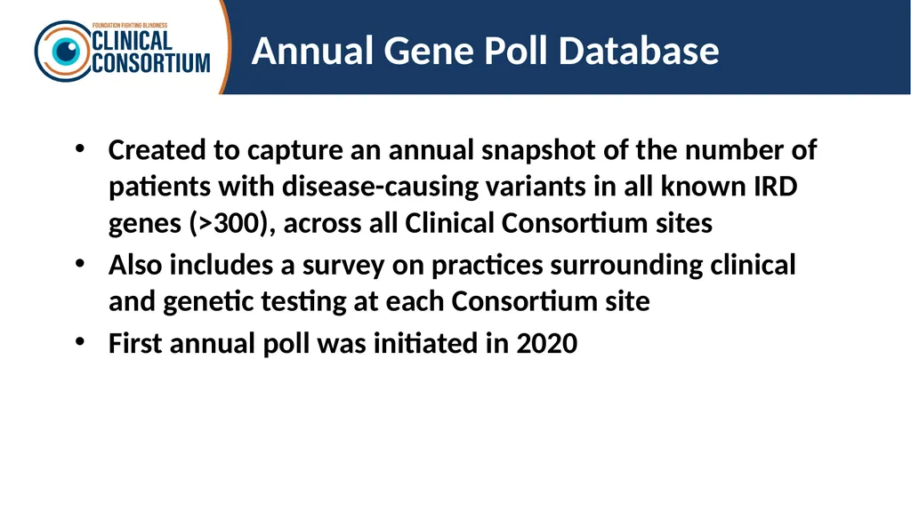
Annual Gene Poll Database Created to capture an annual snapshot of the number of patients with disease-causing variants in all known IRD genes (>300), across all Clinical Consortium sites
Also includes a survey on practices surrounding clinical and genetic testing at each Consortium site
First annual poll was initiated in 2020<br>
Also includes a survey on practices surrounding clinical and genetic testing at each Consortium site
First annual poll was initiated in 2020<br>
24
Manuscripts and Presentations<br>
25
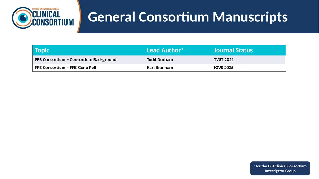
General Consortium Manuscripts *for the FFB Clinical Consortium Investigator Group<br>
26
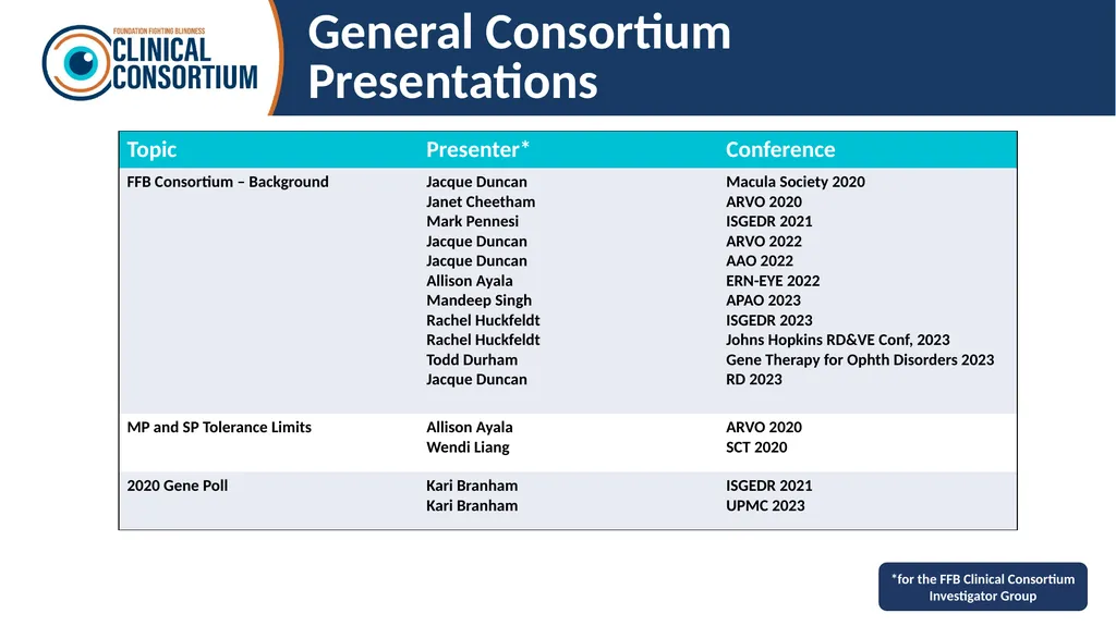
General Consortium Presentations *for the FFB Clinical Consortium Investigator Group<br>
27
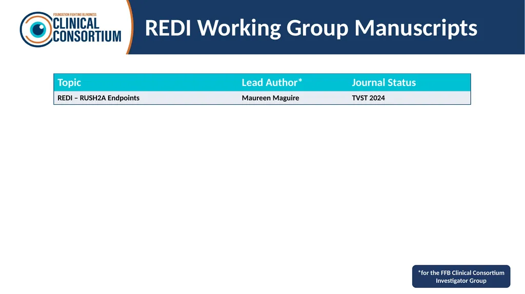
REDI Working Group Manuscripts *for the FFB Clinical Consortium Investigator Group<br>
28
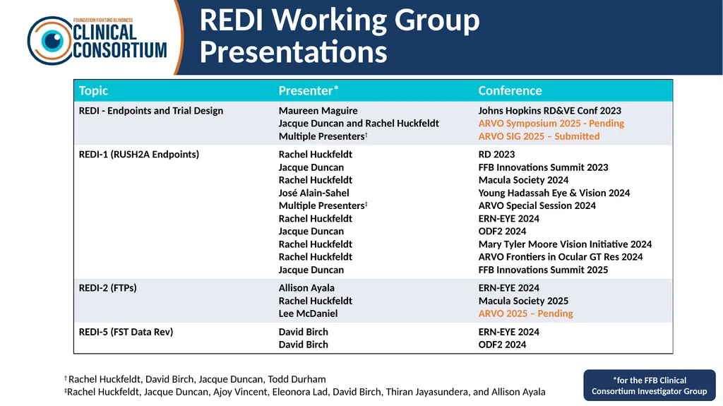
REDI Working Group Presentations *for the FFB Clinical Consortium Investigator Group † Rachel Huckfeldt, David Birch, Jacque Duncan, Todd Durham
‡Rachel Huckfeldt, Jacque Duncan, Ajoy Vincent, Eleonora Lad, David Birch, Thiran Jayasundera, and Allison Ayala<br>
‡Rachel Huckfeldt, Jacque Duncan, Ajoy Vincent, Eleonora Lad, David Birch, Thiran Jayasundera, and Allison Ayala<br>
29
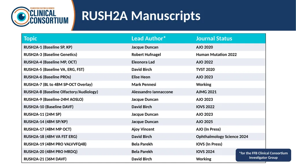
RUSH2A Manuscripts *for the FFB Clinical Consortium Investigator Group<br>
30
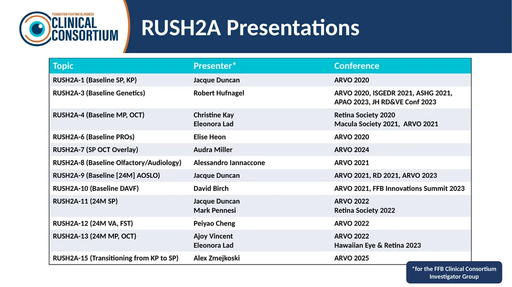
RUSH2A Presentations *for the FFB Clinical Consortium Investigator Group<br>
31
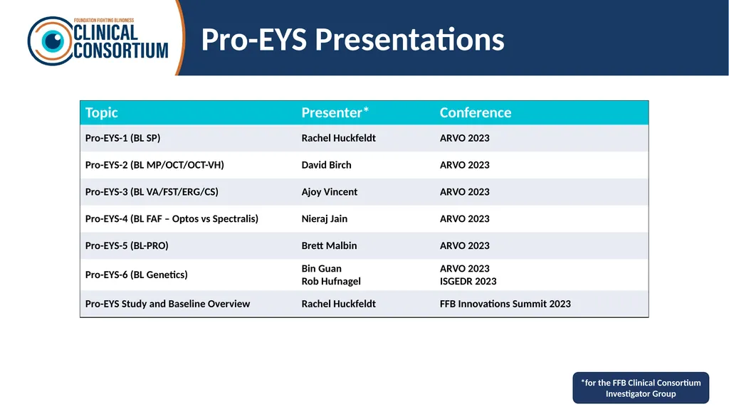
Pro-EYS Presentations *for the FFB Clinical Consortium Investigator Group<br>
32
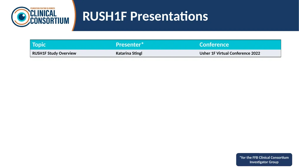
RUSH1F Presentations *for the FFB Clinical Consortium Investigator Group<br>
33
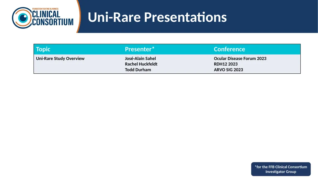
Uni-Rare Presentations *for the FFB Clinical Consortium Investigator Group<br>
34
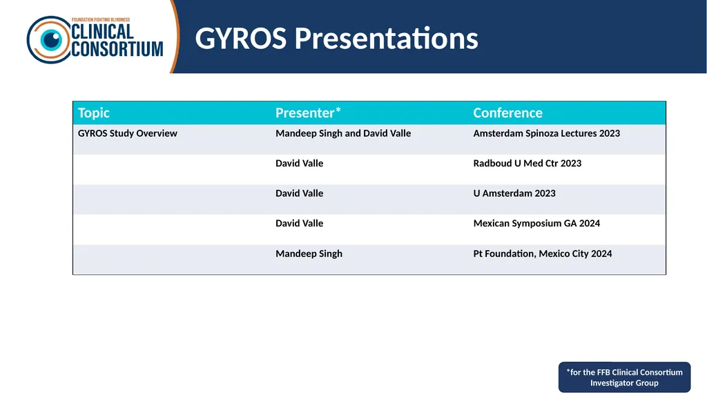
GYROS Presentations *for the FFB Clinical Consortium Investigator Group<br>
35
Results Summaries<br>
36
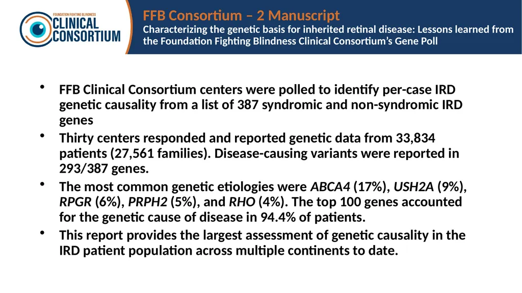
FFB Consortium – 2 Manuscript
Characterizing the genetic basis for inherited retinal disease: Lessons learned from the Foundation Fighting Blindness Clinical Consortium’s Gene Poll FFB Clinical Consortium centers were polled to identify per-case IRD genetic causality from a list of 387 syndromic and non-syndromic IRD genes
Thirty centers responded and reported genetic data from 33,834 patients (27,561 families). Disease-causing variants were reported in 293/387 genes.
The most common genetic etiologies were ABCA4 (17%), USH2A (9%), RPGR (6%), PRPH2 (5%), and RHO (4%). The top 100 genes accounted for the genetic cause of disease in 94.4% of patients.
This report provides the largest assessment of genetic causality in the IRD patient population across multiple continents to date.<br>
Thirty centers responded and reported genetic data from 33,834 patients (27,561 families). Disease-causing variants were reported in 293/387 genes.
The most common genetic etiologies were ABCA4 (17%), USH2A (9%), RPGR (6%), PRPH2 (5%), and RHO (4%). The top 100 genes accounted for the genetic cause of disease in 94.4% of patients.
This report provides the largest assessment of genetic causality in the IRD patient population across multiple continents to date.<br>
37
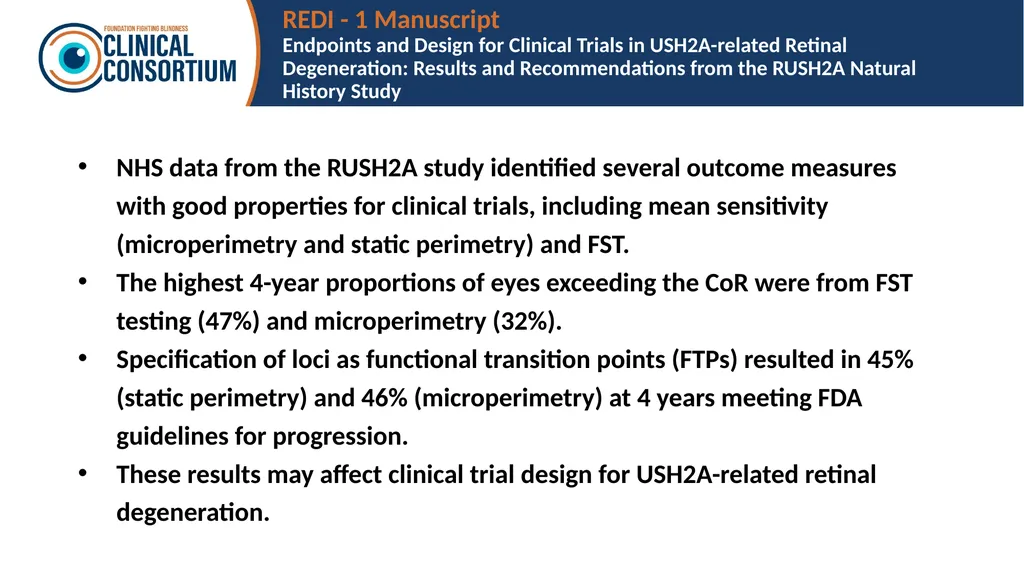
REDI - 1 Manuscript
Endpoints and Design for Clinical Trials in USH2A-related Retinal Degeneration: Results and Recommendations from the RUSH2A Natural History Study NHS data from the RUSH2A study identified several outcome measures with good properties for clinical trials, including mean sensitivity (microperimetry and static perimetry) and FST.
The highest 4-year proportions of eyes exceeding the CoR were from FST testing (47%) and microperimetry (32%).
Specification of loci as functional transition points (FTPs) resulted in 45% (static perimetry) and 46% (microperimetry) at 4 years meeting FDA guidelines for progression.
These results may affect clinical trial design for USH2A-related retinal degeneration.<br>
The highest 4-year proportions of eyes exceeding the CoR were from FST testing (47%) and microperimetry (32%).
Specification of loci as functional transition points (FTPs) resulted in 45% (static perimetry) and 46% (microperimetry) at 4 years meeting FDA guidelines for progression.
These results may affect clinical trial design for USH2A-related retinal degeneration.<br>
38
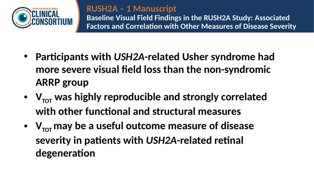
RUSH2A – 1 Manuscript
Baseline Visual Field Findings in the RUSH2A Study: Associated Factors and Correlation with Other Measures of Disease Severity Participants with USH2A-related Usher syndrome had more severe visual field loss than the non-syndromic ARRP group
VTOT was highly reproducible and strongly correlated with other functional and structural measures
VTOT may be a useful outcome measure of disease severity in patients with USH2A-related retinal degeneration<br>
VTOT was highly reproducible and strongly correlated with other functional and structural measures
VTOT may be a useful outcome measure of disease severity in patients with USH2A-related retinal degeneration<br>
39
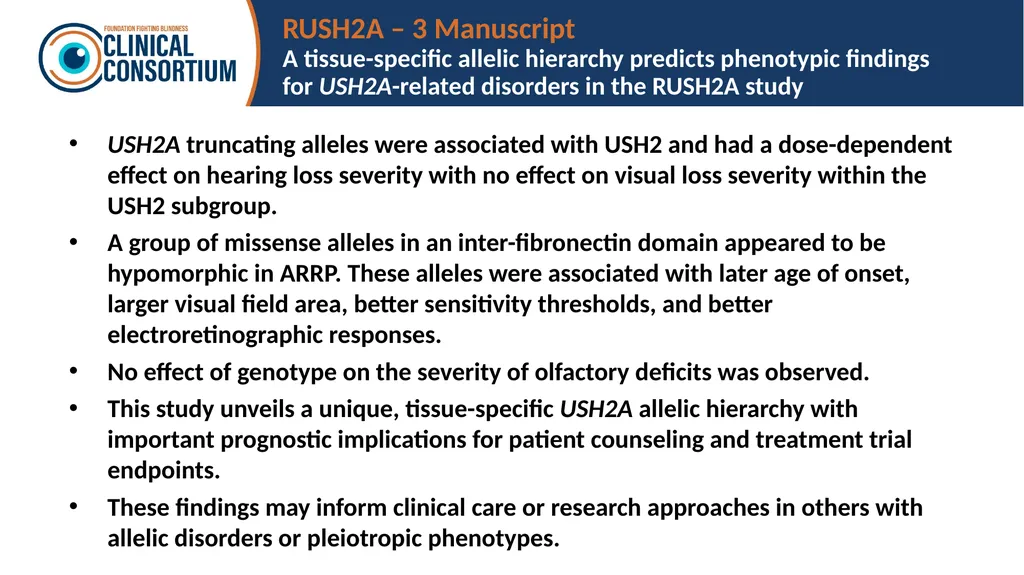
RUSH2A – 3 Manuscript
A tissue-specific allelic hierarchy predicts phenotypic findings for USH2A-related disorders in the RUSH2A study USH2A truncating alleles were associated with USH2 and had a dose-dependent effect on hearing loss severity with no effect on visual loss severity within the USH2 subgroup.
A group of missense alleles in an inter-fibronectin domain appeared to be hypomorphic in ARRP. These alleles were associated with later age of onset, larger visual field area, better sensitivity thresholds, and better electroretinographic responses.
No effect of genotype on the severity of olfactory deficits was observed.
This study unveils a unique, tissue-specific USH2A allelic hierarchy with important prognostic implications for patient counseling and treatment trial endpoints.
These findings may inform clinical care or research approaches in others with allelic disorders or pleiotropic phenotypes.<br>
A group of missense alleles in an inter-fibronectin domain appeared to be hypomorphic in ARRP. These alleles were associated with later age of onset, larger visual field area, better sensitivity thresholds, and better electroretinographic responses.
No effect of genotype on the severity of olfactory deficits was observed.
This study unveils a unique, tissue-specific USH2A allelic hierarchy with important prognostic implications for patient counseling and treatment trial endpoints.
These findings may inform clinical care or research approaches in others with allelic disorders or pleiotropic phenotypes.<br>
40
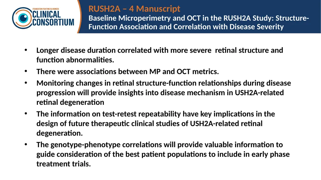
RUSH2A – 4 Manuscript
Baseline Microperimetry and OCT in the RUSH2A Study: Structure-Function Association and Correlation with Disease Severity Longer disease duration correlated with more severe retinal structure and function abnormalities.
There were associations between MP and OCT metrics.
Monitoring changes in retinal structure-function relationships during disease progression will provide insights into disease mechanism in USH2A-related retinal degeneration
The information on test-retest repeatability have key implications in the design of future therapeutic clinical studies of USH2A-related retinal degeneration.
The genotype-phenotype correlations will provide valuable information to guide consideration of the best patient populations to include in early phase treatment trials.<br>
There were associations between MP and OCT metrics.
Monitoring changes in retinal structure-function relationships during disease progression will provide insights into disease mechanism in USH2A-related retinal degeneration
The information on test-retest repeatability have key implications in the design of future therapeutic clinical studies of USH2A-related retinal degeneration.
The genotype-phenotype correlations will provide valuable information to guide consideration of the best patient populations to include in early phase treatment trials.<br>
41
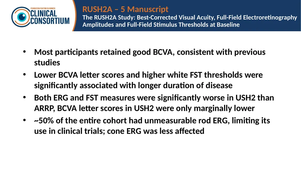
RUSH2A – 5 Manuscript
The RUSH2A Study: Best-Corrected Visual Acuity, Full-Field Electroretinography Amplitudes and Full-Field Stimulus Thresholds at Baseline Most participants retained good BCVA, consistent with previous studies
Lower BCVA letter scores and higher white FST thresholds were significantly associated with longer duration of disease
Both ERG and FST measures were significantly worse in USH2 than ARRP, BCVA letter scores in USH2 were only marginally lower
~50% of the entire cohort had unmeasurable rod ERG, limiting its use in clinical trials; cone ERG was less affected<br>
Lower BCVA letter scores and higher white FST thresholds were significantly associated with longer duration of disease
Both ERG and FST measures were significantly worse in USH2 than ARRP, BCVA letter scores in USH2 were only marginally lower
~50% of the entire cohort had unmeasurable rod ERG, limiting its use in clinical trials; cone ERG was less affected<br>
42
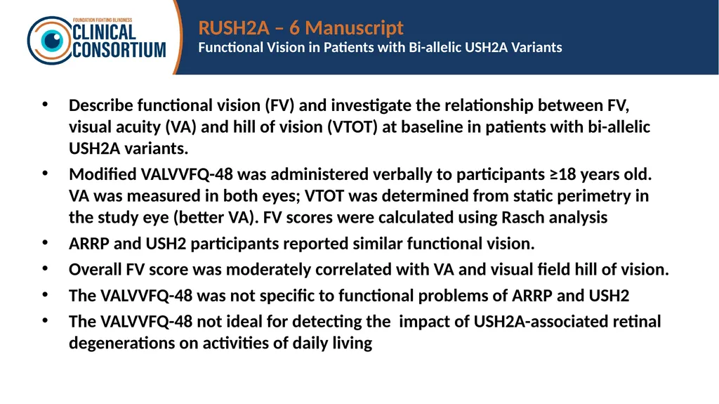
RUSH2A – 6 Manuscript
Functional Vision in Patients with Bi-allelic USH2A Variants Describe functional vision (FV) and investigate the relationship between FV, visual acuity (VA) and hill of vision (VTOT) at baseline in patients with bi-allelic USH2A variants.
Modified VALVVFQ-48 was administered verbally to participants ≥18 years old. VA was measured in both eyes; VTOT was determined from static perimetry in the study eye (better VA). FV scores were calculated using Rasch analysis
ARRP and USH2 participants reported similar functional vision.
Overall FV score was moderately correlated with VA and visual field hill of vision.
The VALVVFQ-48 was not specific to functional problems of ARRP and USH2
The VALVVFQ-48 not ideal for detecting the impact of USH2A-associated retinal degenerations on activities of daily living<br>
Modified VALVVFQ-48 was administered verbally to participants ≥18 years old. VA was measured in both eyes; VTOT was determined from static perimetry in the study eye (better VA). FV scores were calculated using Rasch analysis
ARRP and USH2 participants reported similar functional vision.
Overall FV score was moderately correlated with VA and visual field hill of vision.
The VALVVFQ-48 was not specific to functional problems of ARRP and USH2
The VALVVFQ-48 not ideal for detecting the impact of USH2A-associated retinal degenerations on activities of daily living<br>
43
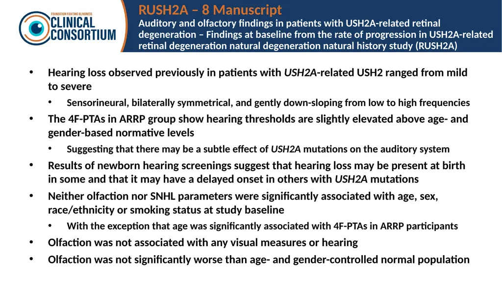
RUSH2A – 8 Manuscript
Auditory and olfactory findings in patients with USH2A-related retinal degeneration – Findings at baseline from the rate of progression in USH2A-related retinal degeneration natural degeneration natural history study (RUSH2A) Hearing loss observed previously in patients with USH2A-related USH2 ranged from mild to severe
Sensorineural, bilaterally symmetrical, and gently down-sloping from low to high frequencies
The 4F-PTAs in ARRP group show hearing thresholds are slightly elevated above age- and gender-based normative levels
Suggesting that there may be a subtle effect of USH2A mutations on the auditory system
Results of newborn hearing screenings suggest that hearing loss may be present at birth in some and that it may have a delayed onset in others with USH2A mutations
Neither olfaction nor SNHL parameters were significantly associated with age, sex, race/ethnicity or smoking status at study baseline
With the exception that age was significantly associated with 4F-PTAs in ARRP participants
Olfaction was not associated with any visual measures or hearing
Olfaction was not significantly worse than age- and gender-controlled normal population<br>
Sensorineural, bilaterally symmetrical, and gently down-sloping from low to high frequencies
The 4F-PTAs in ARRP group show hearing thresholds are slightly elevated above age- and gender-based normative levels
Suggesting that there may be a subtle effect of USH2A mutations on the auditory system
Results of newborn hearing screenings suggest that hearing loss may be present at birth in some and that it may have a delayed onset in others with USH2A mutations
Neither olfaction nor SNHL parameters were significantly associated with age, sex, race/ethnicity or smoking status at study baseline
With the exception that age was significantly associated with 4F-PTAs in ARRP participants
Olfaction was not associated with any visual measures or hearing
Olfaction was not significantly worse than age- and gender-controlled normal population<br>
44
RUSH2A – 9 Manuscript
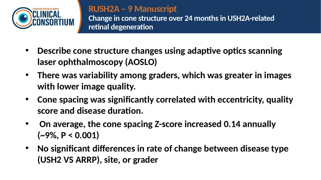
Change in cone structure over 24 months in USH2A-related retinal degeneration Describe cone structure changes using adaptive optics scanning laser ophthalmoscopy (AOSLO)
There was variability among graders, which was greater in images with lower image quality.
Cone spacing was significantly correlated with eccentricity, quality score and disease duration.
On average, the cone spacing Z-score increased 0.14 annually (~9%, P < 0.001)
No significant differences in rate of change between disease type (USH2 VS ARRP), site, or grader<br>
There was variability among graders, which was greater in images with lower image quality.
Cone spacing was significantly correlated with eccentricity, quality score and disease duration.
On average, the cone spacing Z-score increased 0.14 annually (~9%, P < 0.001)
No significant differences in rate of change between disease type (USH2 VS ARRP), site, or grader<br>
45
RUSH2A – 10 Manuscript
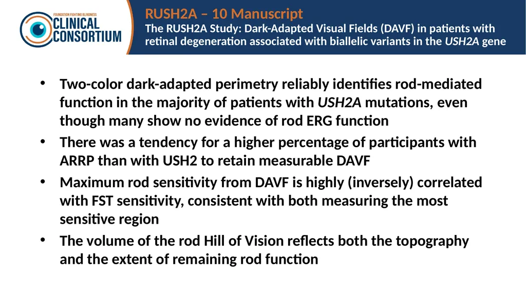
The RUSH2A Study: Dark-Adapted Visual Fields (DAVF) in patients with retinal degeneration associated with biallelic variants in the USH2A gene Two-color dark-adapted perimetry reliably identifies rod-mediated function in the majority of patients with USH2A mutations, even though many show no evidence of rod ERG function
There was a tendency for a higher percentage of participants with ARRP than with USH2 to retain measurable DAVF
Maximum rod sensitivity from DAVF is highly (inversely) correlated with FST sensitivity, consistent with both measuring the most sensitive region
The volume of the rod Hill of Vision reflects both the topography and the extent of remaining rod function<br>
There was a tendency for a higher percentage of participants with ARRP than with USH2 to retain measurable DAVF
Maximum rod sensitivity from DAVF is highly (inversely) correlated with FST sensitivity, consistent with both measuring the most sensitive region
The volume of the rod Hill of Vision reflects both the topography and the extent of remaining rod function<br>
46
RUSH2A – 11 Manuscript
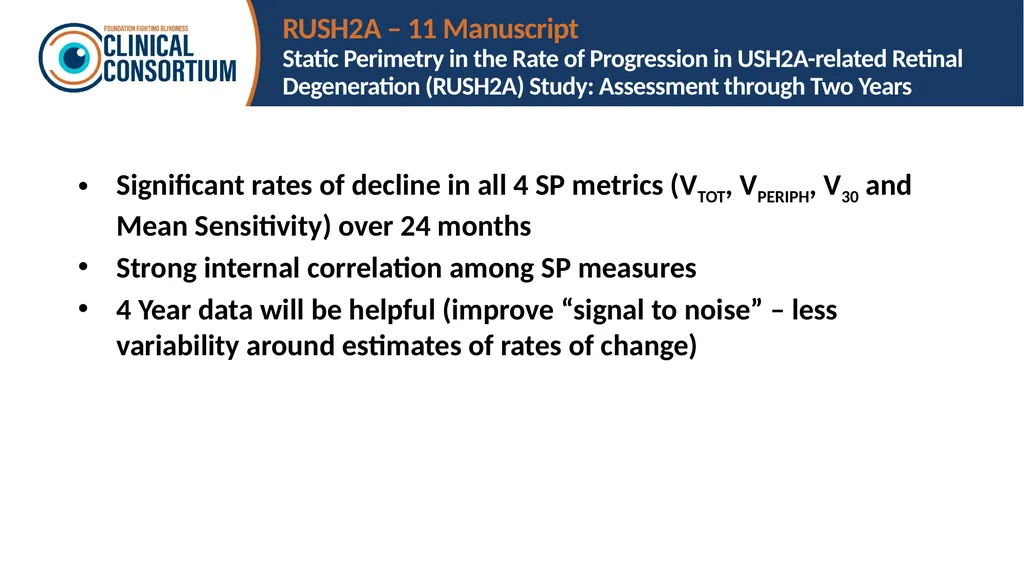
Static Perimetry in the Rate of Progression in USH2A-related Retinal Degeneration (RUSH2A) Study: Assessment through Two Years Significant rates of decline in all 4 SP metrics (VTOT, VPERIPH, V30 and Mean Sensitivity) over 24 months
Strong internal correlation among SP measures
4 Year data will be helpful (improve “signal to noise” – less variability around estimates of rates of change)<br>
Strong internal correlation among SP measures
4 Year data will be helpful (improve “signal to noise” – less variability around estimates of rates of change)<br>
47
RUSH2A – 18 Manuscript
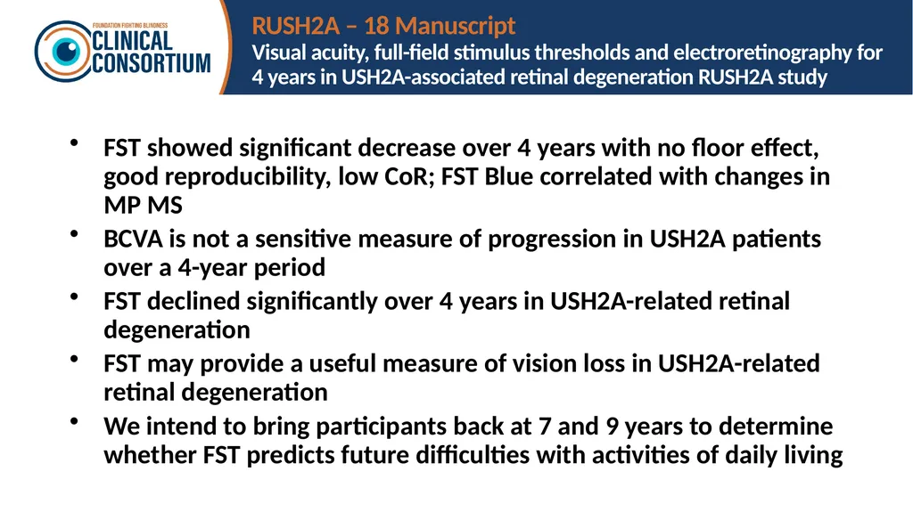
Visual acuity, full-field stimulus thresholds and electroretinography for 4 years in USH2A-associated retinal degeneration RUSH2A study FST showed significant decrease over 4 years with no floor effect, good reproducibility, low CoR; FST Blue correlated with changes in MP MS
BCVA is not a sensitive measure of progression in USH2A patients over a 4-year period
FST declined significantly over 4 years in USH2A-related retinal degeneration
FST may provide a useful measure of vision loss in USH2A-related retinal degeneration
We intend to bring participants back at 7 and 9 years to determine whether FST predicts future difficulties with activities of daily living<br>
BCVA is not a sensitive measure of progression in USH2A patients over a 4-year period
FST declined significantly over 4 years in USH2A-related retinal degeneration
FST may provide a useful measure of vision loss in USH2A-related retinal degeneration
We intend to bring participants back at 7 and 9 years to determine whether FST predicts future difficulties with activities of daily living<br>
48
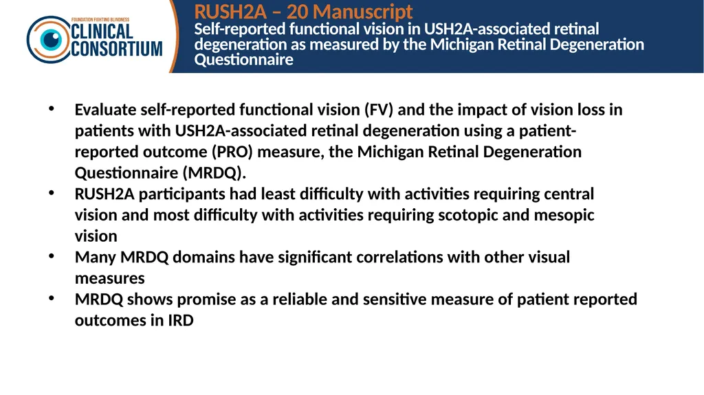
RUSH2A – 20 Manuscript
Self-reported functional vision in USH2A-associated retinal degeneration as measured by the Michigan Retinal Degeneration Questionnaire Evaluate self-reported functional vision (FV) and the impact of vision loss in patients with USH2A-associated retinal degeneration using a patient-reported outcome (PRO) measure, the Michigan Retinal Degeneration Questionnaire (MRDQ).
RUSH2A participants had least difficulty with activities requiring central vision and most difficulty with activities requiring scotopic and mesopic vision
Many MRDQ domains have significant correlations with other visual measures
MRDQ shows promise as a reliable and sensitive measure of patient reported outcomes in IRD<br>
RUSH2A participants had least difficulty with activities requiring central vision and most difficulty with activities requiring scotopic and mesopic vision
Many MRDQ domains have significant correlations with other visual measures
MRDQ shows promise as a reliable and sensitive measure of patient reported outcomes in IRD<br>
49
Where to Find Results of Clinical
Consortium Studies All publications are listed under the PUBLICATIONS tab of the public website (ffb.jaeb.org)
Summary slides of all publications are available under the PRESENTATIONS tab of the public website (ffb.jaeb.org)<br>
Summary slides of all publications are available under the PRESENTATIONS tab of the public website (ffb.jaeb.org)<br>
50
How to Get Involved<br>
51
How to Join the Clinical Consortium Complete a Clinical Site Application
Available on the public website homepage ffb.jaeb.org
Email completed form to ffb@jaeb.org
Site Requirements
Minimum required equipment
Octopus 900 Pro Perimetry
Heidelberg Spectralis OCT
Site PI must meet qualifications (listed on application)
U.S. sites must use the central IRB Additional information about the Consortium, listings of participating centers, and publications can be found on ffb.jaeb.org The Consortium is not accepting new applications at this time<br>
Available on the public website homepage ffb.jaeb.org
Email completed form to ffb@jaeb.org
Site Requirements
Minimum required equipment
Octopus 900 Pro Perimetry
Heidelberg Spectralis OCT
Site PI must meet qualifications (listed on application)
U.S. sites must use the central IRB Additional information about the Consortium, listings of participating centers, and publications can be found on ffb.jaeb.org The Consortium is not accepting new applications at this time<br>
52
How to Submit a Protocol Idea Complete a Protocol Idea Form
Available on the public website (ffb.jaeb.org)
See FAQ on the public website (ffb.jaeb.org) for more information
Email completed form to ffb@jaeb.org<br>
Available on the public website (ffb.jaeb.org)
See FAQ on the public website (ffb.jaeb.org) for more information
Email completed form to ffb@jaeb.org<br>
53
How to Request Use of Clinical Consortium Data It depends on the status of the data and the intended use
See FAQ on the public website (ffb.jaeb.org) for more information
Email completed request forms to ffb@jaeb.org<br>
See FAQ on the public website (ffb.jaeb.org) for more information
Email completed request forms to ffb@jaeb.org<br>