
PFT Interpretation Darrin Hursey, MD Overview



Description: PFT Interpretation Darrin Hursey, MD Overview Indications for performing PFTs Brief review of relevant pulmonary physiology Interpretation scheme Indications Evaluation of chronic dyspnea (looking specifically for asthma or COPD) Monitoring
Related Topics
Download Presentation
"PFT Interpretation Darrin Hursey, MD Overview" is the property of its rightful owner. Permission is granted to download and print the materials on this website for personal, non-commercial use only, and to display it on your personal computer provided you do not modify the materials and that you retain all copyright notices contained in the materials. By downloading content from our website, you accept the terms of this agreement.
Presentation Transcript
slide1. PFT Interpretation Darrin Hursey, MD<br>
slide2. Overview Indications for performing PFTs
Brief review of relevant pulmonary physiology
Interpretation scheme<br>
slide3. Indications Evaluation of chronic dyspnea (looking specifically for asthma or COPD)
Monitoring for response to therapy for asthma or COPD
Pre-operative evaluation<br>
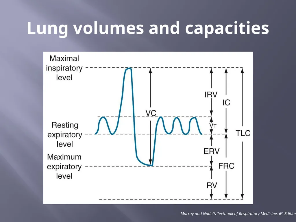
slide5. Lung volumes and capacities Murray and Nadel’s Textbook of Respiratory Medicine, 6th Edition<br>
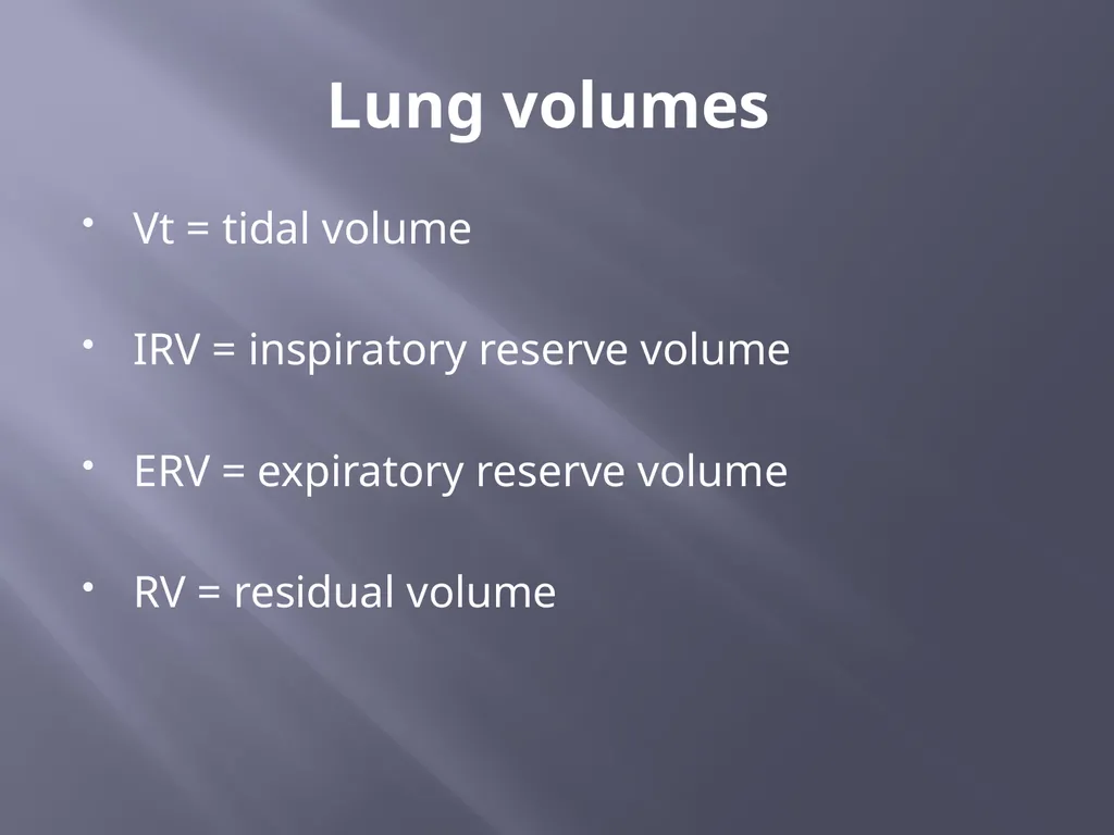
slide6. Lung volumes Vt = tidal volume
IRV = inspiratory reserve volume
ERV = expiratory reserve volume
RV = residual volume<br>
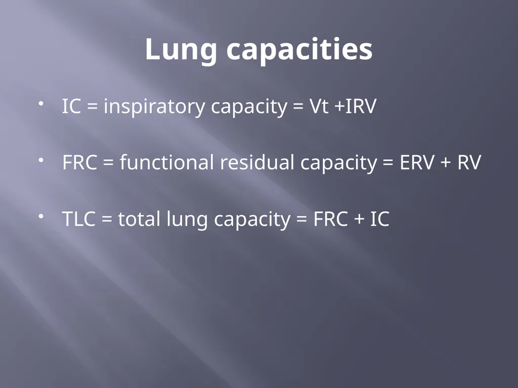
slide7. Lung capacities IC = inspiratory capacity = Vt +IRV
FRC = functional residual capacity = ERV + RV
TLC = total lung capacity = FRC + IC<br>
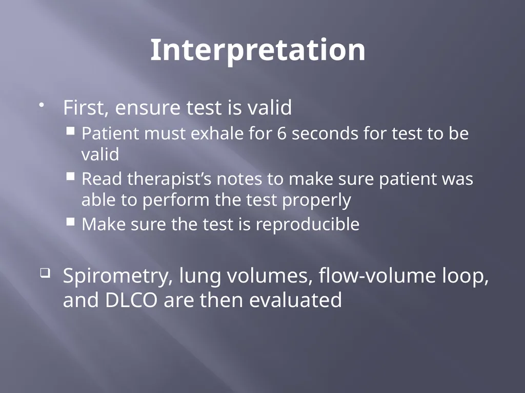
slide8. Interpretation First, ensure test is valid
Patient must exhale for 6 seconds for test to be valid
Read therapist’s notes to make sure patient was able to perform the test properly
Make sure the test is reproducible
Spirometry, lung volumes, flow-volume loop, and DLCO are then evaluated<br>
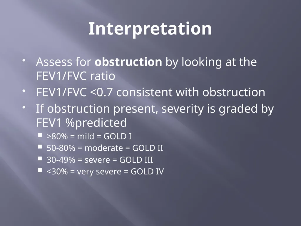
slide9. Interpretation Assess for obstruction by looking at the FEV1/FVC ratio
FEV1/FVC <0.7 consistent with obstruction
If obstruction present, severity is graded by FEV1 %predicted
>80% = mild = GOLD I
50-80% = moderate = GOLD II
30-49% = severe = GOLD III
<30% = very severe = GOLD IV<br>
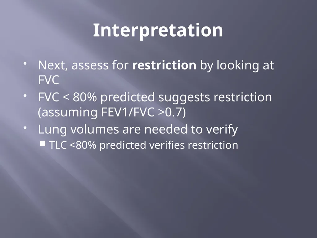
slide10. Interpretation Next, assess for restriction by looking at FVC
FVC < 80% predicted suggests restriction (assuming FEV1/FVC >0.7)
Lung volumes are needed to verify
TLC <80% predicted verifies restriction<br>
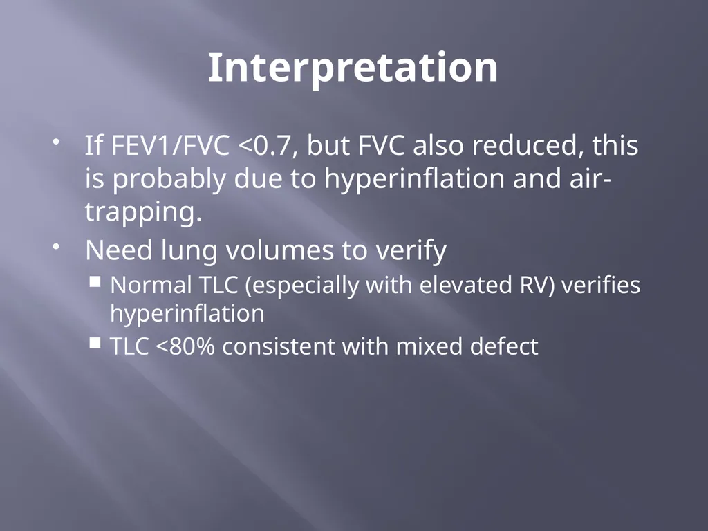
slide11. Interpretation If FEV1/FVC <0.7, but FVC also reduced, this is probably due to hyperinflation and air-trapping.
Need lung volumes to verify
Normal TLC (especially with elevated RV) verifies hyperinflation
TLC <80% consistent with mixed defect<br>
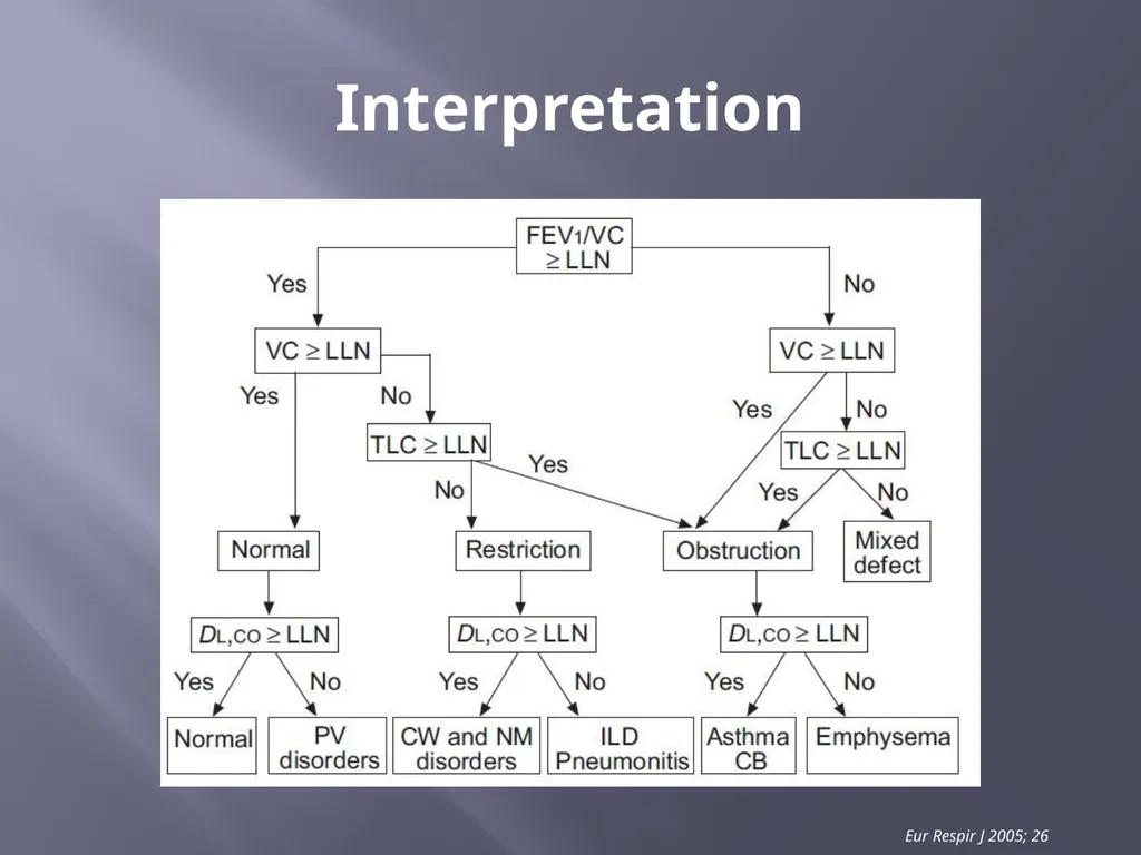
slide12. Interpretation Eur Respir J 2005; 26<br>
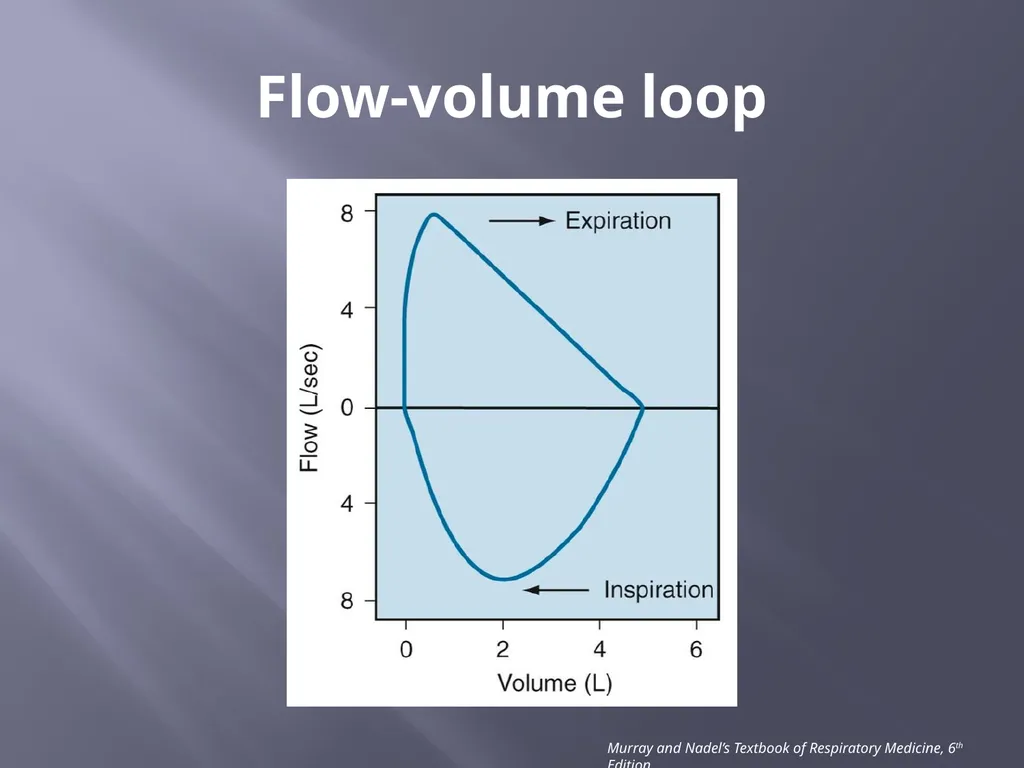
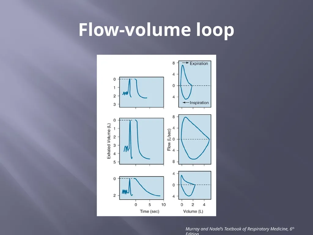
slide13. Flow-volume loop Murray and Nadel’s Textbook of Respiratory Medicine, 6th Edition<br>
slide14. Flow-volume loop Murray and Nadel’s Textbook of Respiratory Medicine, 6th Edition<br>
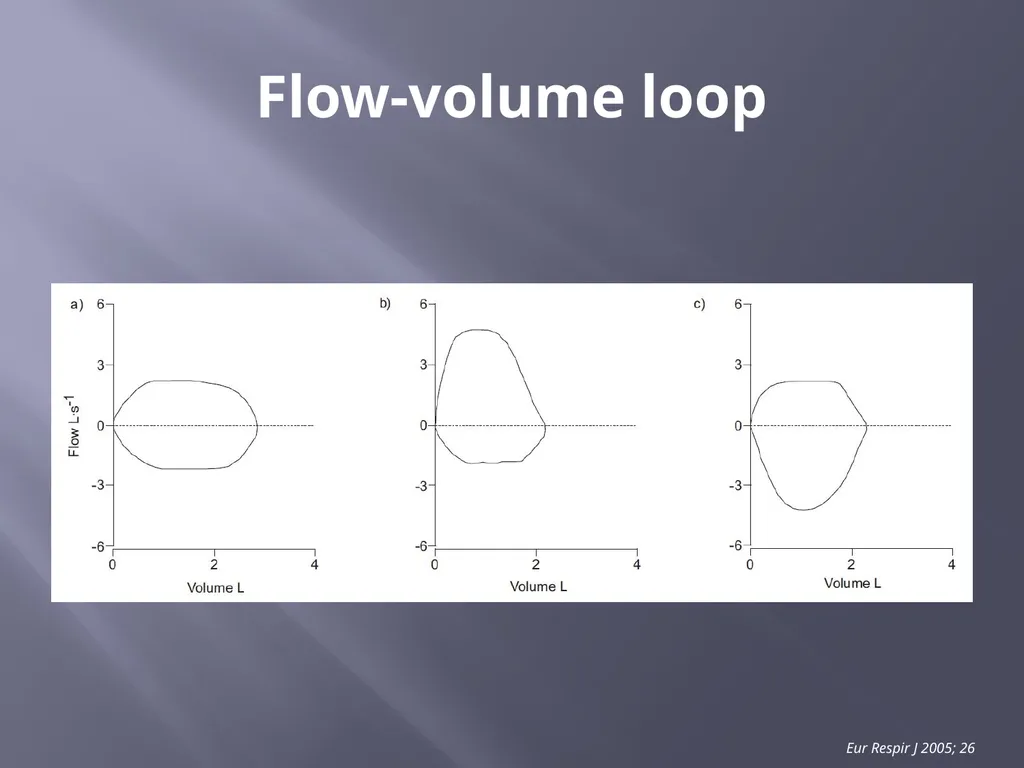
slide15. Flow-volume loop Eur Respir J 2005; 26<br>
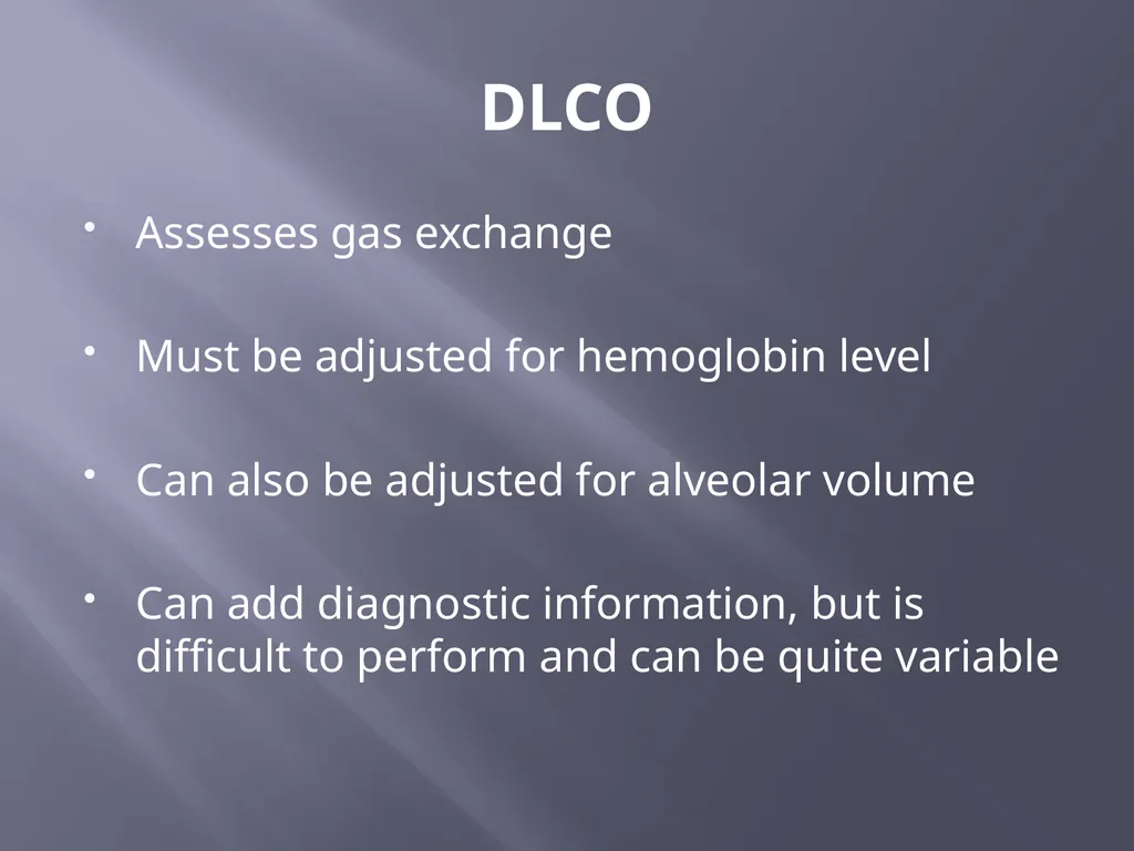
slide16. DLCO Assesses gas exchange
Must be adjusted for hemoglobin level
Can also be adjusted for alveolar volume
Can add diagnostic information, but is difficult to perform and can be quite variable<br>
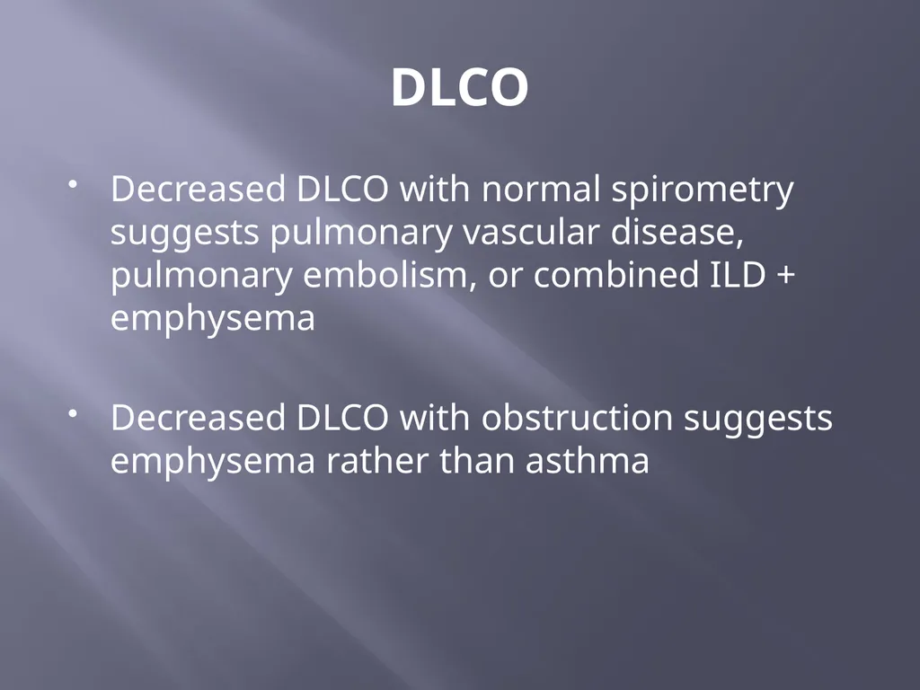
slide17. DLCO Decreased DLCO with normal spirometry suggests pulmonary vascular disease, pulmonary embolism, or combined ILD + emphysema
Decreased DLCO with obstruction suggests emphysema rather than asthma<br>
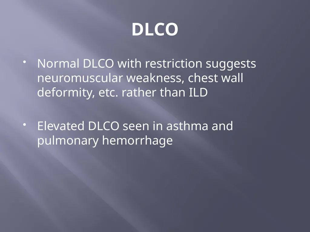
slide18. DLCO Normal DLCO with restriction suggests neuromuscular weakness, chest wall deformity, etc. rather than ILD
Elevated DLCO seen in asthma and pulmonary hemorrhage<br>
slide20. Further reading Broaddus, VC, Mason, RJ, Ernst, JD, King, TE, Lazarus, SC, Murray, FJ, Nadel, JA, Slutsky, AS, Gotway, MB 2015 Murray & Nadel’s Textbook of Respiratory Medicine, 6th edition, Elsevier Saunders, Philadelphia PA.
R. Pellegrino, G. Viegi, V. Brusasco, R.O. Crapo, F. Burgos, R. Casaburi, A. Coates, C.P.M. van der Grinten, P. Gustafsson, J. Hankinson, R. Jensen, D.C. Johnson, N. MacIntyre, R. McKay, M.R. Miller, D. Navajas, O.F. Pedersen and J. Wanger 2005 “Intepretive strategies for lung function tests” Eur Respir J, 26, 948–968<br>
slide2. Overview Indications for performing PFTs
Brief review of relevant pulmonary physiology
Interpretation scheme<br>
slide3. Indications Evaluation of chronic dyspnea (looking specifically for asthma or COPD)
Monitoring for response to therapy for asthma or COPD
Pre-operative evaluation<br>
slide5. Lung volumes and capacities Murray and Nadel’s Textbook of Respiratory Medicine, 6th Edition<br>
slide6. Lung volumes Vt = tidal volume
IRV = inspiratory reserve volume
ERV = expiratory reserve volume
RV = residual volume<br>
slide7. Lung capacities IC = inspiratory capacity = Vt +IRV
FRC = functional residual capacity = ERV + RV
TLC = total lung capacity = FRC + IC<br>
slide8. Interpretation First, ensure test is valid
Patient must exhale for 6 seconds for test to be valid
Read therapist’s notes to make sure patient was able to perform the test properly
Make sure the test is reproducible
Spirometry, lung volumes, flow-volume loop, and DLCO are then evaluated<br>
slide9. Interpretation Assess for obstruction by looking at the FEV1/FVC ratio
FEV1/FVC <0.7 consistent with obstruction
If obstruction present, severity is graded by FEV1 %predicted
>80% = mild = GOLD I
50-80% = moderate = GOLD II
30-49% = severe = GOLD III
<30% = very severe = GOLD IV<br>
slide10. Interpretation Next, assess for restriction by looking at FVC
FVC < 80% predicted suggests restriction (assuming FEV1/FVC >0.7)
Lung volumes are needed to verify
TLC <80% predicted verifies restriction<br>
slide11. Interpretation If FEV1/FVC <0.7, but FVC also reduced, this is probably due to hyperinflation and air-trapping.
Need lung volumes to verify
Normal TLC (especially with elevated RV) verifies hyperinflation
TLC <80% consistent with mixed defect<br>
slide12. Interpretation Eur Respir J 2005; 26<br>
slide13. Flow-volume loop Murray and Nadel’s Textbook of Respiratory Medicine, 6th Edition<br>
slide14. Flow-volume loop Murray and Nadel’s Textbook of Respiratory Medicine, 6th Edition<br>
slide15. Flow-volume loop Eur Respir J 2005; 26<br>
slide16. DLCO Assesses gas exchange
Must be adjusted for hemoglobin level
Can also be adjusted for alveolar volume
Can add diagnostic information, but is difficult to perform and can be quite variable<br>
slide17. DLCO Decreased DLCO with normal spirometry suggests pulmonary vascular disease, pulmonary embolism, or combined ILD + emphysema
Decreased DLCO with obstruction suggests emphysema rather than asthma<br>
slide18. DLCO Normal DLCO with restriction suggests neuromuscular weakness, chest wall deformity, etc. rather than ILD
Elevated DLCO seen in asthma and pulmonary hemorrhage<br>
slide20. Further reading Broaddus, VC, Mason, RJ, Ernst, JD, King, TE, Lazarus, SC, Murray, FJ, Nadel, JA, Slutsky, AS, Gotway, MB 2015 Murray & Nadel’s Textbook of Respiratory Medicine, 6th edition, Elsevier Saunders, Philadelphia PA.
R. Pellegrino, G. Viegi, V. Brusasco, R.O. Crapo, F. Burgos, R. Casaburi, A. Coates, C.P.M. van der Grinten, P. Gustafsson, J. Hankinson, R. Jensen, D.C. Johnson, N. MacIntyre, R. McKay, M.R. Miller, D. Navajas, O.F. Pedersen and J. Wanger 2005 “Intepretive strategies for lung function tests” Eur Respir J, 26, 948–968<br>