Preparation for spinal tumour surgery...embolization, cell saver, monitoring, other teams Jolyn Khoo Where is it? Extradural Most common (60) Metastatic 97: breast, lung, prostate 15-20 each Primary lesions benign 80, malignant 20
"Preparation for spinal tumour" is the property of its rightful owner. Permission is granted to
download and print the materials on this website for personal, non-commercial use only, and to display it
on your personal computer provided you do not modify the materials and that you retain all copyright
notices contained in the materials. By downloading content from our website, you accept the terms of this
agreement.
Presentation Transcript
01
Preparation for spinal tumour surgery...embolization, cell saver, monitoring, other teams Jolyn Khoo<br>
02
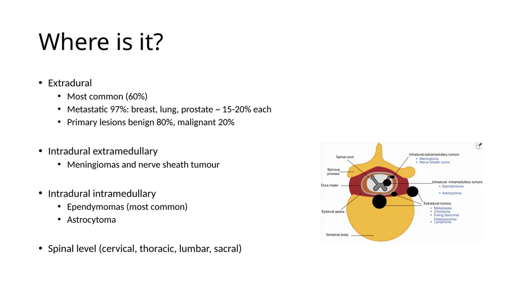
Where is it? Extradural
Most common (60%)
Metastatic 97%: breast, lung, prostate ~ 15-20% each
Primary lesions benign 80%, malignant 20%
Intradural extramedullary
Meningiomas and nerve sheath tumour
What is it? Benign vs malignant (primary or metastatic)
Most likely differentials based on location
Known cancers or syndromes (eg NF1 and 2)
Biopsy – risk of seeding rare
Risk of recurrence, blood loss, stability etc<br>
04
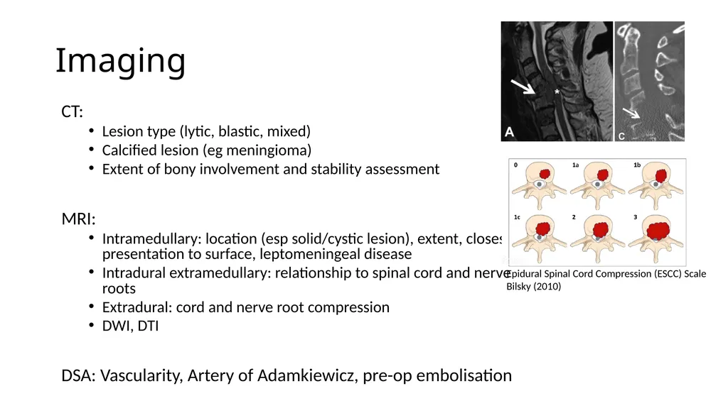
Imaging CT:
Lesion type (lytic, blastic, mixed)
Calcified lesion (eg meningioma)
Extent of bony involvement and stability assessment
MRI:
Intramedullary: location (esp solid/cystic lesion), extent, closest presentation to surface, leptomeningeal disease
Intradural extramedullary: relationship to spinal cord and nerve roots
Extradural: cord and nerve root compression
DWI, DTI
Goal of Surgery? Diagnosis
Maintenance or recovery of neurologic function
Local durable tumour control: en bloc resection, maximal safe resection, separation surgery with plan for adjuvant stereotactic radiation
Spine stability
Pain relief
Improved QoL<br>
06
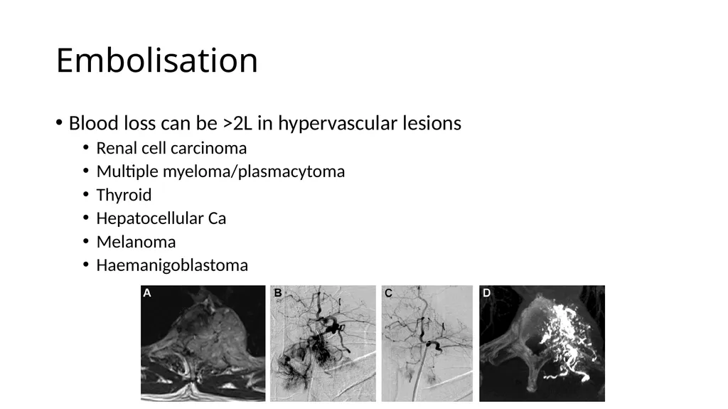
Embolisation Blood loss can be >2L in hypervascular lesions
Renal cell carcinoma
Multiple myeloma/plasmacytoma
Thyroid
Hepatocellular Ca
Melanoma
Haemanigoblastoma<br>
07
Embolisation First described in 1974 by Benati et al
Reduces intraoperative blood loss (30-50%) and need for perioperative transfusions
Improves tumour visualisation
Facilitates tumour removal
Decreases complications and surgical operating time<br>
Embolisation Optimal timing
Retrospective studies suggest surgery same day post embolisation had less intraoperative blood loss
Thought to be due to tumour revascularisation
Post embolisation swelling
Surgery “as soon as possible”
Day after for tumours with mild cord compression
Same day for myelopathy or high grade cord compression<br>
10
Cell Saver Allogenic blood transfusion
Immune mediated complications eg transfusion related acute lung injury
Immunomodulation increasing risk of operative infections (mixed evidence)
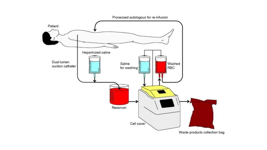
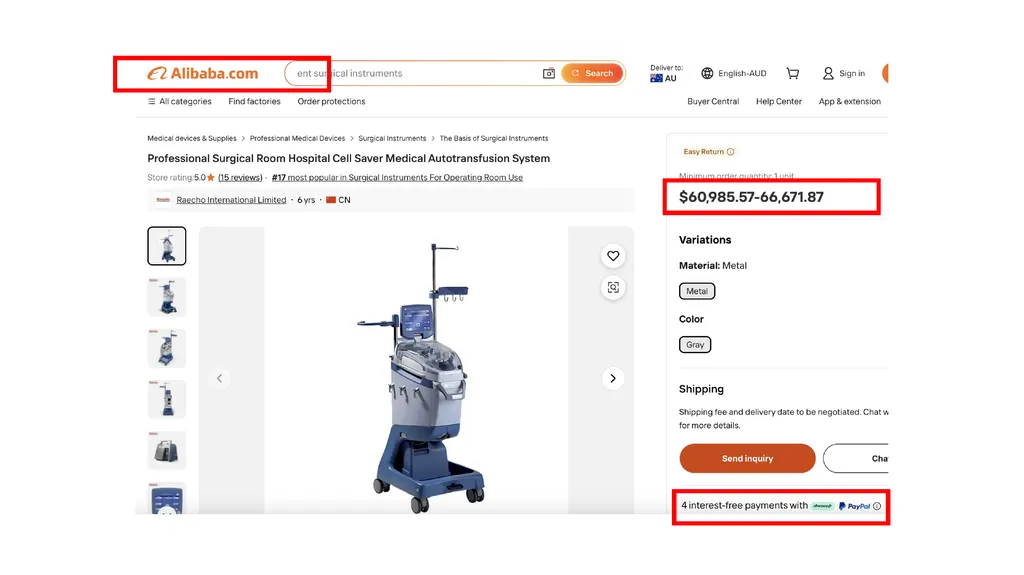
Cell Saver: intraoperative cell salvage and autotransfusion
Use of leukocyte depletion filtration system -> negligible tumorous cells within salvaged blood
Recent prospective study over 4 years of salvaged (n=33) vs allogenic (n=39) blood transfusions in metastatic spine surgery -> no significant difference in overall survival or increased risk of 4 year tumour progression (Hui et al 2026)<br>
11
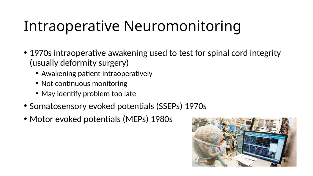
Intraoperative Neuromonitoring 1970s intraoperative awakening used to test for spinal cord integrity (usually deformity surgery)
Awakening patient intraoperatively
Not continuous monitoring
May identify problem too late
Somatosensory evoked potentials (SSEPs) 1970s
Motor evoked potentials (MEPs) 1980s<br>
12
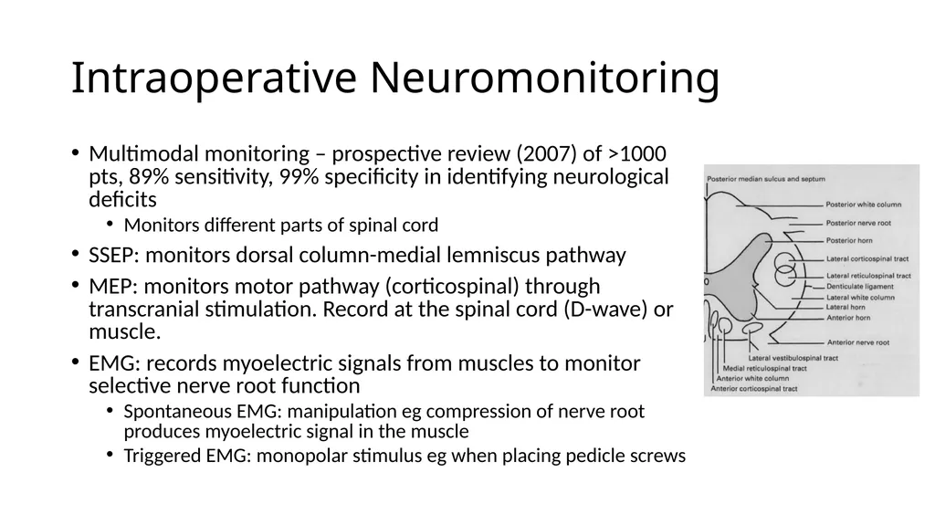
Intraoperative Neuromonitoring Multimodal monitoring – prospective review (2007) of >1000 pts, 89% sensitivity, 99% specificity in identifying neurological deficits
Monitors different parts of spinal cord
SSEP: monitors dorsal column-medial lemniscus pathway
MEP: monitors motor pathway (corticospinal) through transcranial stimulation. Record at the spinal cord (D-wave) or muscle.
EMG: records myoelectric signals from muscles to monitor selective nerve root function
Spontaneous EMG: manipulation eg compression of nerve root produces myoelectric signal in the muscle
Triggered EMG: monopolar stimulus eg when placing pedicle screws<br>
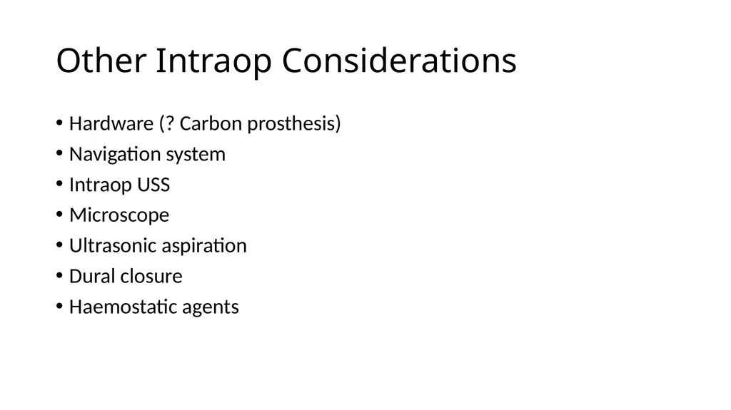
Other Intraop Considerations Hardware (? Carbon prosthesis)
Navigation system
Intraop USS
Microscope
Ultrasonic aspiration
Dural closure
Haemostatic agents<br>
17
References Kumar N, Tan W, Wei W et al. An overview of the tumours affecting the spine – inside to out. Neuro-oncology Practice. 2020. 18;7:i10-i17. doi: 10.1093/nop/npaa049
Soichiro T, Anahita M, Abdullah I et al. Advances in imaging modalities for spinal tumours. Neuro-Oncology Advances. Oct 2024: 6;3. Pg iii13–iii27. https://doi.org/10.1093/noajnl/vdae045
Facchini, G., Parmeggiani, A., Peta, G. et al. The role of percutaneous transarterial embolization in the management of spinal bone tumors: a literature review. Eur Spine J. 2021. 30;2839–2851. https://doi-org.ezproxy.surgeons.org/10.1007/s00586-021-06963-5
Ozkan E, Gupta S. Embolisation of spinal tumours: vascular anatomy, indications, and technique. Techniques in Vascular and Interventional Radiology. 2011. 14:3;129-140
Hui S, Tan J, Tan Y et al. Shaping the future of blood management in metastatic spine tumour surgery: the case for cell-salvaged transfusion with a propensity matched study. North American Spine Society Journal. 2026. https://doi.org/10.1016/j.xnsj.2026.100862
Galloway G, Nuwer M, Lopez J et al. Intraoperative wake-up test. Intraoperative Neurophysiologic Monitoring. 2013.
Sutter M, Eggspuehler A, Grob D et al. The diagnostiv value of multimodal intraoperative monitoring (MIOM) during spine surgery: a prospective study of 1017 patients. European Spine Journal. 2007. 31;16:162-170
Ghatol D, Widrich J. Intraoperative Neurophysiological Monitoring. StatPearls. 2023
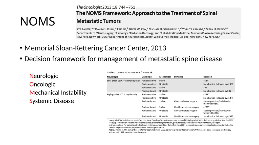
Laufer I, Rubin D, Lis E et al. The NOMS framework: approach to the treatment of spinal metastatic tumours. The Oncologist. 2013.. 18L:744-751<br>