Radiology Case Presentation Sami Natour, MS4 UVA School of Medicine E.R. is a 34 year old female with a past medical history of ADHD who was transferred from an OSH with malaise, altered mental status, and thrombocytopenia Was recently in
"Radiology Case Presentation Sami Natour, MS4 UVA" is the property of its rightful owner. Permission is granted to
download and print the materials on this website for personal, non-commercial use only, and to display it
on your personal computer provided you do not modify the materials and that you retain all copyright
notices contained in the materials. By downloading content from our website, you accept the terms of this
agreement.
Presentation Transcript
01
Radiology Case Presentation Sami Natour, MS4
UVA School of Medicine<br>
02
E.R. is a 34 year old female with a past medical history of ADHD who was transferred from an OSH with malaise, altered mental status, and thrombocytopenia
Was recently in North Carolina for funeral where she was in heavily wooded area. Returned ~1 week ago.
On ride home, developed headache, chills, and somnolence
In days leading up to UVA admission:
- Urgent care clinic suspected herpes zoster; Acyclovir reportedly worsened her symptoms
- Presented twice to OSH after developing N/V. On second admission, found to be febrile and hypotensive to 79/40
Other notable labs: PLT 36,000, AST/ALT/ALP 50/58/137
Started on empiric vancomycin, ceftriaxone, and solumedrol Clinical History<br>
03
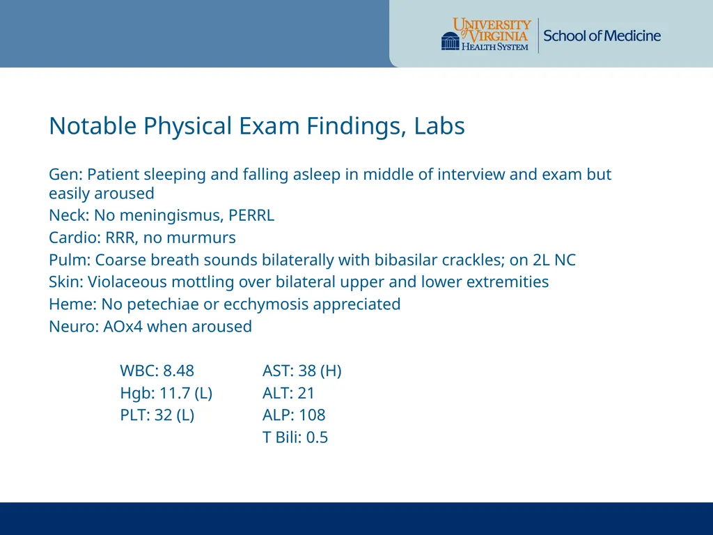
Gen: Patient sleeping and falling asleep in middle of interview and exam but easily aroused
Neck: No meningismus, PERRL
Cardio: RRR, no murmurs
Pulm: Coarse breath sounds bilaterally with bibasilar crackles; on 2L NC
Skin: Violaceous mottling over bilateral upper and lower extremities
Heme: No petechiae or ecchymosis appreciated
Neuro: AOx4 when aroused
Studies ordered at OSH (UVA overread)
- CT Head (No acute intracranial abnormality)
- US Gallbladder (Unremarkable)
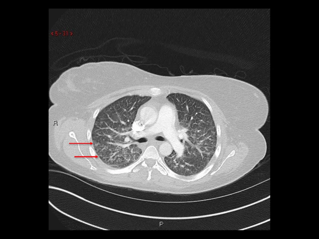
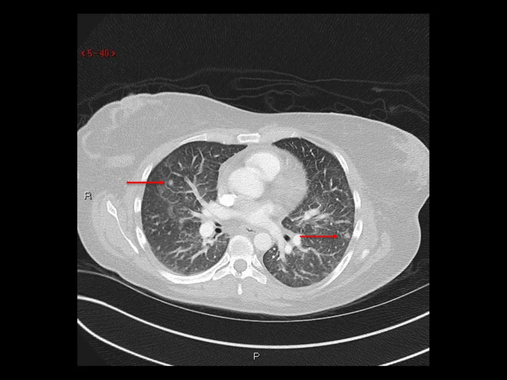
CTPA ordered due to elevated D-Dimer (see following slide) Imaging<br>
05
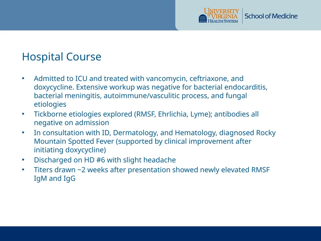
Admitted to ICU and treated with vancomycin, ceftriaxone, and doxycycline. Extensive workup was negative for bacterial endocarditis, bacterial meningitis, autoimmune/vasculitic process, and fungal etiologies
Tickborne etiologies explored (RMSF, Ehrlichia, Lyme); antibodies all negative on admission
In consultation with ID, Dermatology, and Hematology, diagnosed Rocky Mountain Spotted Fever (supported by clinical improvement after initiating doxycycline)
Discharged on HD #6 with slight headache
Titers drawn ~2 weeks after presentation showed newly elevated RMSF IgM and IgG Hospital Course<br>
06
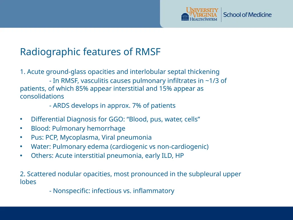
1. Acute ground-glass opacities and interlobular septal thickening
- In RMSF, vasculitis causes pulmonary infiltrates in ~1/3 of patients, of which 85% appear interstitial and 15% appear as consolidations
- ARDS develops in approx. 7% of patients
Differential Diagnosis for GGO: “Blood, pus, water, cells”
Blood: Pulmonary hemorrhage
Pus: PCP, Mycoplasma, Viral pneumonia
Water: Pulmonary edema (cardiogenic vs non-cardiogenic)
Others: Acute interstitial pneumonia, early ILD, HP
2. Scattered nodular opacities, most pronounced in the subpleural upper lobes
- Nonspecific: infectious vs. inflammatory Radiographic features of RMSF<br>
07
1. Faul JL, Doyle RL, Kao PN, Ruoss SJ. Tick-borne pulmonary disease: update on diagnosis and management. Chest. 1999 Jul;116(1):222-30. Review. PubMed PMID: 10424529.
2. Miller WT Jr, Shah RM. Isolated diffuse ground-glass opacity in thoracic CT: causes and clinical presentations. AJR Am J Roentgenol. 2005 Feb;184(2):613-22. Review. PubMed PMID: 15671387.
3. Robert NA. Squire's Fundamentals of Radiology. 6 ed. Cambridge, Massachusetts, and London, England: Harvard University Press; 2004. References<br>