
Scientific Achievement Using the Advanced Light
Description: Scientific Achievement Using the Advanced Light Source, researchers detected nanoscale deposits of elemental copper and iron in brain tissues isolated from Alzheimers disease subjects. Significance and Impact The discovery suggests new
Related Topics
Download Presentation
"Scientific Achievement Using the Advanced Light" is the property of its rightful owner. Permission is granted to download and print the materials on this website for personal, non-commercial use only, and to display it on your personal computer provided you do not modify the materials and that you retain all copyright notices contained in the materials. By downloading content from our website, you accept the terms of this agreement.
Presentation Transcript
slide1. Scientific Achievement
Using the Advanced Light Source, researchers detected nanoscale deposits of elemental copper and iron in brain tissues isolated from Alzheimer’s disease subjects.
Significance and Impact
The discovery suggests new directions of study to determine the role that elemental metals might play in neurochemistry, neurobiology, and the development of neurodegenerative disease. Research Details
Scanning transmission x-ray microscopy (STXM) was performed on amyloid plaque cores isolated from the brain tissues of deceased Alzheimer's patients.
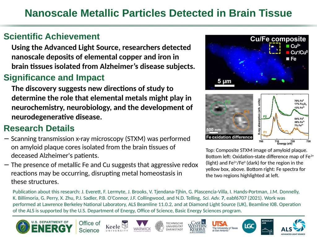
The presence of metallic Fe and Cu suggests that aggressive redox reactions may be occurring, disrupting metal homeostasis in these structures. Nanoscale Metallic Particles Detected in Brain Tissue Publication about this research: J. Everett, F. Lermyte, J. Brooks, V. Tjendana-Tjhin, G. Plascencia-Villa, I. Hands-Portman, J.M. Donnelly, K. Billimoria, G. Perry, X. Zhu, P.J. Sadler, P.B. O’Connor, J.F. Collingwood, and N.D. Telling, Sci. Adv. 7, eabf6707 (2021). Work was performed at Lawrence Berkeley National Laboratory, ALS Beamline 11.0.2, and at Diamond Light Source (UK), Beamline I08. Operation of the ALS is supported by the U.S. Department of Energy, Office of Science, Basic Energy Sciences program. Top: Composite STXM image of amyloid plaque. Bottom left: Oxidation-state difference map of Fe3+ (light) and Fe2+/Fe0 (dark) for the region in the yellow box, above. Bottom right: Fe spectra for the two regions highlighted at left.<br>
Using the Advanced Light Source, researchers detected nanoscale deposits of elemental copper and iron in brain tissues isolated from Alzheimer’s disease subjects.
Significance and Impact
The discovery suggests new directions of study to determine the role that elemental metals might play in neurochemistry, neurobiology, and the development of neurodegenerative disease. Research Details
Scanning transmission x-ray microscopy (STXM) was performed on amyloid plaque cores isolated from the brain tissues of deceased Alzheimer's patients.
The presence of metallic Fe and Cu suggests that aggressive redox reactions may be occurring, disrupting metal homeostasis in these structures. Nanoscale Metallic Particles Detected in Brain Tissue Publication about this research: J. Everett, F. Lermyte, J. Brooks, V. Tjendana-Tjhin, G. Plascencia-Villa, I. Hands-Portman, J.M. Donnelly, K. Billimoria, G. Perry, X. Zhu, P.J. Sadler, P.B. O’Connor, J.F. Collingwood, and N.D. Telling, Sci. Adv. 7, eabf6707 (2021). Work was performed at Lawrence Berkeley National Laboratory, ALS Beamline 11.0.2, and at Diamond Light Source (UK), Beamline I08. Operation of the ALS is supported by the U.S. Department of Energy, Office of Science, Basic Energy Sciences program. Top: Composite STXM image of amyloid plaque. Bottom left: Oxidation-state difference map of Fe3+ (light) and Fe2+/Fe0 (dark) for the region in the yellow box, above. Bottom right: Fe spectra for the two regions highlighted at left.<br>