
Serological tests A serology blood test is
AS
Published · 18 slides · 0 views

1 / 1





Description
Serological tests A serology blood test is performed to detect and measure the levels of antibodies as a result of exposure to a particular bacteria or viruses (antigens), the immune system produces specific antibodies against these
Related Topics
Share
Embed code
Download this presentation From Below
"Serological tests A serology blood test is" is the property of its rightful owner. Permission is granted to download and print the materials on this website for personal, non-commercial use only, and to display it on your personal computer provided you do not modify the materials and that you retain all copyright notices contained in the materials. By downloading content from our website, you accept the terms of this agreement.
Presentation Transcript
01
Serological tests A serology blood test is performed to detect and measure the levels of antibodies as a result of exposure to a particular bacteria or viruses (antigens), the immune system produces specific antibodies against these organism. Antibody levels (antibody titer) help physicians determine whether an infection occurred recently (Acute) or years ago (chronic).
Serological testing is particularly helpful in the diagnosis of rickettsial and viral diseases such as Rocky Mountain spotted fever, influenza, measles, poliomyelitis, and yellow fever, as well as of infectious mononucleosis and rheumatoid arthritis. As a practical mass-screening diagnostic tool, it has proved valuable in the detection of such conditions as syphilis.<br>
Serological testing is particularly helpful in the diagnosis of rickettsial and viral diseases such as Rocky Mountain spotted fever, influenza, measles, poliomyelitis, and yellow fever, as well as of infectious mononucleosis and rheumatoid arthritis. As a practical mass-screening diagnostic tool, it has proved valuable in the detection of such conditions as syphilis.<br>
02
Submitting specimens Whole blood: Refrigerated specimens must be received within 24 hours of the time of collection.
Serum separated from clot: Refrigerated specimens must be received within 48 hours of the time of collection. Frozen specimens must be received within six months of collection.
CSF: Refrigerated specimens must be received within 48 hours from the time of collection. Frozen specimens must be received within six months of collection.<br>
Serum separated from clot: Refrigerated specimens must be received within 48 hours of the time of collection. Frozen specimens must be received within six months of collection.
CSF: Refrigerated specimens must be received within 48 hours from the time of collection. Frozen specimens must be received within six months of collection.<br>
03
Rose Bengal plate test (RBT) for Brucella The Rose Bengal test (RBT) is a simple, rapid slide-type agglutination assay performed with a stained Brucella abortus suspension at pH 3.6–3.7 and plain serum.
It is often used as a screening test in human brucellosis and would be optimal for small laboratories with limited means. False-negative reactions occur especially in the early stages of acute infection.<br>
It is often used as a screening test in human brucellosis and would be optimal for small laboratories with limited means. False-negative reactions occur especially in the early stages of acute infection.<br>
04
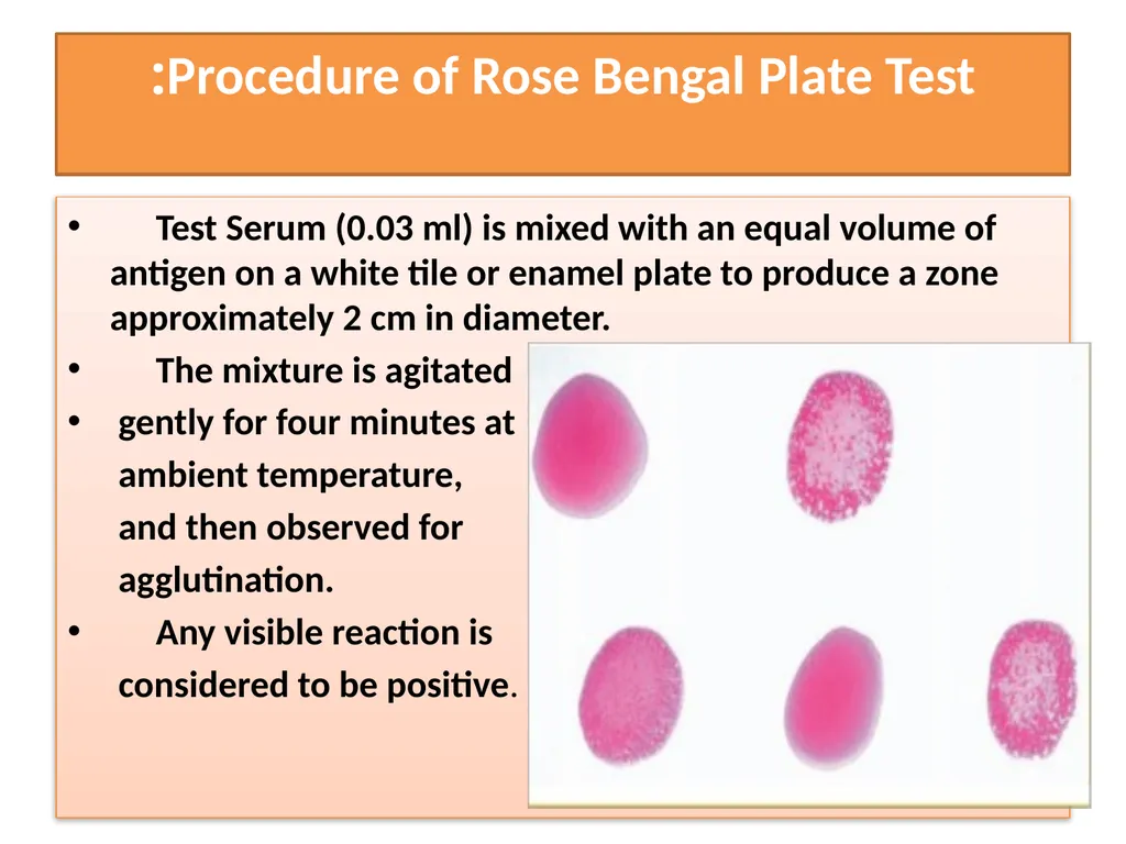
Procedure of Rose Bengal Plate Test: Test Serum (0.03 ml) is mixed with an equal volume of antigen on a white tile or enamel plate to produce a zone approximately 2 cm in diameter.
The mixture is agitated
gently for four minutes at
ambient temperature,
and then observed for
agglutination.
Any visible reaction is
considered to be positive.<br>
The mixture is agitated
gently for four minutes at
ambient temperature,
and then observed for
agglutination.
Any visible reaction is
considered to be positive.<br>
05
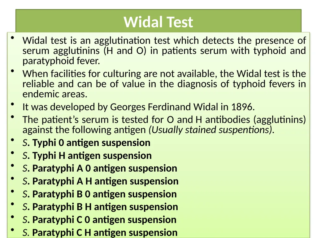
Widal Test Widal test is an agglutination test which detects the presence of serum agglutinins (H and O) in patients serum with typhoid and paratyphoid fever.
When facilities for culturing are not available, the Widal test is the reliable and can be of value in the diagnosis of typhoid fevers in endemic areas.
It was developed by Georges Ferdinand Widal in 1896.
The patient’s serum is tested for O and H antibodies (agglutinins) against the following antigen (Usually stained suspentions).
S. Typhi 0 antigen suspension
S. Typhi H antigen suspension
S. Paratyphi A 0 antigen suspension
S. Paratyphi A H antigen suspension
S. Paratyphi B 0 antigen suspension
S. Paratyphi B H antigen suspension
S. Paratyphi C 0 antigen suspension
S. Paratyphi C H antigen suspension<br>
When facilities for culturing are not available, the Widal test is the reliable and can be of value in the diagnosis of typhoid fevers in endemic areas.
It was developed by Georges Ferdinand Widal in 1896.
The patient’s serum is tested for O and H antibodies (agglutinins) against the following antigen (Usually stained suspentions).
S. Typhi 0 antigen suspension
S. Typhi H antigen suspension
S. Paratyphi A 0 antigen suspension
S. Paratyphi A H antigen suspension
S. Paratyphi B 0 antigen suspension
S. Paratyphi B H antigen suspension
S. Paratyphi C 0 antigen suspension
S. Paratyphi C H antigen suspension<br>
06
Salmonella antibody starts appearing in serum at the end of first week and rise sharply during the 3rd week of endemic fever. In acute typhoid fever, O agglutinins can usually be detected 6–8 days after the onset of fever and H agglutinins after 10–12 days.
It is preferable to test two specimens of sera at an interval of 7 to 10 days to demonstrate a rising antibody titer.
Salmonella antigen suspensions can be used as slide and tube techniques.<br>
It is preferable to test two specimens of sera at an interval of 7 to 10 days to demonstrate a rising antibody titer.
Salmonella antigen suspensions can be used as slide and tube techniques.<br>
07
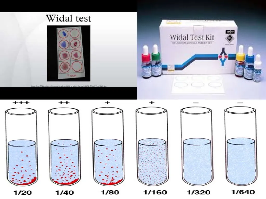
Principle of Widal Test Bacterial suspension which carry antigen will agglutinate on exposure to antibodies to Salmonella organisms. Patients’ suffering from enteric fever would possess antibodies in their sera which can react and agglutinate serial doubling dilutions of killed, coloured Salmonella antigens in an agglutination test.
The main principle of widal test is that if homologous antibody is present in patients serum, it will react with respective antigen in the reagent and gives visible clumping on the test card and agglutination in the tube. The antigens used in the test are “H” and “O” antigens of Salmonella typhi and “H” antigen of S. paratyphi. “O” antigen is a somatic antigen and “H” antigen is flagellar antigen.<br>
The main principle of widal test is that if homologous antibody is present in patients serum, it will react with respective antigen in the reagent and gives visible clumping on the test card and agglutination in the tube. The antigens used in the test are “H” and “O” antigens of Salmonella typhi and “H” antigen of S. paratyphi. “O” antigen is a somatic antigen and “H” antigen is flagellar antigen.<br>
08
VDRL Test What is a VDRL Test?
The venereal disease research laboratory (VDRL) test is designed to assess whether or not you have syphilis, a sexually transmitted infection (STI). Syphilis is caused by the bacteria Treponema pallidum. The bacteria infects by penetrating into the lining of the mouth or genital area.
The VDRL test doesn’t look for the bacteria that cause syphilis. Instead, it checks for the antibodies your body makes in response to antigens produced by cells damaged by the bacteria. Antibodies are a type of protein produced by your immune system to fight off invaders like bacteria or toxins. Testing for these antibodies can let doctors know whether you have syphilis, because it checks for antibodies produced as a result of a syphilis infection.<br>
The venereal disease research laboratory (VDRL) test is designed to assess whether or not you have syphilis, a sexually transmitted infection (STI). Syphilis is caused by the bacteria Treponema pallidum. The bacteria infects by penetrating into the lining of the mouth or genital area.
The VDRL test doesn’t look for the bacteria that cause syphilis. Instead, it checks for the antibodies your body makes in response to antigens produced by cells damaged by the bacteria. Antibodies are a type of protein produced by your immune system to fight off invaders like bacteria or toxins. Testing for these antibodies can let doctors know whether you have syphilis, because it checks for antibodies produced as a result of a syphilis infection.<br>
09
The VDRL test isn’t always accurate. For example, it may give false-negative results if syphilis test was done less than three months, as the body might take this long time to make antibodies. The test is also unreliable in late-stage syphilis. On the other hand, the following can cause false-positive results:
HIV
Lyme disease
malaria
pneumonia (certain types only)
systemic lupus erythematosus
IV drug use
tuberculosis
In some cases, the body may not produce antibodies even if it has been infected with syphilis. This means the VDRL test will be inaccurate.
The antibodies produced as a result of a syphilis infection can stay in the body even after syphilis has been treated. This means you might always have positive results on this test.<br>
HIV
Lyme disease
malaria
pneumonia (certain types only)
systemic lupus erythematosus
IV drug use
tuberculosis
In some cases, the body may not produce antibodies even if it has been infected with syphilis. This means the VDRL test will be inaccurate.
The antibodies produced as a result of a syphilis infection can stay in the body even after syphilis has been treated. This means you might always have positive results on this test.<br>
10
Rheumatoid arthritis Rheumatoid arthritis can be difficult to diagnose because many conditions cause joint stiffness and inflammation and there is no definitive test for the condition.
There is no singular test for diagnosing rheumatoid arthritis. The diagnosis is based on the clinical presentation.
Ultimately, rheumatoid arthritis is diagnosed based on a combination of the presentation of the joints involved, characteristic joint swelling and stiffness in the morning, the presence of blood rheumatoid factor, as well as findings of rheumatoid nodules and radiographic changes (X-ray testing). It is important to understand that there are many forms of joint disease that can mimic rheumatoid arthritis.
(RF) can be found in 80% of patients with rheumatoid arthritis. Patients who are felt to have rheumatoid arthritis and do not have positive rheumatoid factor testing referred to as having "seronegative rheumatoid arthritis<br>
There is no singular test for diagnosing rheumatoid arthritis. The diagnosis is based on the clinical presentation.
Ultimately, rheumatoid arthritis is diagnosed based on a combination of the presentation of the joints involved, characteristic joint swelling and stiffness in the morning, the presence of blood rheumatoid factor, as well as findings of rheumatoid nodules and radiographic changes (X-ray testing). It is important to understand that there are many forms of joint disease that can mimic rheumatoid arthritis.
(RF) can be found in 80% of patients with rheumatoid arthritis. Patients who are felt to have rheumatoid arthritis and do not have positive rheumatoid factor testing referred to as having "seronegative rheumatoid arthritis<br>
11
C-Reactive Protein Test C-reactive protein (CRP) is a substance produced by the liver in response to inflammation. A high level of CRP in the blood is a sign that there may be an inflammatory process occurring in the body. Inflammation itself isn’t typically a problem, but it can indicate a host of other health concerns, including infection, arthritis, kidney failure, and pancreatitis.
A CRP test is a blood test designed to measure the amount of CRP in the blood.
Usually, doctors order the test to determine a person’s risk for heart disease or stroke. Doctors may also order a CRP test after surgery to check for signs of postsurgical infection. They also might use it to monitor inflammatory diseases, including:
pelvic inflammatory disease
inflammatory bowel disease
arthritis
autoimmune diseases, such as lupus
C-reactive protein is measured in milligrams of CRP per liter of blood (mg/L). In general, a low C-reactive protein level is better than a high one, because it indicates less inflammation in the body.<br>
A CRP test is a blood test designed to measure the amount of CRP in the blood.
Usually, doctors order the test to determine a person’s risk for heart disease or stroke. Doctors may also order a CRP test after surgery to check for signs of postsurgical infection. They also might use it to monitor inflammatory diseases, including:
pelvic inflammatory disease
inflammatory bowel disease
arthritis
autoimmune diseases, such as lupus
C-reactive protein is measured in milligrams of CRP per liter of blood (mg/L). In general, a low C-reactive protein level is better than a high one, because it indicates less inflammation in the body.<br>
12
Blood Stream Infections (BSI) Blood stream infection (BSI) is a serious problem that needs immediate attention and treatment. It is a cause of high mortality especially if caused by multidrug resistant bacteria.
Blood culture
Blood is cultured to detect and identify bacteria or other cultivable microorganisms (yeasts, filamentous fungi). The presence of such organisms in the blood is called bacteraemia or fungaemia, and is usually pathological. In healthy subjects, the blood is sterile.<br>
Blood culture
Blood is cultured to detect and identify bacteria or other cultivable microorganisms (yeasts, filamentous fungi). The presence of such organisms in the blood is called bacteraemia or fungaemia, and is usually pathological. In healthy subjects, the blood is sterile.<br>
13
Blood collection Blood should be taken before antibiotics are administered. The best time is when the patient is expected to have chills or a temperature spike. It is recommended that two or preferably three blood cultures be obtained, separated by intervals of approximately 1 hour (or less if treatment cannot be delayed). The advantages of repeated cultures are as follows:
— The chance of missing a transient bacteraemia is reduced.
— The pathogenic role of “saprophytic” isolates (e.g. Staphylococcus epidermidis)is confirmed if they are recovered from multiple venepunctures.
Because the number of bacteria per millilitre of blood is usually low, it is
important to take a reasonable quantity of blood: 10 ml per venepuncture for adults; 2–5 ml may suffice for children.
The skin at the venepuncture site must be meticulously prepared using a bactericidal disinfectant.<br>
— The chance of missing a transient bacteraemia is reduced.
— The pathogenic role of “saprophytic” isolates (e.g. Staphylococcus epidermidis)is confirmed if they are recovered from multiple venepunctures.
Because the number of bacteria per millilitre of blood is usually low, it is
important to take a reasonable quantity of blood: 10 ml per venepuncture for adults; 2–5 ml may suffice for children.
The skin at the venepuncture site must be meticulously prepared using a bactericidal disinfectant.<br>
14
Blood-culture media The blood-culture broth (brain heart infusion broth) and tryptic soy broth (TSB) should be able to support growth of all clinically significant bacteria.
The blood should be mixed with 10 times its volume of broth (5 ml of blood in 50 ml of broth) to dilute any antibiotic present and to reduce the bactericidal effect of human serum.<br>
The blood should be mixed with 10 times its volume of broth (5 ml of blood in 50 ml of broth) to dilute any antibiotic present and to reduce the bactericidal effect of human serum.<br>
15
Incubation time Blood-culture bottles should be incubated at 35–37 C for 7 days. In some cases, incubation may be prolonged for an additional 7 days, e.g. if Brucella or other fastidious organisms are suspected, in cases of endocarditis, or if the patient has received antimicrobials. A sterile culture usually shows a layer of sedimented red blood covered by a pale yellow transparent broth. Growth is evidenced by:
— a floccular deposit on top of the blood layer
— uniform or subsurface turbidity
— haemolysis
— coagulation of the broth
— a surface pellicle
— production of gas
— white grains on the surface or deep in the blood layer.<br>
— a floccular deposit on top of the blood layer
— uniform or subsurface turbidity
— haemolysis
— coagulation of the broth
— a surface pellicle
— production of gas
— white grains on the surface or deep in the blood layer.<br>
16
Whenever visible growth appears, the bottle should be opened aseptically, a
small amount of broth removed with a sterile loop or Pasteur pipette, and a
Gram-stained smear examined for the presence of microorganisms.
Subcultures are performed by streaking a loopful on appropriate media:
MacConkey agar, Blood agar and chocolate agar.
Some microorganisms may grow without producing turbidity or visible alteration of the broth. Other organisms, e.g. pneumococci, tend to undergo autolysis and die very rapidly. For this reason some laboratories perform routine subcultures on chocolate agar after 18–24 hours of incubation.<br>
small amount of broth removed with a sterile loop or Pasteur pipette, and a
Gram-stained smear examined for the presence of microorganisms.
Subcultures are performed by streaking a loopful on appropriate media:
MacConkey agar, Blood agar and chocolate agar.
Some microorganisms may grow without producing turbidity or visible alteration of the broth. Other organisms, e.g. pneumococci, tend to undergo autolysis and die very rapidly. For this reason some laboratories perform routine subcultures on chocolate agar after 18–24 hours of incubation.<br>
17
Common causes of bacteraemia:<br>