
Sigmoid colon Dr. Dima Hamarsheh
LV
Published · 52 slides · 0 views
1 / 1









Description
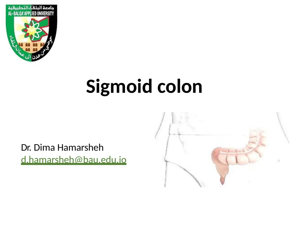
Sigmoid colon Dr. Dima Hamarsheh d.hamarshehbau.edu.jo The sigmoid colon is 10 to 15 in. (25 to 38 cm) long and begins as a continuation of the descending colon in front of the left pelvic brim. Below, it becomes continuous with the rectum
Related Topics
Share
Embed code
Download this presentation From Below
"Sigmoid colon Dr. Dima Hamarsheh" is the property of its rightful owner. Permission is granted to download and print the materials on this website for personal, non-commercial use only, and to display it on your personal computer provided you do not modify the materials and that you retain all copyright notices contained in the materials. By downloading content from our website, you accept the terms of this agreement.
Presentation Transcript
01
Sigmoid colon Dr. Dima Hamarsheh
d.hamarsheh@bau.edu.jo<br>
d.hamarsheh@bau.edu.jo<br>
02
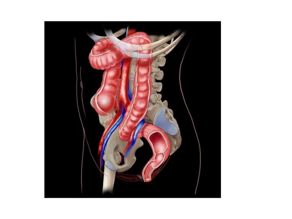
The sigmoid colon is 10 to 15 in. (25 to 38
cm) long
and begins as a continuation of the descending colon in front of the left pelvic brim.
Below, it becomes continuous with the rectum in front of the 3rd sacral vertebra.
The sigmoid colon is mobile and hangs down into the pelvic cavity in the form of a loop.
The sigmoid colon is attached to the posterior pelvic wall by the fan-shaped sigmoid mesocolon.<br>
cm) long
and begins as a continuation of the descending colon in front of the left pelvic brim.
Below, it becomes continuous with the rectum in front of the 3rd sacral vertebra.
The sigmoid colon is mobile and hangs down into the pelvic cavity in the form of a loop.
The sigmoid colon is attached to the posterior pelvic wall by the fan-shaped sigmoid mesocolon.<br>
03
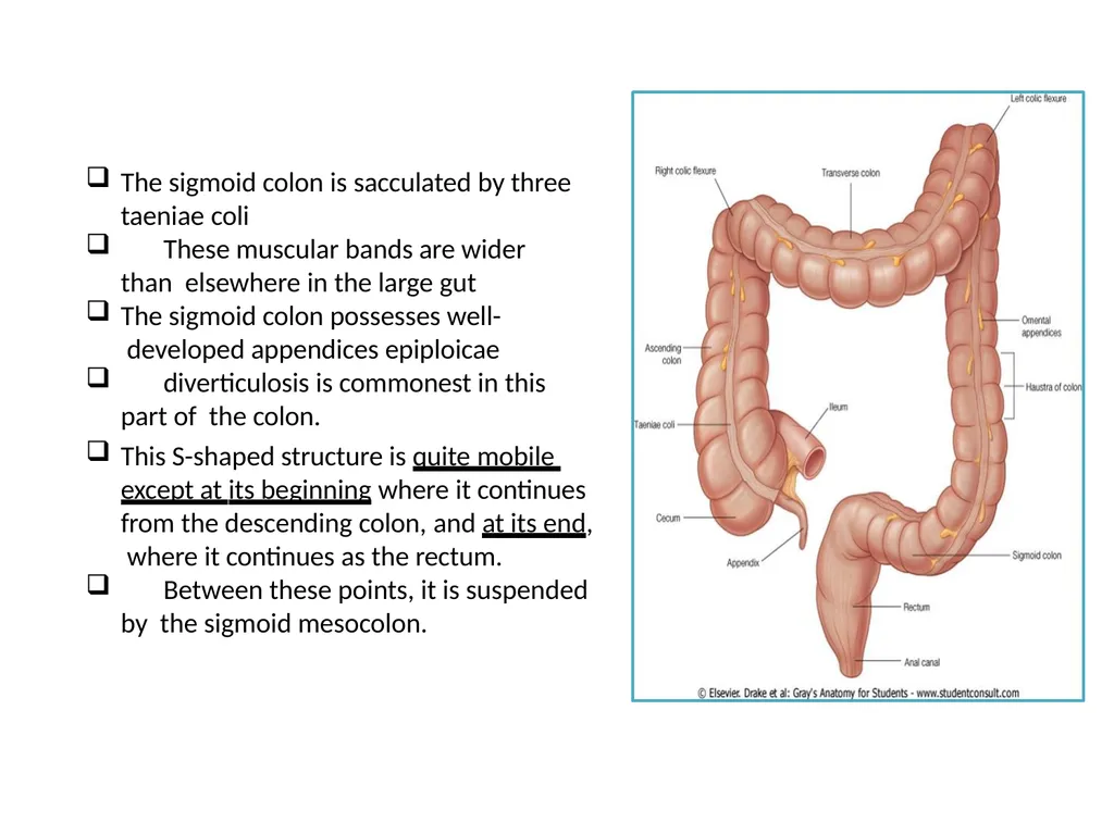
The sigmoid colon is sacculated by three taeniae coli
These muscular bands are wider than elsewhere in the large gut
The sigmoid colon possesses well- developed appendices epiploicae
diverticulosis is commonest in this part of the colon.
This S-shaped structure is quite mobile except at its beginning where it continues from the descending colon, and at its end, where it continues as the rectum.
Between these points, it is suspended by the sigmoid mesocolon.<br>
These muscular bands are wider than elsewhere in the large gut
The sigmoid colon possesses well- developed appendices epiploicae
diverticulosis is commonest in this part of the colon.
This S-shaped structure is quite mobile except at its beginning where it continues from the descending colon, and at its end, where it continues as the rectum.
Between these points, it is suspended by the sigmoid mesocolon.<br>
04
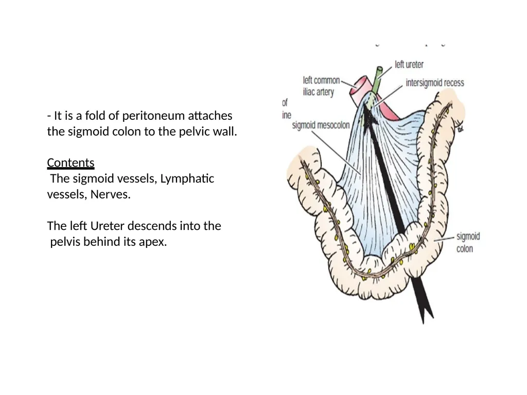
- It is a fold of peritoneum attaches the sigmoid colon to the pelvic wall.
Contents
The sigmoid vessels, Lymphatic
vessels, Nerves.
The left Ureter descends into the pelvis behind its apex.<br>
Contents
The sigmoid vessels, Lymphatic
vessels, Nerves.
The left Ureter descends into the pelvis behind its apex.<br>
05
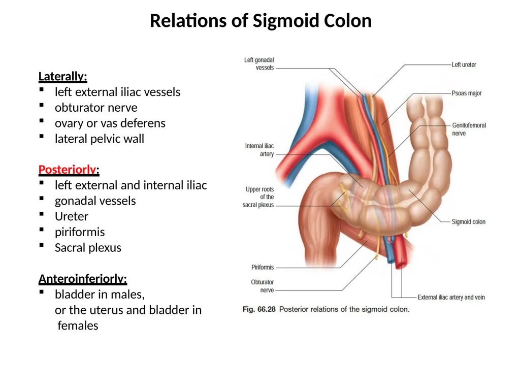
Laterally:
left external iliac vessels
obturator nerve
ovary or vas deferens
lateral pelvic wall
Posteriorly:
left external and internal iliac
gonadal vessels
Ureter
piriformis
Sacral plexus
Anteroinferiorly:
bladder in males,
or the uterus and bladder in females Relations of Sigmoid Colon<br>
left external iliac vessels
obturator nerve
ovary or vas deferens
lateral pelvic wall
Posteriorly:
left external and internal iliac
gonadal vessels
Ureter
piriformis
Sacral plexus
Anteroinferiorly:
bladder in males,
or the uterus and bladder in females Relations of Sigmoid Colon<br>
06
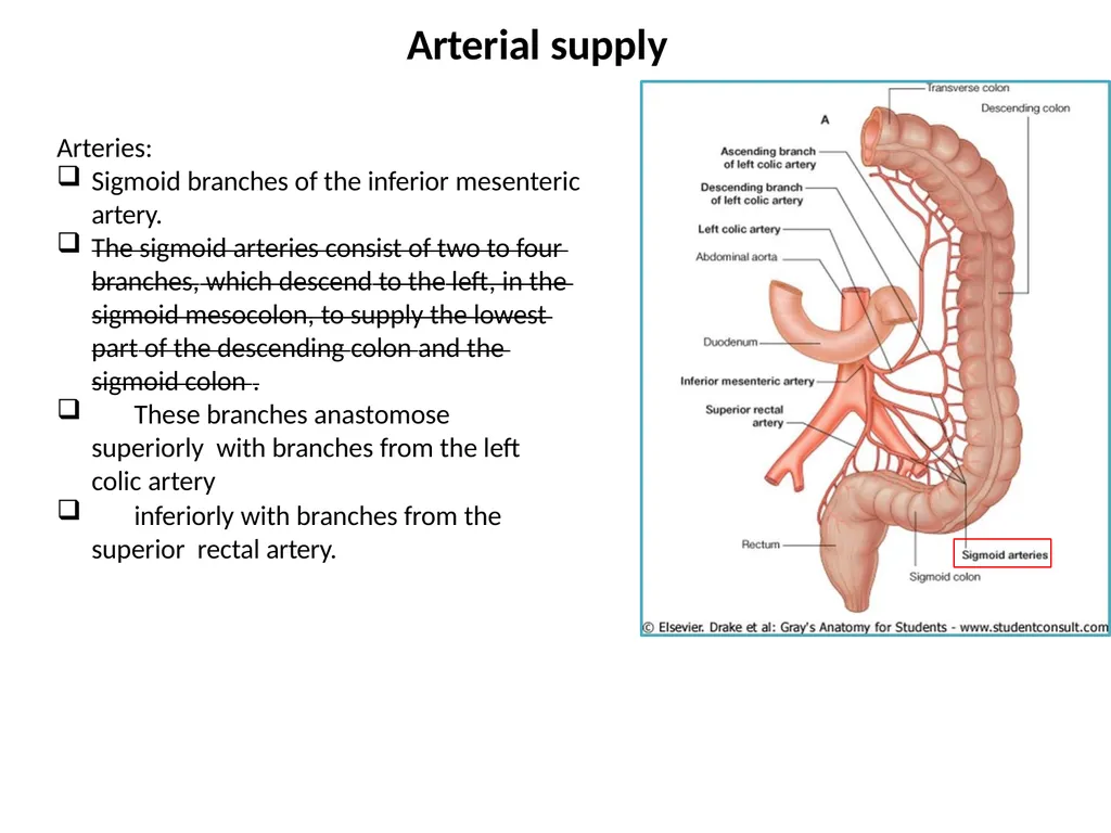
Arteries:
Sigmoid branches of the inferior mesenteric
artery.
The sigmoid arteries consist of two to four branches, which descend to the left, in the sigmoid mesocolon, to supply the lowest part of the descending colon and the sigmoid colon .
These branches anastomose superiorly with branches from the left colic artery
inferiorly with branches from the superior rectal artery. Arterial supply<br>
Sigmoid branches of the inferior mesenteric
artery.
The sigmoid arteries consist of two to four branches, which descend to the left, in the sigmoid mesocolon, to supply the lowest part of the descending colon and the sigmoid colon .
These branches anastomose superiorly with branches from the left colic artery
inferiorly with branches from the superior rectal artery. Arterial supply<br>
07
Veins:
The veins drain into the inferior mesenteric vein, which joins the portal venous system<br>
The veins drain into the inferior mesenteric vein, which joins the portal venous system<br>
08
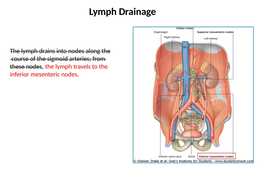
The lymph drains into nodes along the course of the sigmoid arteries; from these nodes, the lymph travels to the inferior mesenteric nodes. Lymph Drainage<br>
09
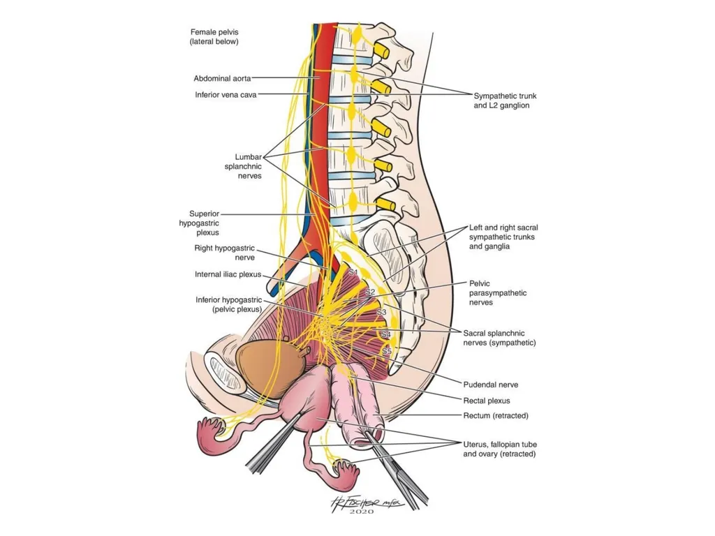
The sympathetic and parasympathetic nerves from the inferior hypogastric plexuses.
The parasympathetic supply is derived
from the pelvic splanchnic nerves Nerve Supply<br>
The parasympathetic supply is derived
from the pelvic splanchnic nerves Nerve Supply<br>
10
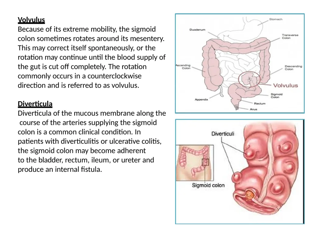
Volvulus
Because of its extreme mobility, the sigmoid colon sometimes rotates around its mesentery. This may correct itself spontaneously, or the rotation may continue until the blood supply of the gut is cut off completely. The rotation commonly occurs in a counterclockwise direction and is referred to as volvulus.
Diverticula
Diverticula of the mucous membrane along the course of the arteries supplying the sigmoid colon is a common clinical condition. In patients with diverticulitis or ulcerative colitis, the sigmoid colon may become adherent
to the bladder, rectum, ileum, or ureter and produce an internal fistula.<br>
Because of its extreme mobility, the sigmoid colon sometimes rotates around its mesentery. This may correct itself spontaneously, or the rotation may continue until the blood supply of the gut is cut off completely. The rotation commonly occurs in a counterclockwise direction and is referred to as volvulus.
Diverticula
Diverticula of the mucous membrane along the course of the arteries supplying the sigmoid colon is a common clinical condition. In patients with diverticulitis or ulcerative colitis, the sigmoid colon may become adherent
to the bladder, rectum, ileum, or ureter and produce an internal fistula.<br>
11
Rectum<br>
12
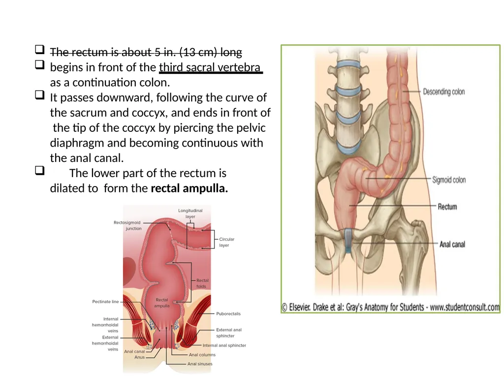
The rectum is about 5 in. (13 cm) long
begins in front of the third sacral vertebra as a continuation colon.
It passes downward, following the curve of the sacrum and coccyx, and ends in front of the tip of the coccyx by piercing the pelvic diaphragm and becoming continuous with the anal canal.
The lower part of the rectum is dilated to form the rectal ampulla.<br>
begins in front of the third sacral vertebra as a continuation colon.
It passes downward, following the curve of the sacrum and coccyx, and ends in front of the tip of the coccyx by piercing the pelvic diaphragm and becoming continuous with the anal canal.
The lower part of the rectum is dilated to form the rectal ampulla.<br>
13
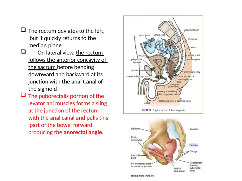
The rectum deviates to the left, but it quickly returns to the median plane .
On lateral view, the rectum follows the anterior concavity of the sacrum before bending downward and backward at its junction with the anal Canal of the sigmoid .
The puborectalis portion of the levator ani muscles forms a sling at the junction of the rectum with the anal canal and pulls this part of the bowel forward, producing the anorectal angle.<br>
On lateral view, the rectum follows the anterior concavity of the sacrum before bending downward and backward at its junction with the anal Canal of the sigmoid .
The puborectalis portion of the levator ani muscles forms a sling at the junction of the rectum with the anal canal and pulls this part of the bowel forward, producing the anorectal angle.<br>
14
The peritoneum covers the anterior and lateral surfaces of the first third of the rectum and only the anterior surface of the middle third, leaving the lower third devoid of peritoneum. Peritoneal covering of Rectum<br>
15
The muscular coat of the rectum is arranged in the usual outer longitudinal and inner circular layers of smooth muscle.
The three teniae coli of the sigmoid colon come together so that the longitudinal
fibers form a broad band on the anterior and posterior surfaces of the rectum The muscular coat<br>
The three teniae coli of the sigmoid colon come together so that the longitudinal
fibers form a broad band on the anterior and posterior surfaces of the rectum The muscular coat<br>
16
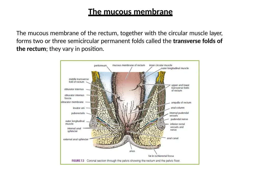
The mucous membrane of the rectum, together with the circular muscle layer, forms two or three semicircular permanent folds called the transverse folds of the rectum; they vary in position. The mucous membrane<br>
17
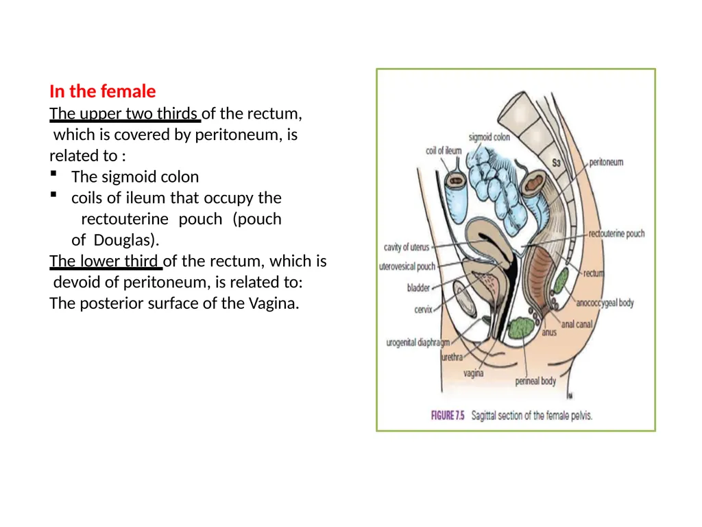
In the female The upper two thirds of the rectum, which is covered by peritoneum, is related to :
The sigmoid colon
coils of ileum that occupy the rectouterine pouch (pouch of Douglas).
The lower third of the rectum, which is devoid of peritoneum, is related to:
The posterior surface of the Vagina.<br>
The sigmoid colon
coils of ileum that occupy the rectouterine pouch (pouch of Douglas).
The lower third of the rectum, which is devoid of peritoneum, is related to:
The posterior surface of the Vagina.<br>
18
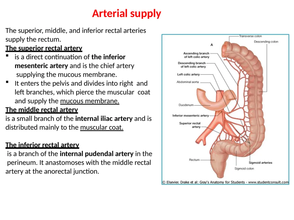
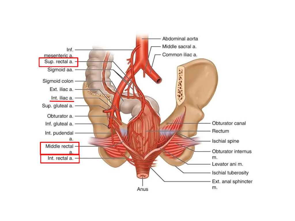
The superior, middle, and inferior rectal arteries supply the rectum.
The superior rectal artery
is a direct continuation of the inferior mesenteric artery and is the chief artery supplying the mucous membrane.
It enters the pelvis and divides into right and left branches, which pierce the muscular coat and supply the mucous membrane.
The middle rectal artery
is a small branch of the internal iliac artery and is
distributed mainly to the muscular coat.
The inferior rectal artery
is a branch of the internal pudendal artery in the perineum. It anastomoses with the middle rectal artery at the anorectal junction. Arterial supply<br>
The superior rectal artery
is a direct continuation of the inferior mesenteric artery and is the chief artery supplying the mucous membrane.
It enters the pelvis and divides into right and left branches, which pierce the muscular coat and supply the mucous membrane.
The middle rectal artery
is a small branch of the internal iliac artery and is
distributed mainly to the muscular coat.
The inferior rectal artery
is a branch of the internal pudendal artery in the perineum. It anastomoses with the middle rectal artery at the anorectal junction. Arterial supply<br>
19
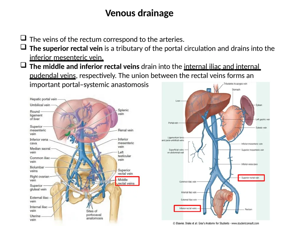
The veins of the rectum correspond to the arteries.
The superior rectal vein is a tributary of the portal circulation and drains into the inferior mesenteric vein.
The middle and inferior rectal veins drain into the internal iliac and internal pudendal veins, respectively. The union between the rectal veins forms an important portal–systemic anastomosis Venous drainage<br>
The superior rectal vein is a tributary of the portal circulation and drains into the inferior mesenteric vein.
The middle and inferior rectal veins drain into the internal iliac and internal pudendal veins, respectively. The union between the rectal veins forms an important portal–systemic anastomosis Venous drainage<br>
20
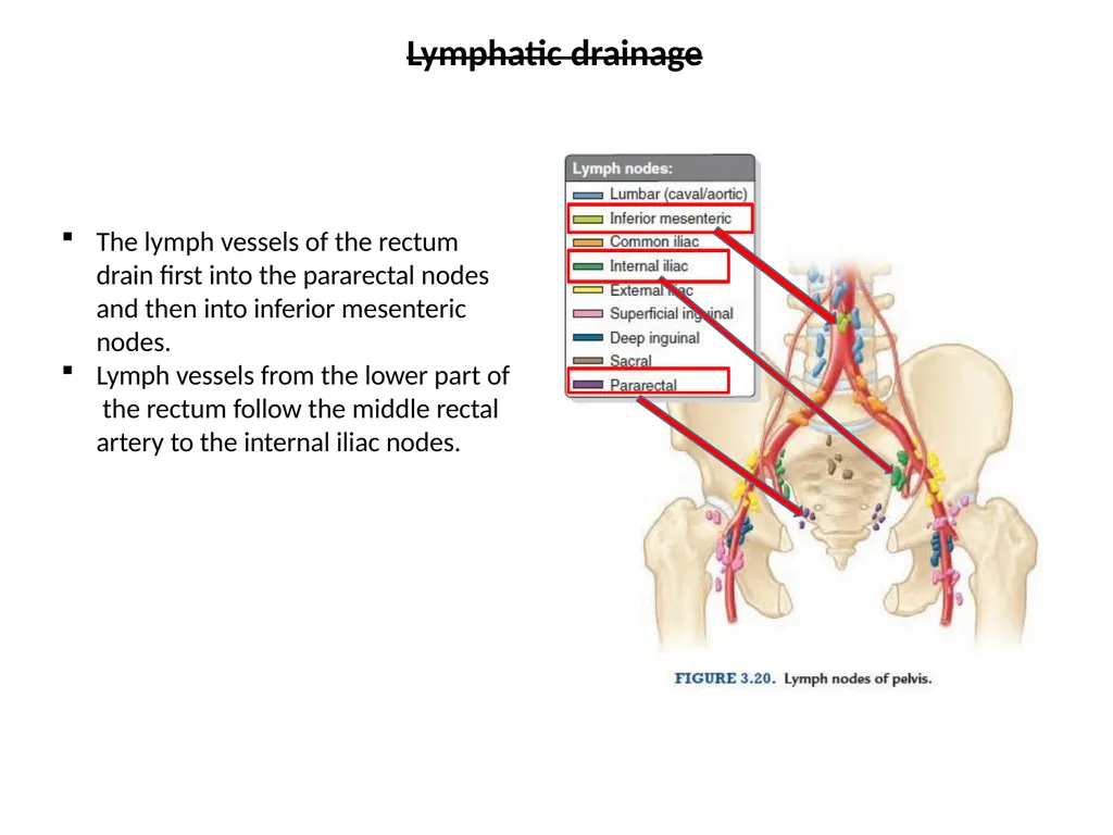
The lymph vessels of the rectum drain first into the pararectal nodes and then into inferior mesenteric nodes.
Lymph vessels from the lower part of the rectum follow the middle rectal artery to the internal iliac nodes. Lymphatic drainage<br>
Lymph vessels from the lower part of the rectum follow the middle rectal artery to the internal iliac nodes. Lymphatic drainage<br>
21
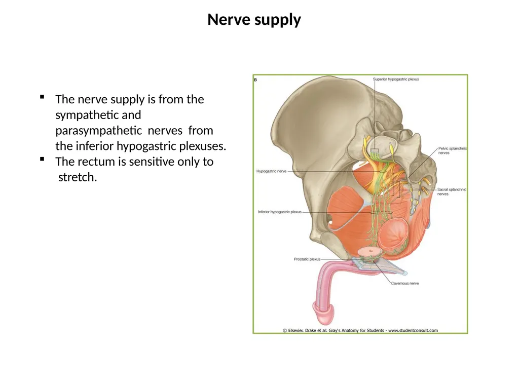
The nerve supply is from the sympathetic and parasympathetic nerves from the inferior hypogastric plexuses.
The rectum is sensitive only to stretch. Nerve supply<br>
The rectum is sensitive only to stretch. Nerve supply<br>
22
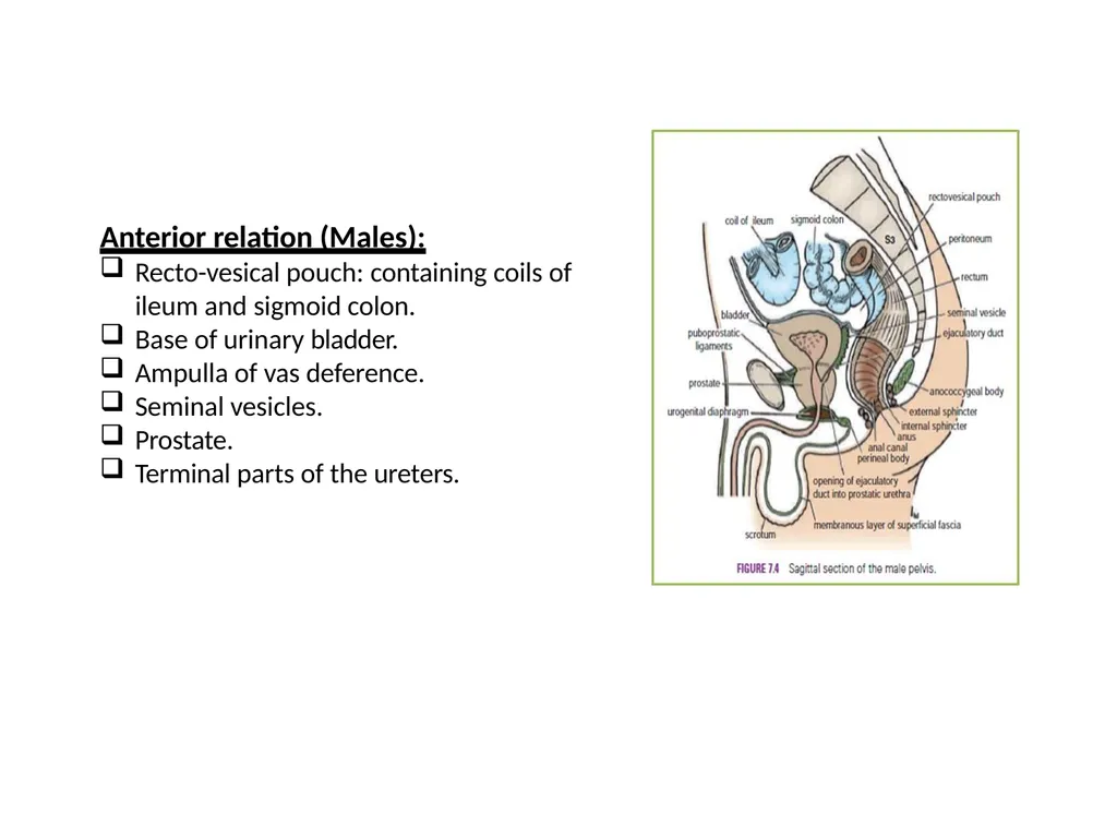
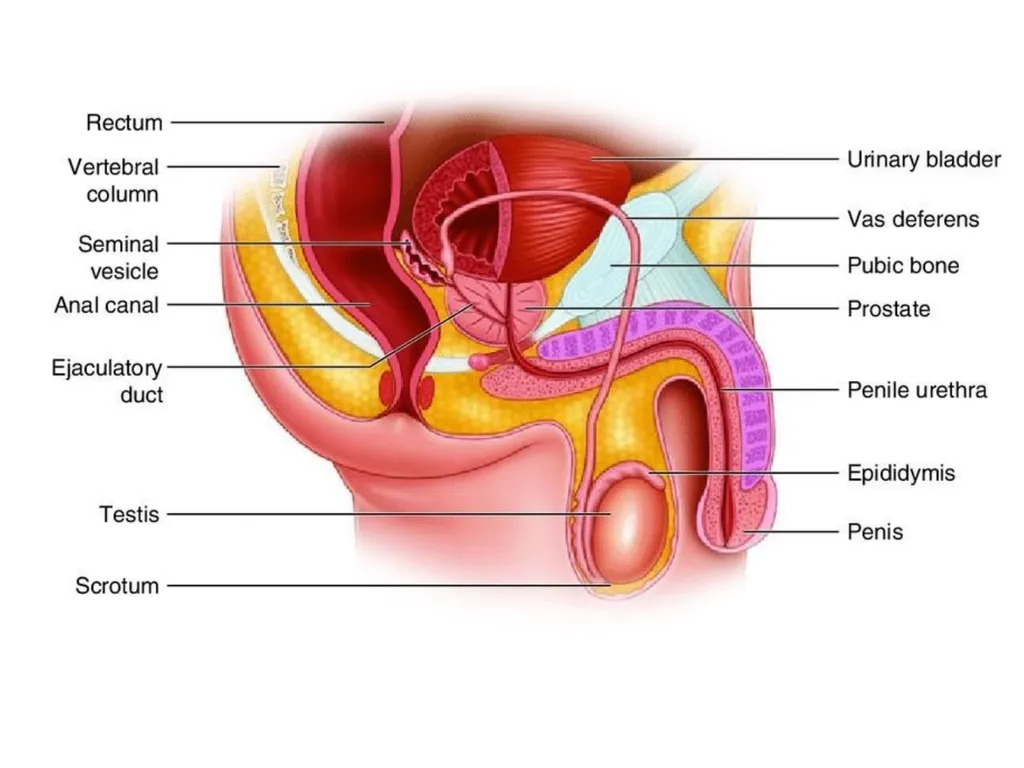
Anterior relation (Males): Recto-vesical pouch: containing coils of ileum and sigmoid colon.
Base of urinary bladder.
Ampulla of vas deference.
Seminal vesicles.
Prostate.
Terminal parts of the ureters.<br>
Base of urinary bladder.
Ampulla of vas deference.
Seminal vesicles.
Prostate.
Terminal parts of the ureters.<br>
23
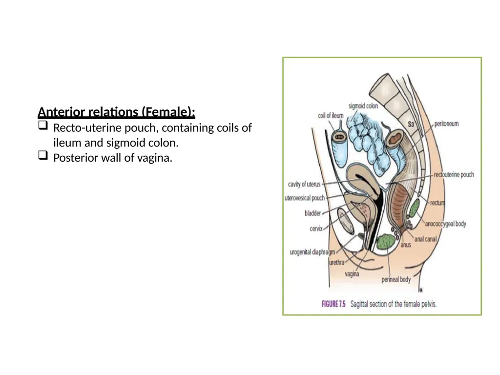
Anterior relations (Female): Recto-uterine pouch, containing coils of ileum and sigmoid colon.
Posterior wall of vagina.<br>
Posterior wall of vagina.<br>
24
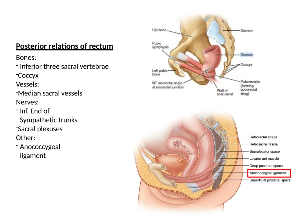
Bones:
Inferior three sacral vertebrae
Coccyx Vessels:
Median sacral vessels Nerves:
Inf. End of Sympathetic trunks
Sacral plexuses Other:
Anococcygeal ligament Posterior relations of rectum<br>
Inferior three sacral vertebrae
Coccyx Vessels:
Median sacral vessels Nerves:
Inf. End of Sympathetic trunks
Sacral plexuses Other:
Anococcygeal ligament Posterior relations of rectum<br>
25
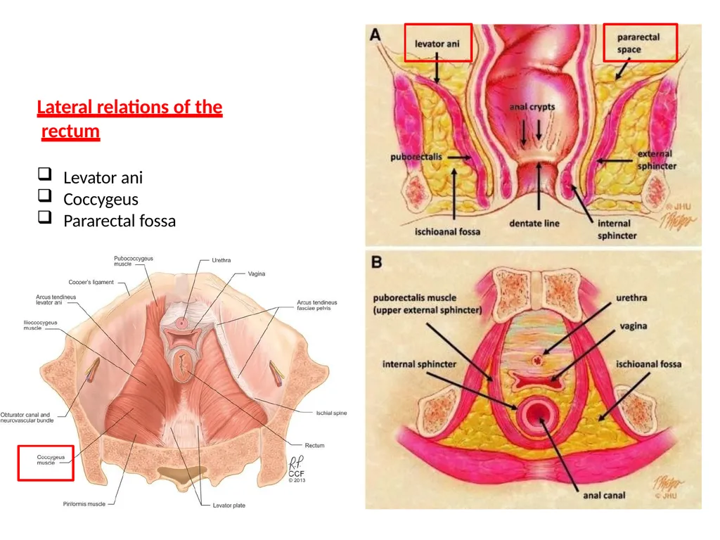
Lateral relations of the rectum Levator ani
Coccygeus
Pararectal fossa<br>
Coccygeus
Pararectal fossa<br>
26
Anal canal<br>
27
The anal canal is about 1.5 in. (4 cm) long
passes downward and backward
from the rectal ampulla to the anus.<br>
passes downward and backward
from the rectal ampulla to the anus.<br>
28
■■ Posteriorly:
The anococcygeal body, which is a mass of fibrous tissue lying between the anal canal and the coccyx.
■■ Laterally:
The fat-filled ischiorectal fossae.
■■ Anteriorly:
In male
The perineal body
The urogenital diaphragm
The membranous part of the urethra
The bulb of the penis.
In female
The perineal body
The urogenital diaphragm
The lower part of the vagina Relations<br>
The anococcygeal body, which is a mass of fibrous tissue lying between the anal canal and the coccyx.
■■ Laterally:
The fat-filled ischiorectal fossae.
■■ Anteriorly:
In male
The perineal body
The urogenital diaphragm
The membranous part of the urethra
The bulb of the penis.
In female
The perineal body
The urogenital diaphragm
The lower part of the vagina Relations<br>
29
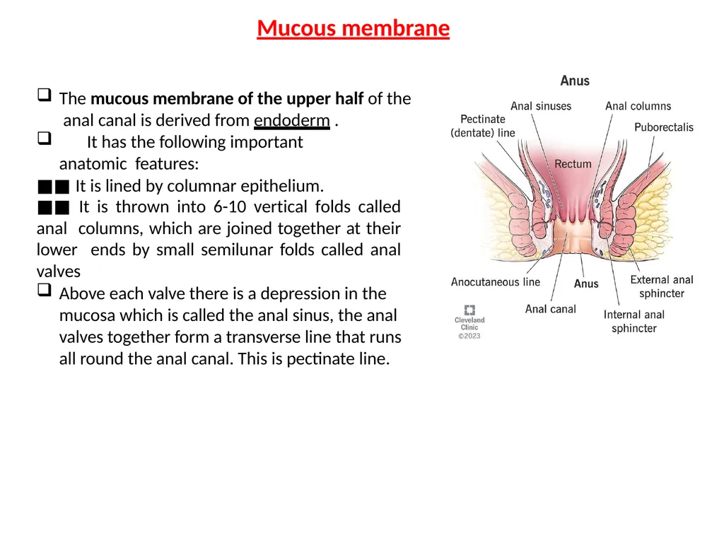
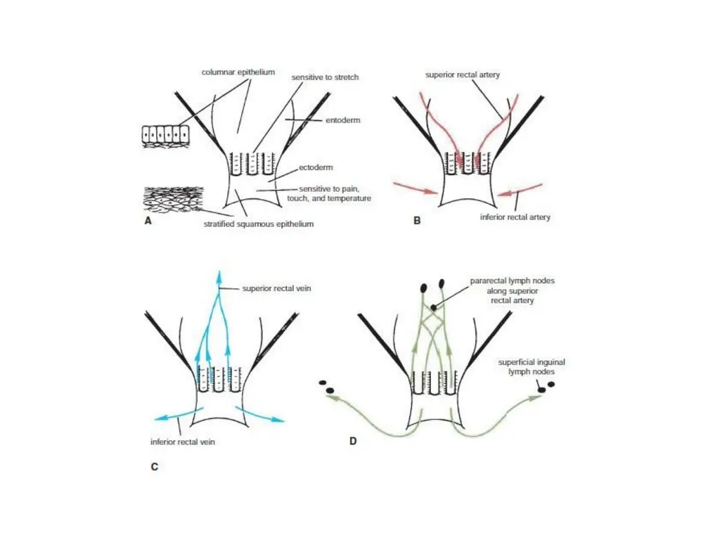
The mucous membrane of the upper half of the anal canal is derived from endoderm .
It has the following important anatomic features:
■■ It is lined by columnar epithelium.
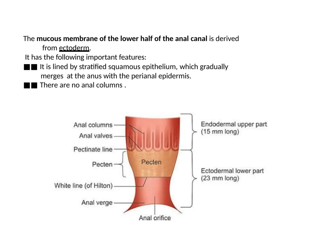
■■ It is thrown into 6-10 vertical folds called anal columns, which are joined together at their lower ends by small semilunar folds called anal valves
Above each valve there is a depression in the mucosa which is called the anal sinus, the anal valves together form a transverse line that runs all round the anal canal. This is pectinate line. Mucous membrane<br>
It has the following important anatomic features:
■■ It is lined by columnar epithelium.
■■ It is thrown into 6-10 vertical folds called anal columns, which are joined together at their lower ends by small semilunar folds called anal valves
Above each valve there is a depression in the mucosa which is called the anal sinus, the anal valves together form a transverse line that runs all round the anal canal. This is pectinate line. Mucous membrane<br>
30
■■ The nerve supply is the same as that for the rectal mucosa and is derived from the autonomic hypogastric plexuses. It is sensitive only to stretch.
■■ The arterial supply is that of the hindgut—namely, the superior rectal artery, a branch of the inferior mesenteric artery .
■■ The venous drainage is mainly by the superior rectal vein, a tributary of the
inferior mesenteric vein, and the portal vein .
■■ The lymphatic drainage is mainly upward along the superior rectal artery to the pararectal nodes and then eventually to the inferior mesenteric nodes<br>
■■ The arterial supply is that of the hindgut—namely, the superior rectal artery, a branch of the inferior mesenteric artery .
■■ The venous drainage is mainly by the superior rectal vein, a tributary of the
inferior mesenteric vein, and the portal vein .
■■ The lymphatic drainage is mainly upward along the superior rectal artery to the pararectal nodes and then eventually to the inferior mesenteric nodes<br>
31
The mucous membrane of the lower half of the anal canal is derived from ectoderm.
It has the following important features:
■■ It is lined by stratified squamous epithelium, which gradually merges at the anus with the perianal epidermis.
■■ There are no anal columns .<br>
It has the following important features:
■■ It is lined by stratified squamous epithelium, which gradually merges at the anus with the perianal epidermis.
■■ There are no anal columns .<br>
32
■■ The nerve supply is from the somatic inferior rectal nerve; it is thus
sensitive to pain, temperature, touch, and pressure.
■■ The arterial supply is the inferior rectal artery, a branch of the internal pudendal artery .
■■ The venous drainage is by the inferior rectal vein, a tributary of the
internal pudendal vein, which drains into the internal iliac vein
■■ The lymph drainage is downward to the medial group of superficial inguinal nodes .<br>
sensitive to pain, temperature, touch, and pressure.
■■ The arterial supply is the inferior rectal artery, a branch of the internal pudendal artery .
■■ The venous drainage is by the inferior rectal vein, a tributary of the
internal pudendal vein, which drains into the internal iliac vein
■■ The lymph drainage is downward to the medial group of superficial inguinal nodes .<br>
33
The middle part of anal canal
It is termed as transitional zone or pecten, it is also lined by mucous
membrane.
-The mucosa has a bluish appearance because of a dense venous plexus that lies beneath.
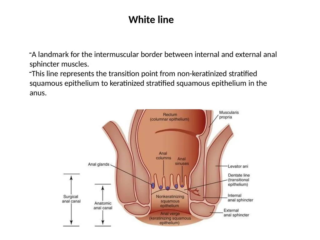
The lower limit of the pecten often has a whitish appearance because of which it is referred to as the white line or Hilton’s line, is situated at the level the interval between the subcutaneous part of external anal sphincter and the lower border of internal anal sphincter.<br>
It is termed as transitional zone or pecten, it is also lined by mucous
membrane.
-The mucosa has a bluish appearance because of a dense venous plexus that lies beneath.
The lower limit of the pecten often has a whitish appearance because of which it is referred to as the white line or Hilton’s line, is situated at the level the interval between the subcutaneous part of external anal sphincter and the lower border of internal anal sphincter.<br>
34
A landmark for the intermuscular border between internal and external anal sphincter muscles.
This line represents the transition point from non-keratinized stratified squamous epithelium to keratinized stratified squamous epithelium in the anus. White line<br>
This line represents the transition point from non-keratinized stratified squamous epithelium to keratinized stratified squamous epithelium in the anus. White line<br>
35
As in the upper parts of the intestinal tract, it is divided into an outer longitudinal and an inner circular layer of smooth Muscle.
The anal canal has an involuntary internal sphincter and a voluntary external sphincter.
The internal sphincter is formed from a thickening of the smooth muscle of the circular coat at the upper end of the anal canal. Muscle Coat<br>
The anal canal has an involuntary internal sphincter and a voluntary external sphincter.
The internal sphincter is formed from a thickening of the smooth muscle of the circular coat at the upper end of the anal canal. Muscle Coat<br>
36
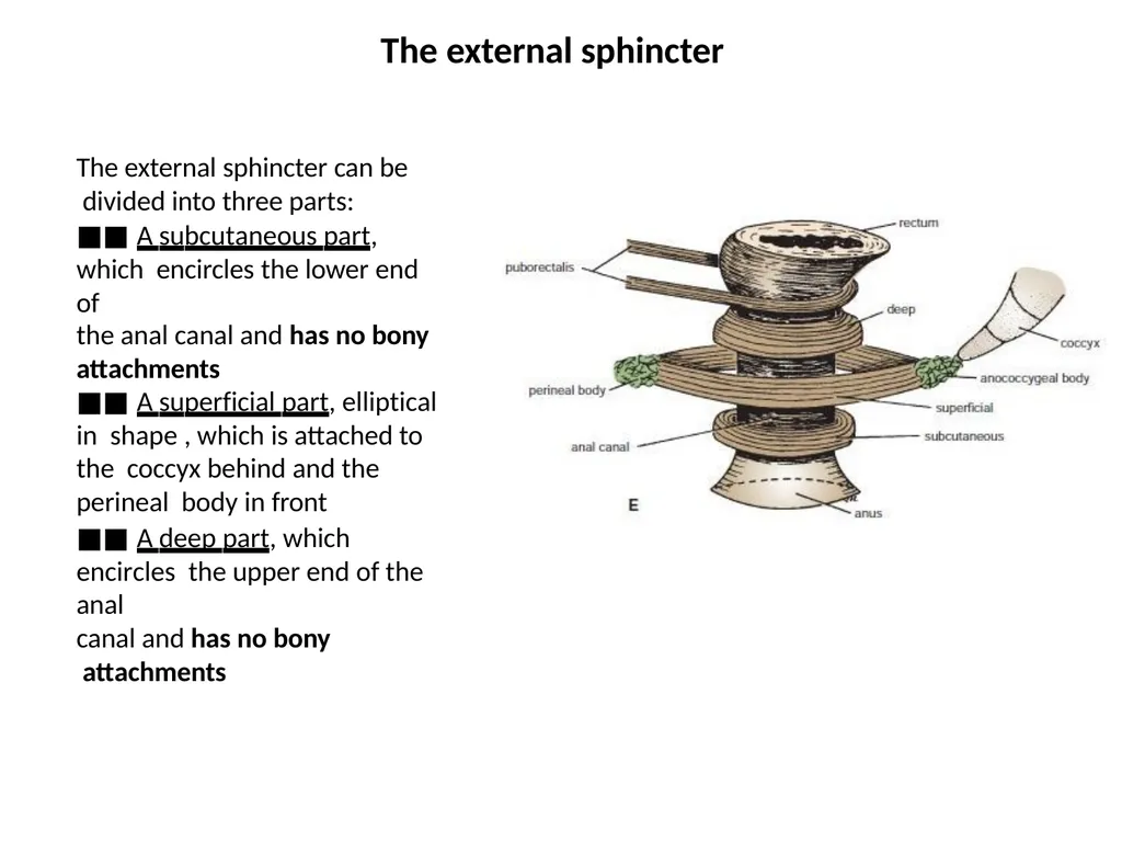
The external sphincter can be divided into three parts:
■■ A subcutaneous part, which encircles the lower end of
the anal canal and has no bony
attachments
■■ A superficial part, elliptical in shape , which is attached to the coccyx behind and the perineal body in front
■■ A deep part, which encircles the upper end of the anal
canal and has no bony attachments The external sphincter<br>
■■ A subcutaneous part, which encircles the lower end of
the anal canal and has no bony
attachments
■■ A superficial part, elliptical in shape , which is attached to the coccyx behind and the perineal body in front
■■ A deep part, which encircles the upper end of the anal
canal and has no bony attachments The external sphincter<br>
37
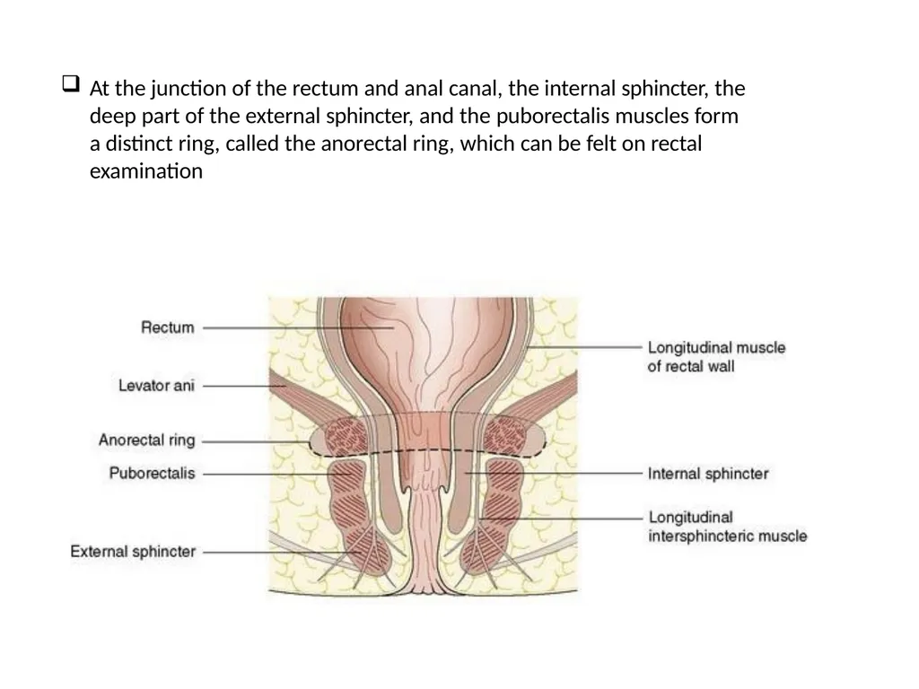
At the junction of the rectum and anal canal, the internal sphincter, the deep part of the external sphincter, and the puborectalis muscles form a distinct ring, called the anorectal ring, which can be felt on rectal examination<br>
38
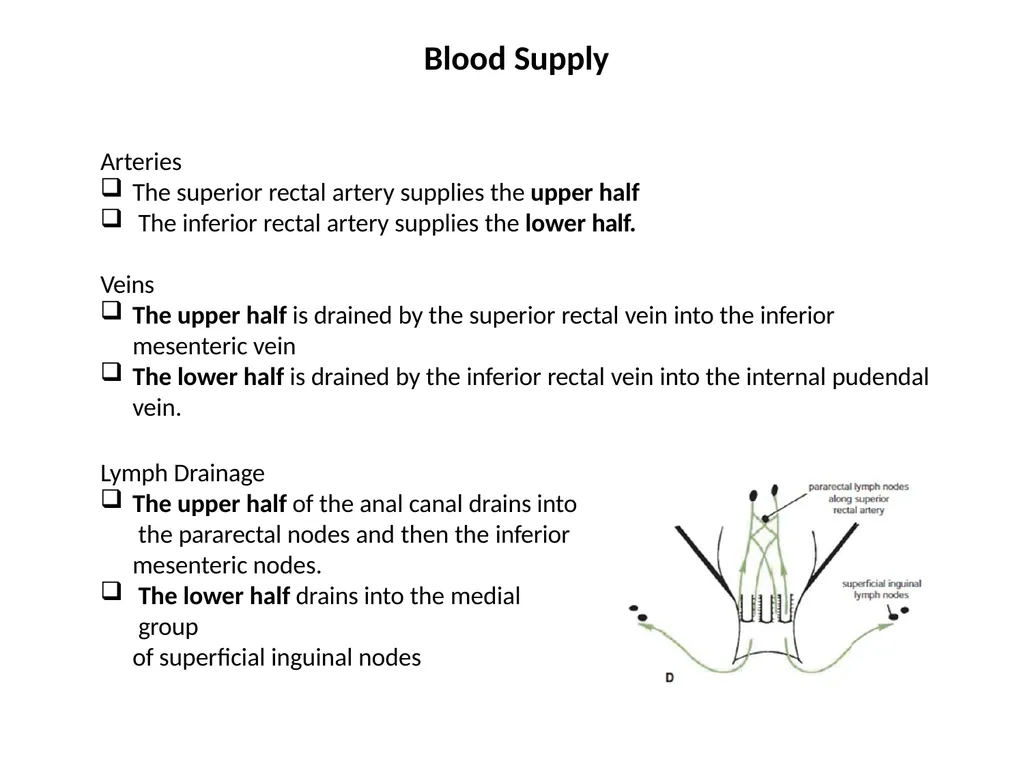
Arteries
The superior rectal artery supplies the upper half
The inferior rectal artery supplies the lower half.
Veins
The upper half is drained by the superior rectal vein into the inferior mesenteric vein
The lower half is drained by the inferior rectal vein into the internal pudendal vein.
Lymph Drainage
The upper half of the anal canal drains into the pararectal nodes and then the inferior mesenteric nodes.
The lower half drains into the medial group
of superficial inguinal nodes Blood Supply<br>
The superior rectal artery supplies the upper half
The inferior rectal artery supplies the lower half.
Veins
The upper half is drained by the superior rectal vein into the inferior mesenteric vein
The lower half is drained by the inferior rectal vein into the internal pudendal vein.
Lymph Drainage
The upper half of the anal canal drains into the pararectal nodes and then the inferior mesenteric nodes.
The lower half drains into the medial group
of superficial inguinal nodes Blood Supply<br>
39
The mucous membrane of the upper half is sensitive to stretch and is innervated by sensory fibers that ascend through the hypogastric plexuses.
The lower half is sensitive to pain, temperature, touch, and pressure and is innervated by the inferior rectal nerves.
The involuntary internal sphincter is supplied by sympathetic fibers from the inferior hypogastric plexuses.
The voluntary external sphincter is supplied by the inferior rectal nerve, a branch of the pudendal nerve, and the perineal branch of the fourth sacral nerve. Nerve Supply<br>
The lower half is sensitive to pain, temperature, touch, and pressure and is innervated by the inferior rectal nerves.
The involuntary internal sphincter is supplied by sympathetic fibers from the inferior hypogastric plexuses.
The voluntary external sphincter is supplied by the inferior rectal nerve, a branch of the pudendal nerve, and the perineal branch of the fourth sacral nerve. Nerve Supply<br>
40
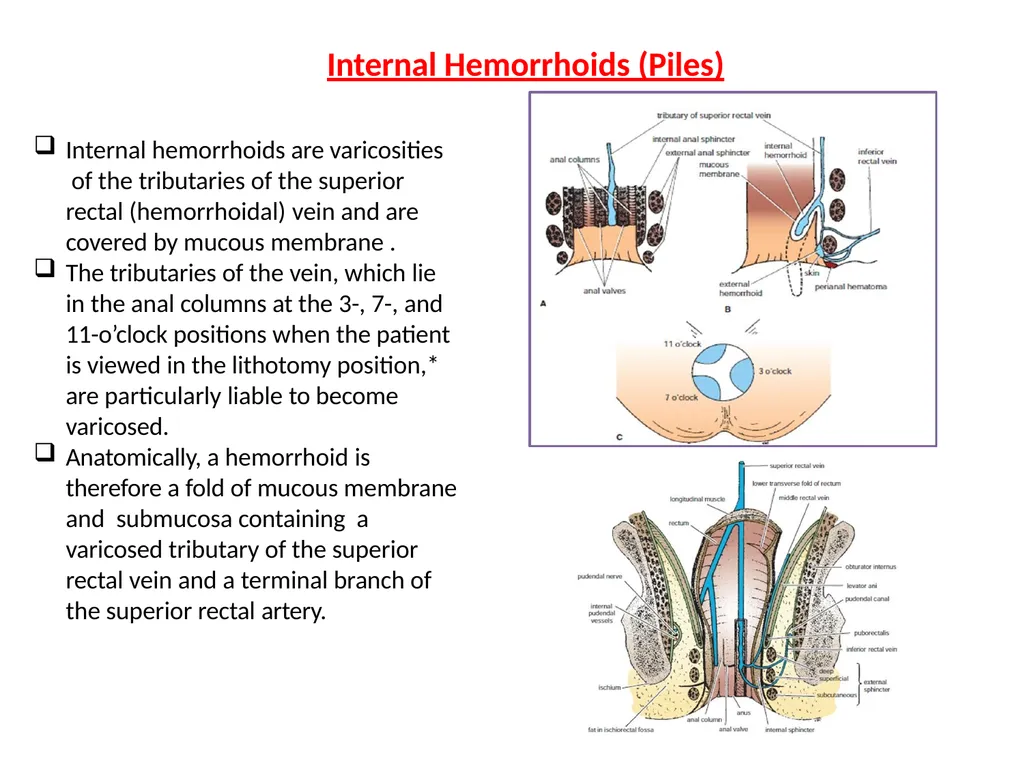
Internal hemorrhoids are varicosities of the tributaries of the superior rectal (hemorrhoidal) vein and are covered by mucous membrane .
The tributaries of the vein, which lie in the anal columns at the 3-, 7-, and 11-o’clock positions when the patient is viewed in the lithotomy position,* are particularly liable to become varicosed.
Anatomically, a hemorrhoid is therefore a fold of mucous membrane and submucosa containing a varicosed tributary of the superior rectal vein and a terminal branch of the superior rectal artery. Internal Hemorrhoids (Piles)<br>
The tributaries of the vein, which lie in the anal columns at the 3-, 7-, and 11-o’clock positions when the patient is viewed in the lithotomy position,* are particularly liable to become varicosed.
Anatomically, a hemorrhoid is therefore a fold of mucous membrane and submucosa containing a varicosed tributary of the superior rectal vein and a terminal branch of the superior rectal artery. Internal Hemorrhoids (Piles)<br>
41
internal hemorrhoids are initially contained within the anal canal (first degree).
As they enlarge, they extrude from the canal on defecation but return at the end of the act (second degree).
With further elongation, they prolapse on defecation and remain outside the anus (third degree).
Because internal hemorrhoids occur in the upper half of the anal canal, where the mucous membrane is innervated by autonomic afferent nerves, they are painless and are sensitive only to stretch.
This may explain why large internal hemorrhoids give rise to an aching sensation rather than acute pain. Internal Hemorrhoids (Piles)<br>
As they enlarge, they extrude from the canal on defecation but return at the end of the act (second degree).
With further elongation, they prolapse on defecation and remain outside the anus (third degree).
Because internal hemorrhoids occur in the upper half of the anal canal, where the mucous membrane is innervated by autonomic afferent nerves, they are painless and are sensitive only to stretch.
This may explain why large internal hemorrhoids give rise to an aching sensation rather than acute pain. Internal Hemorrhoids (Piles)<br>
42
The causes of internal hemorrhoids are many.
They frequently occur in members of the same family, which suggests a congenital weakness of the vein walls
Chronic constipation is a common predisposing factor.
Pregnancy hemorrhoids
Portal hypertension as a result of cirrhosis of the liver can also cause hemorrhoids.
cancerous tumors of the rectum The causes of internal hemorrhoids<br>
They frequently occur in members of the same family, which suggests a congenital weakness of the vein walls
Chronic constipation is a common predisposing factor.
Pregnancy hemorrhoids
Portal hypertension as a result of cirrhosis of the liver can also cause hemorrhoids.
cancerous tumors of the rectum The causes of internal hemorrhoids<br>
43
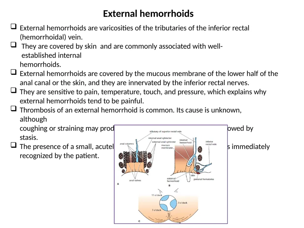
External hemorrhoids are varicosities of the tributaries of the inferior rectal (hemorrhoidal) vein.
They are covered by skin and are commonly associated with well-established internal
hemorrhoids.
External hemorrhoids are covered by the mucous membrane of the lower half of the anal canal or the skin, and they are innervated by the inferior rectal nerves.
They are sensitive to pain, temperature, touch, and pressure, which explains why external hemorrhoids tend to be painful.
Thrombosis of an external hemorrhoid is common. Its cause is unknown, although
coughing or straining may produce distention of the hemorrhoid followed by stasis.
The presence of a small, acutely tender swelling at the anal margin is immediately recognized by the patient. External hemorrhoids<br>
They are covered by skin and are commonly associated with well-established internal
hemorrhoids.
External hemorrhoids are covered by the mucous membrane of the lower half of the anal canal or the skin, and they are innervated by the inferior rectal nerves.
They are sensitive to pain, temperature, touch, and pressure, which explains why external hemorrhoids tend to be painful.
Thrombosis of an external hemorrhoid is common. Its cause is unknown, although
coughing or straining may produce distention of the hemorrhoid followed by stasis.
The presence of a small, acutely tender swelling at the anal margin is immediately recognized by the patient. External hemorrhoids<br>
44
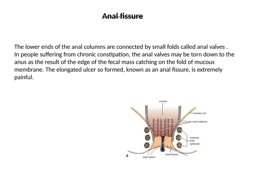
The lower ends of the anal columns are connected by small folds called anal valves .
In people suffering from chronic constipation, the anal valves may be torn down to the anus as the result of the edge of the fecal mass catching on the fold of mucous membrane. The elongated ulcer so formed, known as an anal fissure, is extremely painful. Anal fissure<br>
In people suffering from chronic constipation, the anal valves may be torn down to the anus as the result of the edge of the fecal mass catching on the fold of mucous membrane. The elongated ulcer so formed, known as an anal fissure, is extremely painful. Anal fissure<br>
45
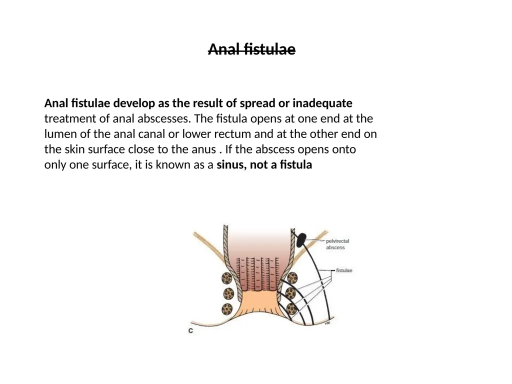
Anal fistulae develop as the result of spread or inadequate treatment of anal abscesses. The fistula opens at one end at the lumen of the anal canal or lower rectum and at the other end on the skin surface close to the anus . If the abscess opens onto only one surface, it is known as a sinus, not a fistula Anal fistulae<br>
46
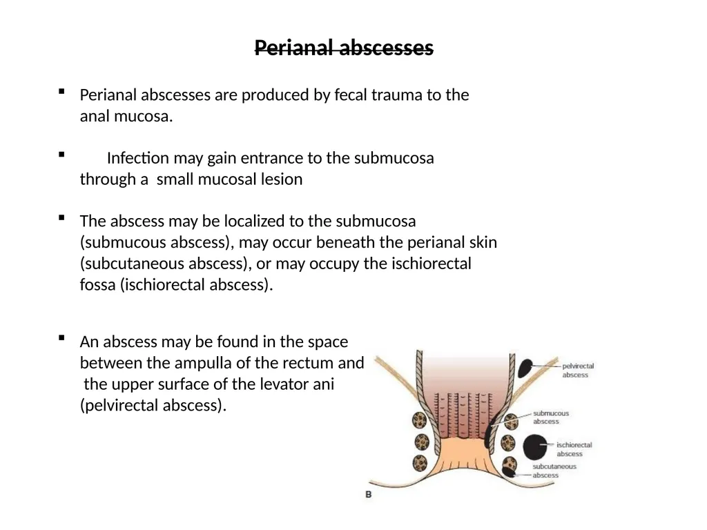
Perianal abscesses Perianal abscesses are produced by fecal trauma to the
anal mucosa.
Infection may gain entrance to the submucosa through a small mucosal lesion
The abscess may be localized to the submucosa (submucous abscess), may occur beneath the perianal skin (subcutaneous abscess), or may occupy the ischiorectal fossa (ischiorectal abscess).
An abscess may be found in the space between the ampulla of the rectum and the upper surface of the levator ani (pelvirectal abscess).<br>
anal mucosa.
Infection may gain entrance to the submucosa through a small mucosal lesion
The abscess may be localized to the submucosa (submucous abscess), may occur beneath the perianal skin (subcutaneous abscess), or may occupy the ischiorectal fossa (ischiorectal abscess).
An abscess may be found in the space between the ampulla of the rectum and the upper surface of the levator ani (pelvirectal abscess).<br>
47
Thank you<br>