
SOFT PALATE It is a movable,muscular fold

Description: SOFT PALATE It is a movable,muscular fold suspended from the posterior border of the hard palate It separates the nasopharynx from the oropharynx,the cross road between the food and air passages The soft palate has two surfaces-ANTERIOR
Related Topics
Download Presentation
"SOFT PALATE It is a movable,muscular fold" is the property of its rightful owner. Permission is granted to download and print the materials on this website for personal, non-commercial use only, and to display it on your personal computer provided you do not modify the materials and that you retain all copyright notices contained in the materials. By downloading content from our website, you accept the terms of this agreement.
Presentation Transcript
slide1. SOFT PALATE It is a movable,muscular fold suspended from the posterior border of the hard palate
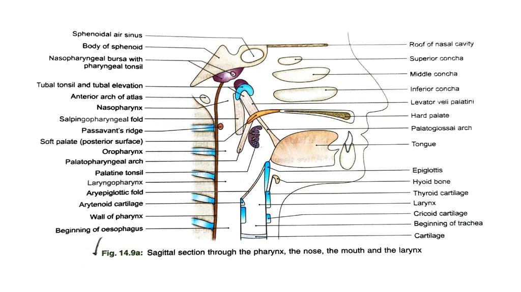
It separates the nasopharynx from the oropharynx,the cross road between the food and air passages
The soft palate has two surfaces-ANTERIOR & POSTERIOR
The soft palate has two borders- SUPERIOR & INFERIOR
The ant surface is concave and is marked by a median raphe
The post surface is convex,and is continuous superiorly with the floor of the nasal cavity<br>
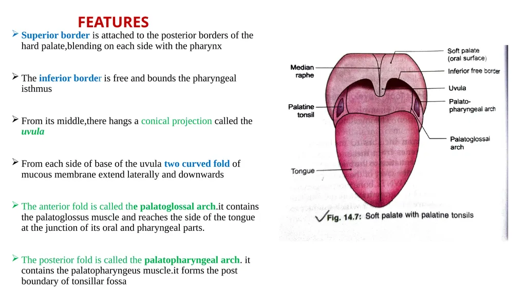
slide2. FEATURES Superior border is attached to the posterior borders of the hard palate,blending on each side with the pharynx
The inferior border is free and bounds the pharyngeal isthmus
From its middle,there hangs a conical projection called the uvula
From each side of base of the uvula two curved fold of mucous membrane extend laterally and downwards
The anterior fold is called the palatoglossal arch.it contains the palatoglossus muscle and reaches the side of the tongue at the junction of its oral and pharyngeal parts.
The posterior fold is called the palatopharyngeal arch. it contains the palatopharyngeus muscle.it forms the post boundary of tonsillar fossa<br>
slide3. STRUCTURE The soft palate is a fold of mucous membrane containing the following parts
The palatine aponeurosis which is the flattened tendon of the tensor veli palatini forms the fibrous basis of the palate. near the median plane,the aponeurosis splits to enclose the musculus uvulae
The levator veli palatini and the palatopharyngeus lie on the superior surface of the palatine aponeurosis
The palatoglossus lies on the inferior or anterior surface of the palatine aponeurosis
Neumerous mucous glands, and some taste buds are present
Note- The muscles of the soft palate are derived from 1st, 4th, and 6th branchial arches and are supplied by mandibular and vagoaccessory complex<br>
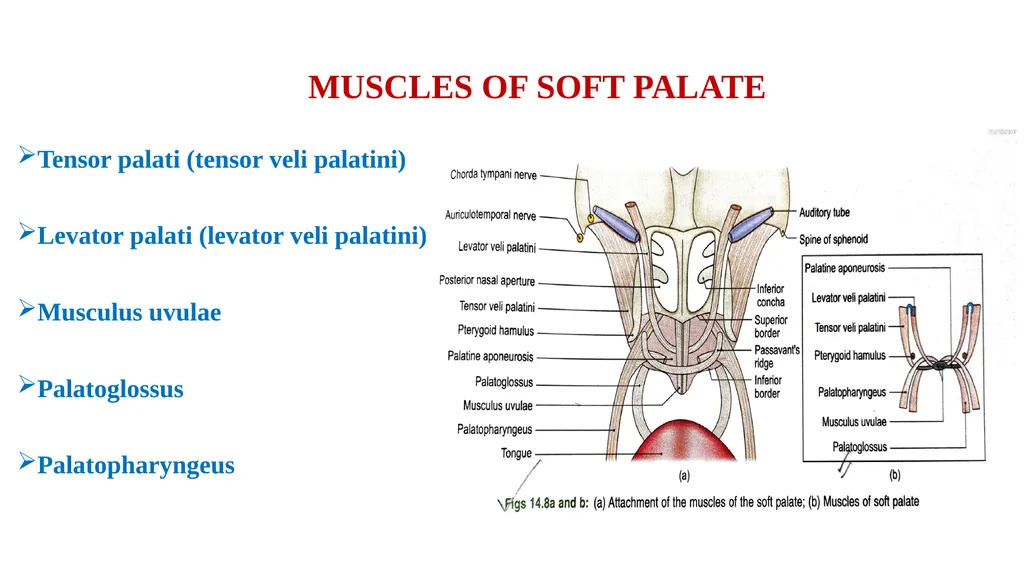
slide4. MUSCLES OF SOFT PALATE Tensor palati (tensor veli palatini)
Levator palati (levator veli palatini)
Musculus uvulae
Palatoglossus
Palatopharyngeus<br>
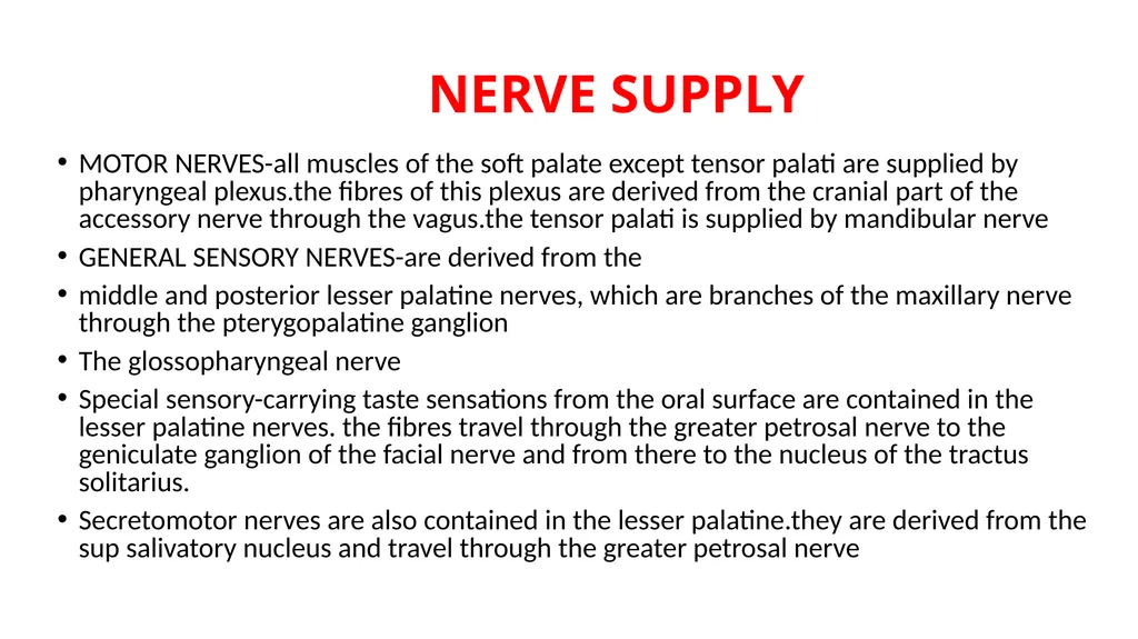
slide5. NERVE SUPPLY MOTOR NERVES-all muscles of the soft palate except tensor palati are supplied by pharyngeal plexus.the fibres of this plexus are derived from the cranial part of the accessory nerve through the vagus.the tensor palati is supplied by mandibular nerve
GENERAL SENSORY NERVES-are derived from the
middle and posterior lesser palatine nerves, which are branches of the maxillary nerve through the pterygopalatine ganglion
The glossopharyngeal nerve
Special sensory-carrying taste sensations from the oral surface are contained in the lesser palatine nerves. the fibres travel through the greater petrosal nerve to the geniculate ganglion of the facial nerve and from there to the nucleus of the tractus solitarius.
Secretomotor nerves are also contained in the lesser palatine.they are derived from the sup salivatory nucleus and travel through the greater petrosal nerve<br>
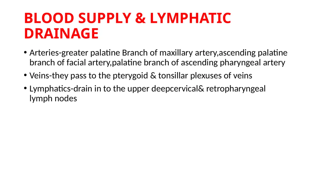
slide6. BLOOD SUPPLY & LYMPHATIC DRAINAGE Arteries-greater palatine Branch of maxillary artery,ascending palatine branch of facial artery,palatine branch of ascending pharyngeal artery
Veins-they pass to the pterygoid & tonsillar plexuses of veins
Lymphatics-drain in to the upper deepcervical& retropharyngeal lymph nodes<br>
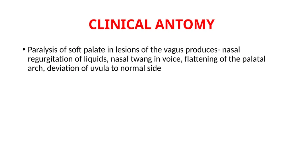
slide7. CLINICAL ANTOMY Paralysis of soft palate in lesions of the vagus produces- nasal regurgitation of liquids, nasal twang in voice, flattening of the palatal arch, deviation of uvula to normal side<br>
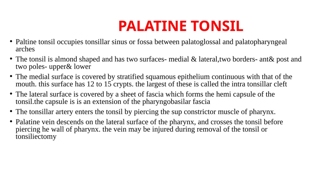
slide8. PALATINE TONSIL Paltine tonsil occupies tonsillar sinus or fossa between palatoglossal and palatopharyngeal arches
The tonsil is almond shaped and has two surfaces- medial & lateral,two borders- ant& post and two poles- upper& lower
The medial surface is covered by stratified squamous epithelium continuous with that of the mouth. this surface has 12 to 15 crypts. the largest of these is called the intra tonsillar cleft
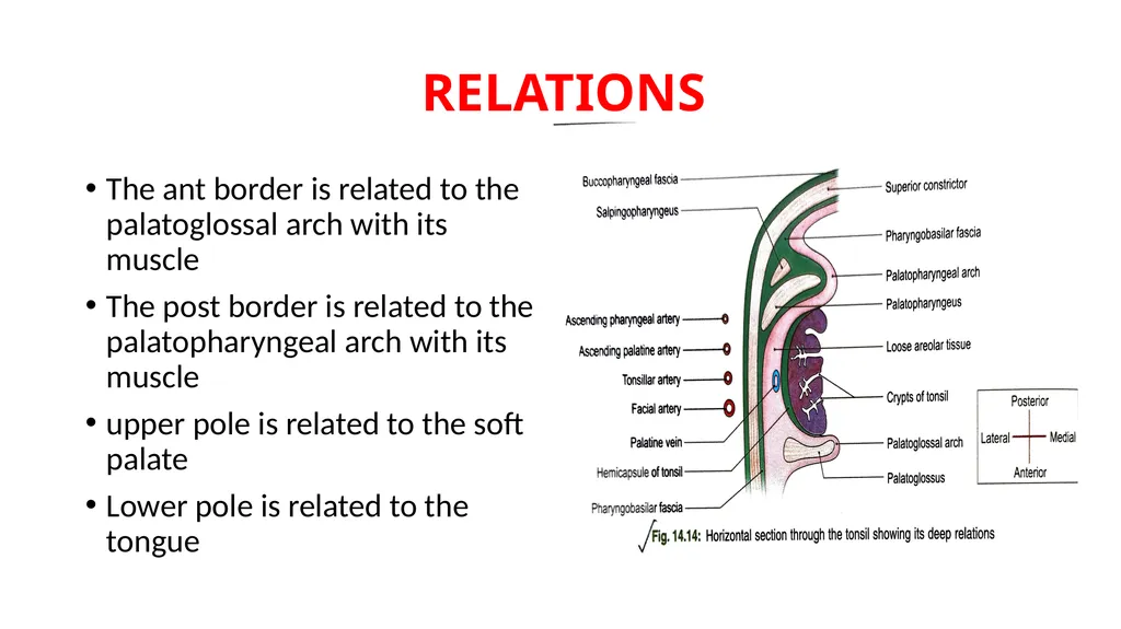
The lateral surface is covered by a sheet of fascia which forms the hemi capsule of the tonsil.the capsule is is an extension of the pharyngobasilar fascia
The tonsillar artery enters the tonsil by piercing the sup constrictor muscle of pharynx.
Palatine vein descends on the lateral surface of the pharynx, and crosses the tonsil before piercing he wall of pharynx. the vein may be injured during removal of the tonsil or tonsiliectomy<br>
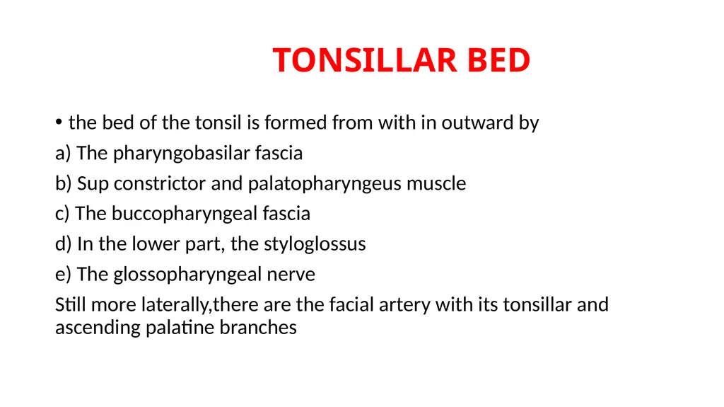
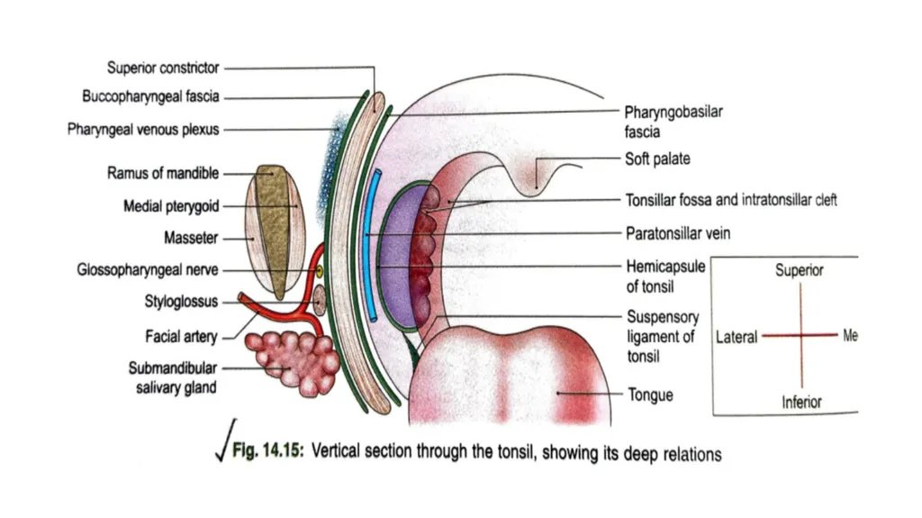
slide10. TONSILLAR BED the bed of the tonsil is formed from with in outward by
a) The pharyngobasilar fascia
b) Sup constrictor and palatopharyngeus muscle
c) The buccopharyngeal fascia
d) In the lower part, the styloglossus
e) The glossopharyngeal nerve
Still more laterally,there are the facial artery with its tonsillar and ascending palatine branches<br>
slide11. RELATIONS The ant border is related to the palatoglossal arch with its muscle
The post border is related to the palatopharyngeal arch with its muscle
upper pole is related to the soft palate
Lower pole is related to the tongue<br>
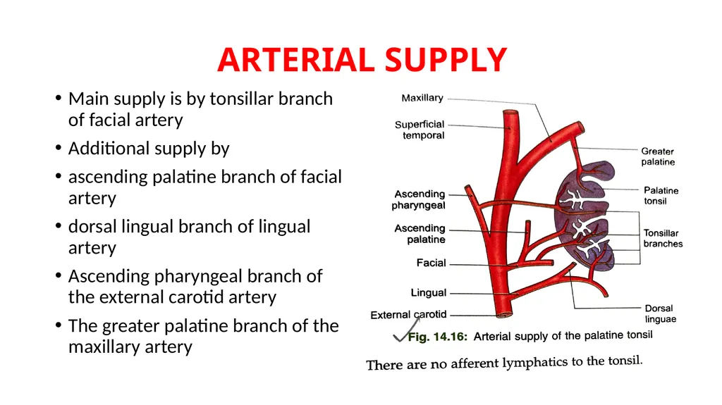
slide13. ARTERIAL SUPPLY Main supply is by tonsillar branch of facial artery
Additional supply by
ascending palatine branch of facial artery
dorsal lingual branch of lingual artery
Ascending pharyngeal branch of the external carotid artery
The greater palatine branch of the maxillary artery<br>
slide14. VENOUS DRAINAGE & LYMPHATIC DRAINAGE One or more veins leave the lower part of deep urface of the tonsil,pierce thesup constrictor,and join the palatine ,pharyngeal,or facial veins
Lymphatic drainage-Lymphatics pass to jugulodigastric node
There are no afferent lymphatics to the tonsil
NERVE SUPPLY-Glossopharyngeal and lesser palatine nerves<br>
slide15. CLINICAL ANATOMY The tonsils are large in children. they retrogress after puberty.
The tonsils are frequently sites of infection,especially in children.infection may spread to surrounding tissue forming a peritonsillar abscess.
enlarged and infected tonsils often require surgical removal.the operation is called tonsillectomy
Tonsillitis may cause referred pain in the ear as glossopharyngeal nerve supplies both these area
Suppuration in the peritonsillar area is called quinsy.<br>
It separates the nasopharynx from the oropharynx,the cross road between the food and air passages
The soft palate has two surfaces-ANTERIOR & POSTERIOR
The soft palate has two borders- SUPERIOR & INFERIOR
The ant surface is concave and is marked by a median raphe
The post surface is convex,and is continuous superiorly with the floor of the nasal cavity<br>
slide2. FEATURES Superior border is attached to the posterior borders of the hard palate,blending on each side with the pharynx
The inferior border is free and bounds the pharyngeal isthmus
From its middle,there hangs a conical projection called the uvula
From each side of base of the uvula two curved fold of mucous membrane extend laterally and downwards
The anterior fold is called the palatoglossal arch.it contains the palatoglossus muscle and reaches the side of the tongue at the junction of its oral and pharyngeal parts.
The posterior fold is called the palatopharyngeal arch. it contains the palatopharyngeus muscle.it forms the post boundary of tonsillar fossa<br>
slide3. STRUCTURE The soft palate is a fold of mucous membrane containing the following parts
The palatine aponeurosis which is the flattened tendon of the tensor veli palatini forms the fibrous basis of the palate. near the median plane,the aponeurosis splits to enclose the musculus uvulae
The levator veli palatini and the palatopharyngeus lie on the superior surface of the palatine aponeurosis
The palatoglossus lies on the inferior or anterior surface of the palatine aponeurosis
Neumerous mucous glands, and some taste buds are present
Note- The muscles of the soft palate are derived from 1st, 4th, and 6th branchial arches and are supplied by mandibular and vagoaccessory complex<br>
slide4. MUSCLES OF SOFT PALATE Tensor palati (tensor veli palatini)
Levator palati (levator veli palatini)
Musculus uvulae
Palatoglossus
Palatopharyngeus<br>
slide5. NERVE SUPPLY MOTOR NERVES-all muscles of the soft palate except tensor palati are supplied by pharyngeal plexus.the fibres of this plexus are derived from the cranial part of the accessory nerve through the vagus.the tensor palati is supplied by mandibular nerve
GENERAL SENSORY NERVES-are derived from the
middle and posterior lesser palatine nerves, which are branches of the maxillary nerve through the pterygopalatine ganglion
The glossopharyngeal nerve
Special sensory-carrying taste sensations from the oral surface are contained in the lesser palatine nerves. the fibres travel through the greater petrosal nerve to the geniculate ganglion of the facial nerve and from there to the nucleus of the tractus solitarius.
Secretomotor nerves are also contained in the lesser palatine.they are derived from the sup salivatory nucleus and travel through the greater petrosal nerve<br>
slide6. BLOOD SUPPLY & LYMPHATIC DRAINAGE Arteries-greater palatine Branch of maxillary artery,ascending palatine branch of facial artery,palatine branch of ascending pharyngeal artery
Veins-they pass to the pterygoid & tonsillar plexuses of veins
Lymphatics-drain in to the upper deepcervical& retropharyngeal lymph nodes<br>
slide7. CLINICAL ANTOMY Paralysis of soft palate in lesions of the vagus produces- nasal regurgitation of liquids, nasal twang in voice, flattening of the palatal arch, deviation of uvula to normal side<br>
slide8. PALATINE TONSIL Paltine tonsil occupies tonsillar sinus or fossa between palatoglossal and palatopharyngeal arches
The tonsil is almond shaped and has two surfaces- medial & lateral,two borders- ant& post and two poles- upper& lower
The medial surface is covered by stratified squamous epithelium continuous with that of the mouth. this surface has 12 to 15 crypts. the largest of these is called the intra tonsillar cleft
The lateral surface is covered by a sheet of fascia which forms the hemi capsule of the tonsil.the capsule is is an extension of the pharyngobasilar fascia
The tonsillar artery enters the tonsil by piercing the sup constrictor muscle of pharynx.
Palatine vein descends on the lateral surface of the pharynx, and crosses the tonsil before piercing he wall of pharynx. the vein may be injured during removal of the tonsil or tonsiliectomy<br>
slide10. TONSILLAR BED the bed of the tonsil is formed from with in outward by
a) The pharyngobasilar fascia
b) Sup constrictor and palatopharyngeus muscle
c) The buccopharyngeal fascia
d) In the lower part, the styloglossus
e) The glossopharyngeal nerve
Still more laterally,there are the facial artery with its tonsillar and ascending palatine branches<br>
slide11. RELATIONS The ant border is related to the palatoglossal arch with its muscle
The post border is related to the palatopharyngeal arch with its muscle
upper pole is related to the soft palate
Lower pole is related to the tongue<br>
slide13. ARTERIAL SUPPLY Main supply is by tonsillar branch of facial artery
Additional supply by
ascending palatine branch of facial artery
dorsal lingual branch of lingual artery
Ascending pharyngeal branch of the external carotid artery
The greater palatine branch of the maxillary artery<br>
slide14. VENOUS DRAINAGE & LYMPHATIC DRAINAGE One or more veins leave the lower part of deep urface of the tonsil,pierce thesup constrictor,and join the palatine ,pharyngeal,or facial veins
Lymphatic drainage-Lymphatics pass to jugulodigastric node
There are no afferent lymphatics to the tonsil
NERVE SUPPLY-Glossopharyngeal and lesser palatine nerves<br>
slide15. CLINICAL ANATOMY The tonsils are large in children. they retrogress after puberty.
The tonsils are frequently sites of infection,especially in children.infection may spread to surrounding tissue forming a peritonsillar abscess.
enlarged and infected tonsils often require surgical removal.the operation is called tonsillectomy
Tonsillitis may cause referred pain in the ear as glossopharyngeal nerve supplies both these area
Suppuration in the peritonsillar area is called quinsy.<br>